
Clinical Image
Austin Med Sci. 2018; 3(1): 1023.
Case Images of a Subhepatic Appendix
Sivakumar J*
1Werribee Mercy Hospital, Melbourne, Australia
*Corresponding author: Sivakumar J, Werribee Mercy Hospital, Melbourne, Australia
Received: May 22, 2018; Accepted: May 29, 2018; Published: June 06, 2018
Clinical Image
This case describes a 35 year old male who presented with right upper quadrant pain. An initial ultrasound featured a normal gallbladder, and a tubular structure also located in this area. A noncontrast computed tomography scan showed the caecum located much higher than usual, with the lower pole lying just below the level of the umbilicus. The retrocaecal appendix was, inflamed with small appendicoliths, and significant stranding. The appendix itself is about 10cm in length, with the tip of the appendix about 3cm distal to the tip of the right lobe of the liver (Figures 1-3).
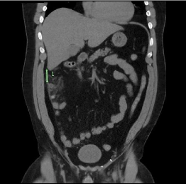
Figure 1: Coronal view of the abdomen.
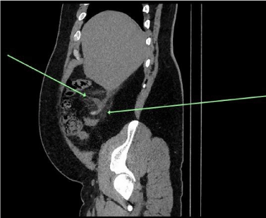
Figure 2: Saggital view of the abdomen.
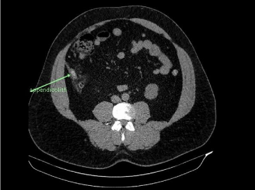
Figure 3: Axial view of the abdomen with appendicolith present.