
Abstract
Organ displacement severity across the esophageal hiatus determines how severe a hiatal hernia is. Rare Type IV paraesophageal hernias entail the protrusion of additional abdominal organs in addition to the stomach. We discuss the case of a 13-year-old boy who experienced intestinal obstruction and had a recurring hiatal hernia. A paraesophageal hiatal hernia with colonic and small bowel herniation was identified through clinical examination and imaging. Surgery was used to strengthen the anti-reflux system, reduce the hernia contents, and fix the hernia defect. The patient’s postoperative course was successful. This instance emphasizes the significance of quick paraesophageal hernia diagnosis and care to avoid consequences. An early surgical intervention can enhance results and reduce possibly fatal consequences.
Keywords: Hiatal hernia; Bowel in chest; Paraeosophageal hernia
Introduction
Hiatal Hernias (HH) are classified based on the position of the Gastroesophageal Junction (GEJ), the extent of herniation of the stomach, and the presence of other organs in the hernia sac. Type IV Paraesophageal Hernias (PEH) is uncommon, accounting for only 2%-5% of all cases [1] and are characterized by the displacement of not only the stomach but also other organs such as the colon, spleen, and small bowel into the chest cavity. Although some individuals with this condition may not experience any symptoms, it can lead to serious complications, including perforation, bleeding, and obstruction. Diagnosis of paraesophageal hernia usually involves a combination of clinical examination and imaging tests. In this article, we present a unique case of colonic obstruction in the chest.
Observation
This concerns a 13-year-old child who underwent surgery for hiatal hernia in 2013, and then underwent another surgery in 2019 due to recurrence. The patient presented with a five-day history of bowel obstruction, characterized by the cessation of bowel movements and gas, along with vomiting and abdominal pain. Upon clinical examination, the patient was in poor general condition, conscious, and hemodynamically stable, but was experiencing shortness of breath with a pulse oxygen saturation level of 92% on room air. The abdomen was distended and painful. After initial stabilization, including oxygen therapy and peripheral venous access, intravenous fluid resuscitation, the patient was taken to the radiology department where an upright abdominal X-ray was performed (Figure 1), revealing air-fluid levels in the abdomen and thorax. The patient was then admitted to the emergency operating room and underwent rapid sequence induction followed by intubation and ventilation. The previous supraumbilical midline incision was reopened, revealing marked distension of the colon and small intestine (Figure 2), along with a paraesophageal hiatal hernia and the ascent of the small bowel, transverse colon, and part of the left colon, as well as a tight adhesion. The adhesion was released, the hernia contents were reduced, and the anti-reflux system was reinforced with a Nissen fundoplication and hiatal narrowing (Figure 3). The patient was transferred to the pediatric intensive care unit postoperatively, where they remained for four days without complications, and then transferred to the surgical ward.
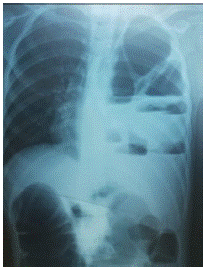
Figure 1: An upright abdominal X-ray revealing air-fluid levels in the abdomen and thorax.
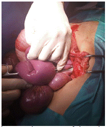
Figure 2: Image showing distension of the colon and small intestine.
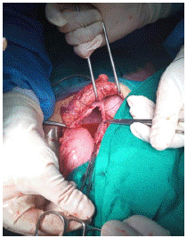
Figure 3: Surgical reduction of Hiatal Hernia and closure of Hiatal orifice.
Discussion
The position of the Gastroesophageal Junction (GEJ), the size of the herniated stomach, and the presence of other organs in the hernia sac are used to classify Hiatal Hernias (HH) into four categories. Over 95% of all HH are of type I, sometimes referred to as sliding hernias, which is the most common kind [2]. As Paraesophageal Hernias (PEH), types II to IV of HH account for 5% of total HH [3,4]. Rarely occurring Type II PEH is characterized by the herniation of the gastric fundus through the diaphragmatic hiatus in conjunction with a regularly positioned GEJ. The most typical PEH is Type III, which occurs when the GEJ and fundus are displaced over the diaphragm. Type III PEH, which involves displacement of the GEJ together with the fundus above the diaphragm, is the most common PEH. Type IV encompasses type II or III PEH, along with additional herniation of other abdominal viscera, such as colon, small intestine, spleen, and pancreas [2,4,5]. This chapter primarily focuses on type IV PEH. The presence of other organs herniated along with the stomach can affect the clinical presentation, course and treatment of the hernia. For instance, if the transverse colon or small bowels are also herniated, it could cause chronic symptoms such as constipation or intermittent partial bowel obstruction. Alternatively, it may be the primary cause of acute obstruction [4,6,7], in this case, patients may present with severe epigastric pain. If the obstruction persists, ischemia, necrosis, perforation, and septic shock may occur. The presence of severe epigastric tenderness, chest pain, and dyspnea is a reason to consider a differential diagnosis of type IV PEH. Chest X-ray often identifies a PEH as retrocardiac air-fluid level in the stomach or intrathoracic bowel. Computed tomography is a useful tool to evaluate the widening of the esophageal hiatus, size, content, and position of the PEH. PEH warrants additional attention compared to the more common sliding hiatal hernia due to potentially life-threatening complications such as strangulation, necrosis, or perforation that may develop [8]. Immediate and intensive surgical intervention is crucial to prevent deterioration.
Acute occlusion of the hiatal hernia is a medical emergency requiring immediate surgical intervention. It presents with sudden onset of chest and abdominal pain, dyspnea, and a lack of bowel movements or passing gas. The symptoms can progress rapidly, leading to respiratory distress in the patient, as in the case of our patient. Repair options for hiatal hernia includes laparoscopic approach, open laparotomy, or open thoracotomy. Traditional methods of treating acute hiatal hernia occlusion have often required open surgery, with high rates of complications, particularly in elderly patients [9]. However, there are no clear guidelines on choosing between laparoscopic or open intervention for the emergency treatment of hiatal hernia. A meta-analysis suggested that open emergency surgery is indicated when there are clear clinical signs of acute ischemia, obstruction, or perforation [10].
While laparoscopy offers benefits such as shorter hospitalization and faster recovery, but with a higher recurrence rate [10,11], the choice between this technique and the open approach remains a topic of debate [12]. Some studies suggest that laparoscopic repair should be attempted whenever possible, even in emergency situations. However, these same studies acknowledge that conversion to an open procedure may be necessary if ischemia or perforation is detected [9].
The non-elective repair of a Strangulated Hernia (SH) is linked with an elevated risk of major adverse events and mortality in contrast to elective repair. The risk of experiencing major adverse events increases by 1.7 times and mortality by 2.7 times [13]. Additionally, a different study found that patients receiving emergency surgery for SH had an 11 times greater chance of mortality than those undergoing elective repair [3].
Moreover, further research has indicated that the timing of the surgical procedure plays a significant role in morbidity and mortality. Individuals who underwent early repair had lower rates of morbidity and mortality than those who received delayed intervention [14].
Conclusion
In conclusion, paraesophageal hernias are a relatively uncommon type of hiatal hernia that can lead to serious complications such as bowel obstruction. Prompt diagnosis and management are essential to prevent morbidity and mortality. As demonstrated in this case report, a combination of clinical examination and imaging studies is necessary for the diagnosis of paraesophageal hernias. Furthermore, surgical intervention is often required to reduce the hernia contents, repair the hernia defect, and prevent recurrence. Timely and appropriate management can improve outcomes and prevent potentially life-threatening complications.
References
- Maish MS, Demeester SR. Paraesophageal hiatal hernia. In: Cameron J, editor. Current surgical therapy. 8th ed. Philadelphia: Elsevier Mosby. 2004; 38-42.
- Banimostafavi ES, Tayebi M. Large hiatal hernia with pancreatic body herniation: case-report. Ann Med Surg (Lond). 2018; 28: 20-2.
- Augustin T, Schneider E, Alaedeen D, Kroh M, Aminian A, Reznick D, et al. Emergent surgery does not independently predict 30-day mortality after paraesophageal hernia repair: results from the ACS NSQIP database. J Gastrointest Surg. 2015; 19: 2097-104.
- Biswas S, Gogna S, Patel P. Jejunal diverticular perforation causing small bowel obstruction in a Type 4 hiatal hernia: A rare case report of a nonagenarian patient and review of relevant literature. Case Rep Surg. 2017; 2017: 8412927.
- Maiti A, Saha D, Basu A. Type 4 hiatal hernia. Am J Med Sci. 2016; 351: e5.
- Hsu CT, Hsiao PJ, Chiu CC, Chan JS, Lin YF, et al. Terminal ileum gangrene secondary to a type IV paraesophageal hernia. World J Gastroenterol. 2016; 22: 2642-6.
- Tagaya N, Tachibana M, Kijima H, Kakihara Y, Hamada K, et al. Laparoscopic treatment of paraesophageal hiatal hernia with incarceration of the pancreas and jejunum. Surg Laparosc Endosc Percutan Tech. 2007; 17: 313-6.
- Kissane NA, Rattner DW. Paraesophageal and other complex diaphragmatic hernias. In: Yeo CJ, editor. Shackelford’s surgery of the alimentary tract. Amsterdam: Elsevier Medicine. 2013; 494-508.
- Shaikh I, Macklin P, Driscoll P, de Beaux A, Couper G, Paterson-Brown S. Surgical management of emergency and elective giant paraesophageal hiatus hernias. J Laparoendosc Adv Surg Tech A. 2013; 23: 100-5.
- Bawahab M, Mitchell P, Church N, Debru E. Management of acute paraesophageal hernia. Surg Endosc. 2009; 23: 255-9.
- White BC, Jeansonne LO, Morgenthal CB, Zagorski S, Davis SS, et al. Do recurrences after paraesophageal hernia repair matter?: ten-year follow-up after laparoscopic repair. Surg Endosc. 2008; 22: 1107-11.
- Patel HJ, Tan BB, Yee J, Orringer MB, Iannettoni MD. A 25-year experience with open primary transthoracic repair of paraesophageal hiatal hernia. J Thorac Cardiovasc Surg. 2004; 127: 843-9.
- Tam V, Luketich JD, Winger DG, Sarkaria IS, Levy RM, et al. Non-elective paraesophageal hernia repair portends worse outcomes in comparable patients: a propensity-adjusted analysis. J Gastrointest Surg. 2017; 21: 137-45.
- Bhayani NH, Kurian AA, Sharata AM, Reavis KM, Dunst CM, et al. Wait only to resuscitate: early surgery for acutely presenting paraesophageal hernias yields better outcomes. Surg Endosc. 2013; 27: 267-71.