Case Report
Austin J Urol. 2025; 11(1): 1085.
Encrusted Pyelitis Caused by Corynebacterium Striatum: Case Report and Literature Review
Oukouhou A¹*, Rbiaa A¹, Jamali M¹, Ameur O², Sobhi M¹, Salama W¹, Harchaoui A¹, Hamedoune L¹, Tetou M¹, Mrabti M¹, Bahri A¹, Alami M¹ and Ameur A¹
¹Department of Urology, Mohammed V Military Instruction Hospital, Rabat, Morocco
²Department of bacteriology, Mohammed V Military Instruction Hospital, Rabat, Morocco
*Corresponding author: Oukouhou Abdelhakim, Department of Urology, Mohammed V Military Instruction Hospital, Rabat, Morocco Email: mao.hakim@gmail.com
Received: February 26, 2025; Accepted: March 17, 2025; Published: March 20, 2025;
Abstract
Introduction: This case highlights the diagnostic and therapeutic challenges of encrusted pyelitis (EP), a rare, destructive complication of urinary tract infections characterized by calcifications. While Corynebacterium urealyticum is classically implicated, this report details a unique case caused by Corynebacterium striatum—a rarely documented pathogen in EP— expanding the microbiological spectrum of the disease. The case underscores the importance of early multidisciplinary management in immunocompromised hosts and enriches surgical literature by emphasizing evolving antimicrobial resistance patterns and atypical pathogens.
Case presentation: A 34-year-old woman with a history of ureteroscopy presented with febrile renal colic and septic shock. Laboratory findings included leukocytosis (15,700/μl), elevated CRP (448 mg/L), and procalcitonin (200 μg/L). CT imaging revealed encrusted pyelitis, a 42 mm renal abscess, and a 28 mm obstructive pyelic calculus.
Diagnoses, Interventions, and Outcomes: EP caused by C. striatum was confirmed via pyelic/abscess cultures. Initial management included percutaneous abscess drainage, ureteral stenting, and empirical imipenem/ aminoglycoside therapy, later adjusted to vancomycin based on sensitivities. Clinical and biochemical improvement followed, with plans for subsequent endoscopic calculus removal.
Conclusion: This case reinforces that EP should be suspected in immunocompromised patients with alkaline UTIs and calcifications on imaging, even with atypical organisms like C. striatum. Successful outcomes require a triad of targeted antibiotics (e.g., vancomycin), urinary acidification, and prompt obstruction relief. Clinicians must communicate closely with microbiologists for pathogen identification and advocate for early imaging to prevent irreversible renal damage. Future efforts should address standardized therapies and novel urease inhibitors to mitigate rising resistance.
Keywords: Encrusted pyelitis; Double J stent; Corynebacterium; Renal abscess; Case report
Introduction
Encrusted pyelitis (EP), also referred to as encrusted pyelitis, is a rare and severe complication of chronic urinary tract infections (UTIs) characterized by inflammatory mineral deposits (calcifications) in the renal pelvis and ureters. First described in the early 20th century, EP is strongly associated with Corynebacterium urealyticum, a ureasplitting, gram-positive bacillus that alkalinizes urine, promoting the precipitation of struvite. The condition primarily affects immunocompromised individuals, including transplant recipients, diabetics, and those with prolonged antibiotic use or urinary stasis. Despite its rarity, EP carries significant morbidity due to diagnostic challenges and the risk of irreversible renal damage if untreated. This article reports the case of an encrusted pyelitis caused by Corynebacterium striatum, and synthesizes current evidence on EP, focusing on its pathophysiology, clinical nuances, and evolving management strategies.
Case Presentation
This is the case of a 34-year-old female patient, with a history of right rigid ureteroscopy in 2019, admitted to the emergency department for management of febrile renal colic evolving over 3 days, complicated by rapidly progressing septic shock.
On admission, the patient was febrile, tachycardic, and tachypneic.
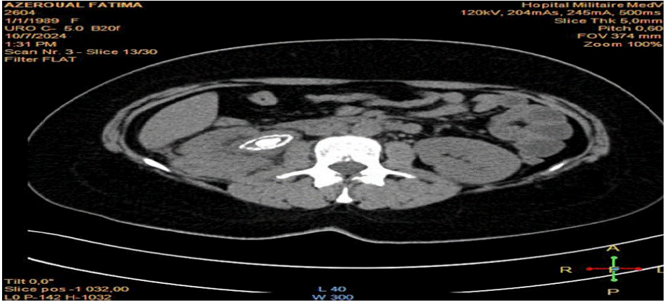
The initial blood tests showed a white blood cell count of 15,700/ μl, a CRP of 448 mg/L, and a procalcitonin level of 200g/l. The CT urogram showed encrusting pyelitis complicated by a 42mm posterior mid-renal abscess, with a 28mm obstructive pyelic calculus (Figure 1).

Figure 1: CT scan of the patient showing the calculus (red arrow), encrusted pyelitis (yellow arrow), and the renal abscess (blue arrow).
The patient underwent placement of a right double J stent with pyelic sampling and percutaneous drainage of the abscess collection, as well as empirical parenteral dual antibiotic therapy with imipenem and aminoglycoside.
Culture of the bacteriological samples from the pyelic and abscess isolates identified Corynebacterium striatum, sensitive to vancomycin.
The postoperative course was marked by clinical and biological improvement after antibiotic adjustment, at the 1 month follow up the patient was asymptomatic, the percutaneous drainage of the abscess was removed because it was no longer bringing pus and it was no longer visible in the control CT scan.
Upon discharge, the patient expressed satisfaction with the treatment provided; however, she exhibited difficulty in accepting the prolonged course of treatment that lies ahead.
The patient is scheduled later for an endoscopic procedure.
Discussion
EP is exceedingly rare, with most data derived from case reports and small series. It predominantly affects immunocompromised hosts, such as renal transplant recipients (30–40% of reported cases), diabetics, and patients with indwelling urinary catheters or prior urologic surgery. A French study noted C. urealyticum as the causative agent in 86% of EP cases, with a mortality rate of 8% in untreated patients [1].
C. urealyticum, a urease-producing organism, hydrolyzes urea into ammonia, elevating urinary pH (>7.5) and facilitating crystallization of struvite and apatite. These deposits incite chronic inflammation, leading to mucosal ulceration and calcification. Predisposing factors include urinary stasis, nephrolithiasis, and immunosuppression [2,3]. Less commonly, Proteus mirabilis or Klebsiella species may contribute to this state, in our case the responsible bacteria are Corynebacterium striatum.
Symptoms are nonspecific: flank pain (70%), hematuria (50%), and dysuria (30%). Fever is uncommon due to the organism’s low virulence. Notably, 20–30% of patients present with acute kidney injury due to obstructive uropathy [4]. A history of recurrent UTIs or recent urologic instrumentation is common.
The diagnosis relies primarily on non-contrast CT scans that reveal linear calcifications in the renal pelvis/ureters (90% sensitivity) (Meria et al., 2010), the scannographic image is evocative and should raise suspicion of the diagnosis. Urine studies show alkaline pH (>7.5). Urine cultures require lipid-enriched media and prolonged incubation (5–7 days) for C. urealyticum isolation. Because of the difficulty of this cultures, a clear communication between treating doctors and microbiologist is important.
The treatment of this entity consists in the association of Antimicrobial Therapy: Vancomycin or teicoplanin for 4–6 weeks remains first-line. Oral fluoroquinolones (e.g., ciprofloxacin) are adjuncts but resistance is rising [4].
• Urine Acidification: Ascorbic acid to maintain pH <6.5 aids dissolution of deposits.
• Surgical Intervention: Ureteral stenting or percutaneous nephrostomy relieves obstruction. Nephrectomy is reserved for nonfunctioning kidneys.
Early treatment achieves resolution in 80–90% of cases. Delayed diagnosis risks chronic kidney disease (15–20%) or sepsis. Relapse occurs in 10% due to incomplete eradication [3,5].
Conclusion
Incrusted pyelitis is a rare but serious condition requiring high clinical suspicion, particularly in immunocompromised patients with alkaline UTIs and calcifications on imaging. Multidisciplinary management—combining targeted antibiotics, urinary acidification, and timely decompression—is critical to preserving renal function. Future research should standardize treatment duration and explore novel urease inhibitors.
Consent
The patient gave informed consent for this publication after explaining the implications and the importance of the study.
Ethical Approval
According to Articles 2 and 3 of the Internal Regulations of the Biomedical Research Ethics Committee (CERB), this article is exempt from the Ethics Committee's approval.
References
- Soriano F & Tauch A. Corynebacterium urealyticum: A Comprehensive Review of an Underestimated Pathogen. Clinical Microbiology Reviews. 2018; 31: e00021-18.
- Meria P, et al. Encrusted Pyelitis of the Renal Transplant. European Urology. 2010; 58: 636–642.
- Aguade M, et al. Challenges in Diagnosing Encrusted Pyelitis: A Case Series. Nephrology Dialysis Transplantation. 2015; 30: 1356–1360.
- Sánchez-Martín FM, et al. Long-term Outcomes of Medical and Surgical Management in Encrusted Pyelitis. Journal of Urology. 2020; 204: 256–262.
- Wein AJ, et al. Campbell-Walsh-Wein Urology (12th ed.). Elsevier. 2020.