
Special Article – Surgery Case Reports
Austin J Surg. 2019; 6(23): 1225.
High-Grade Primary Angiosarcoma of the Breast with MYC Amplification: Case-Report of a 16-Year-Old Patient and Review of the Literature
Maillard C1*, Duhoux FP2, Galant C3, Libbrecht L3, Lengelé B4, Coyette M4, Fellah L5 and Berlière M1
1Department of Gynaecology, Breast Clinic of King Albert II Cancer Institute, Catholic University of Louvain-La- Neuve, Belgium
2Department of Oncology, Breast Clinic of King Albert II Cancer Institute, Catholic University of Louvain-La- Neuve, Belgium
3Department of Anatomopathology, Breast Clinic of King Albert II Cancer Institute, Catholic University of Louvain- La-Neuve, Belgium
4Department of Plastic Surgery, Breast Clinic of King Albert II Cancer Institute, Catholic University of Louvain- La-Neuve, Belgium
5Department of Radiology, Breast Clinic of King Albert II Cancer Institute, Catholic University of Louvain-La- Neuve, Belgium
*Corresponding author: Maillard C, Breast Clinic of King Albert II Cancer Institute, Catholic University of Louvain-La-Neuve/Cliniques Universitaires Saint-Luc, Avenue Hippocrate 10, 1200 Brussels, Belgium; mailid: maillardcv@gmail.com
Received: September 17, 2019; Accepted: October 25, 2019; Published: November 01, 2019
Abstract
Primary angiosarcoma of the breast is a rare but aggressive disease with a poor 5-year survival. An early and precise diagnosis has to be made in order to improve prognosis. A large vascular breast mass should therefore always be considered as an angiosarcoma until proven otherwise. Referral to tertiary care centre and multidisciplinary management are strongly recommended. There is no standard therapeutic approach but surgery remains the mainstay of angiosarcoma treatment. Patients need a close follow-up because recurrence is frequent and often precocious.
We report the rare case of a 16-year-old patient presenting a primary angiosarcoma of the breast with MYC amplification. Such cases should always be reported, as MYC amplification could in a close future be routinely used as a marker of disease aggressiveness and possibly as a therapeutic target.
Keywords: Primary angiosarcoma of the breast; MYC amplification; Radical surgery; Adjuvant therapy
Abbreviations
PASB: Primary Angiosarcoma of The Breast; ASB: Angiosarcoma of the Breast; RT: Radio Therapy; SASB: Secondary Angiosarcoma of the Breast; FNA: Fine Needle Aspiration; CNB: Core Needle Biopsy; MRI: Magnetic Resonance Imaging; LN: Lymph Node; FISH: Fluorescence In Situ Hybridization; PASH: Pseudoangiomatous Stromal Hyperplasia; FDG: Fluoro Deoxy Glucose; SUVmax: Maximum Standardized Uptake Value; OS: Overall Survival; DFS: Disease Free Survival
Introduction
We here report a rare case of Primary Angiosarcoma of the Breast (PASB) with MYC amplification in a young patient. Angiosarcoma of the Breast (ASB) are malignant vascular neoplasia of the breast parenchyma. They are a rare entity that represents less than 0.5% of all breast cancers [1, 2]. Most ASB occur after previous radiotherapy (RT) or chronic arm lymphoedema but 20% of them are primary lesions [3]. ASB metastasize hematogenously, mainly to liver, lungs, lymph nodes, bone marrow, contralateral breast followed by ovaries, kidney, omentum, adrenal gland, stomach, pancreas, peritoneum, oesophagus and skin [2,3]. Primary and secondary ASB (SASB) differ in clinical presentation and age of diagnosis. PASB usually affects young women, from 20 to 50 years old [1,4] with a median age at onset of 40 in comparison to 70 for SASB [5]. Some authors have tried to use the MYC amplification to discriminate primary from SASB [6]. MYC dysregulation is a well-known oncogenic pathway generating an oncogenic transcription factor that affects diverse cellular pathways, such as angiogenesis [7,8].
Case Description
A 16-year-old female patient was referred to our tertiary care centre for evaluation of a painless mass of the right breast, rapidly enlarging over the last six months. The right breast examination showed two bluish cutaneous nodes associated with a solid 10cm mass fixed to the pectoral muscle causing breast asymmetry (Figure 1). We noted as well a suspicious right axillary lymphadenopathy. Left breast was unremarkable. Overall, patient was allegedly considered in good health. She had no breast trauma history nor personal or family history of breast nor ovarian cancer.
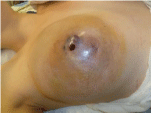
Figure 1: Ten cm enlarging right breast mass with bluish cutaneous nodules.
Breast ultrasound revealed a hypoechoic heterogeneous mass of over 7cm associated with haemorrhagic lesions. A Fine Needle Aspiration (FNA) and a Core Needle Biopsy (CNB) were performed and anatomopathological analysis confirmed the diagnosis of angiosarcoma. Routine blood analysis as well as serum CEA and CA15.3 levels were within normal limits. Magnetic Resonance Imaging (MRI) showed an enhancing voluminous (≃12 cm) mixed mass with spiculated and liquid margins in contact with the skin and partly invading the nipple. It also showed posterior adhesion to the pectoral muscle with disappearance of the greasy edges as well as one right axillary lymphadenopathy that was confirmed metastatic following FNA and cytologic analysis (Figure 2). Further metastatic work-up through PET-scan was found negative.
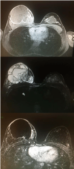
    Figure 2: MRI showing the large encompassing vascular mass in the right
breast.
In collaboration with plastic surgeons, a right modified radical mastectomy combined with axillary lymph node dissection was performed, associated with pectoral muscle resection. The large exposure of the ribcage being thereafter was covered by a pedicled Latissimus dorsi flap and a skin graft (Figure 3).
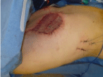
    Figure 3: Right modified radical mastectomy with muscle flap of the latissimus
dorsi and skin graft.
Microscopic examination of the mass showed an angiosarcoma of 15x11x5 cm with well and poorly differentiated areas and a large zone of haemorrhagic necrosis in its centre. There was no malignant infiltration of the pectoral muscle nor the cutaneous tissue. Resection margins were found tumour-free (Figures 4 & 5). The histopathological analysis of the 26 dissected axillary lymph nodes (LN) showed two congestive and hyperplastic LN but none invaded. The pTNM stadification was pT2b pN0 M0. We performed further immunohistochemical analyses and FISH assays showing no amplification of CIC but a MYC amplification (Figures 6 & 7). Constitutional genetic analysis did not show any mutation, deletion nor duplication in the BRCA1, BRCA2, PALB2, TP53 and CHEK2 genes.
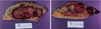
Figure 4: Macroscopic examination of the right breast angiosarcoma.
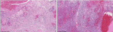
    Figure 5: Hematoxylin and eosin stain on an-ill differenciated part of the right
breast’s angiosarcoma under low magnification.
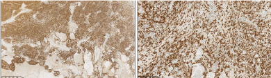
Figure 6: Positive MYC immunohistochemistry.
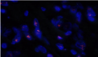
Figure 7: MYC amplification confirmed by FISH assay.
After discussion, our multidisciplinary oncologic team proposed an adjuvant treatment based comprising radiotherapy (RT) of the right chest wall (30 sessions for a total of 60 Grays) and prophylactic irradiation of the lymph nodes area (40 Grays). An alternating followup by clinical exam and CT-scan was proposed every 3 months. At 12 months of follow-up, our patient was free of any evidence of recurrence. At 16 months of follow-up, she showed bilateral lung and pleural metastases that were treated by chemotherapy (Adriamycinolaratumab) with good therapeutic response after six months but further progression of the disease within the next three months under olaratumab alone. Weekly chemotherapy with gemcitabine was then administered for three months but did not prevent further disease progression. Targeted treatment with pazopanib was then initiated and led to partial response after 3 months of therapy.
Discussion
ASB are rare entities. They represent 0.04 – 0.2% of all primary breast tumours and 8% of breast sarcomas [1-4], which corresponds to an incidence of 17 cases per million women [3]. Most ASB occur after previous RT treatment or chronic arm lymphoedema (Steward- Treves syndrom) but 20% occur de novo [5]. Compared to SASB, PASB usually occur in younger women (20-50 year-old) [1,4] with a median age at onset fluctuating between 53 years in the Kunkiel et al series [9] and 41 years in the Nascimento et al series [10] with a range from 15 to 82 years in these studies, in comparison to a median of 70 for SASB [5].
Diagnosis of PASB is not easy due to the insidious onset of the disease. We usually find a painless palpable mass with a fast growing rate associated with purplish, ecchymosis-like skin, without nipple retraction nor axillary node enlargement [3,4,9,11]. Tumour size generally exceeds 4 cm in diameter [10-13]. The differential diagnosis includes benign hemangioma, angiolipoma, fibromatosis, pseudoangiomatous stromal hyperplasia (PASH), cystosarcoma phyllodes, stromal sarcoma, metaplastic carcinoma, fibrosarcoma, liposarcoma, squamous cell carcinoma with sarcomatoid features, myoepithelioma and reactive spindle cell proliferative lesions [4,9].
Imaging of angiosarcoma is often limited. Mammograms frequently show non-specific changes because angioscarcomas do not have a proper pathological pattern and are found in young women with high density breast parenchyma, which may hinder the mass visualisation [3,14]. Sonography and doppler sonography are more helpful, showing angiosarcoma as a hypervascular heterogeneous hyperechoic mass associated with a disruption of normal breast architecture. MRI is usually more specific. ASB appear hypointense on T1-weighted and hyperintense on T2-weighted images while a rapid initial intense enhancement is followed by wash-out [2,3,5,15]. MRI is also useful in determining the tumour extent. The role of PET-Scan in ASB is poorly defined. As established by Cassou-Mounat et al. [16], it could help in differentiating PASB of SASB through a difference of maximum standardized uptake value (SUVmax). In our case for instance, in accordance with this study’s results, the lesion showed intense FDG avidity.
A FNA or a CNB must be performed in order to provide a histological diagnosis, even if Chen et al reported a false negative rate up to 37% [17]. If FNA or CNB is insufficient, surgical resection with microscopic examination can be performed. To avoid wrong or delayed diagnosis, as it is at best difficult, a large vascular breast mass should always be considered as an angiosarcoma until proven otherwise [4].
The neoplasm is of vascular origin. Donnell et al. [18] described three histologic grades (Table 1). Immunohistochemistry exams (CD31, CD 34, F VIII, FLI 1) may help to identify angiosarcoma [3,5,9]. There is no histological difference between PASB and RTinduced ASB. Even if these types of tumours are morphologically indistinguishable, several studies suggest that pathogenetic differences exist and propose to use MYC amplification to discriminate primary from secondary lesions in patients without relevant clinical history. Several studies showed MYC amplification in 100% of post-radiation breast angiosarcoma and nearly none in PASB [6,19-23]. However, in a molecular study investigating CIC, MYC, KDR, PLCG1 and FLT4 gene abnormalities in angiosarcomas of the breast and other somatic soft tissues, Huang et al found MYC amplification in 7% of primary angiosarcomas, in comparison to 91% of secondary angiosarcomas. Similarly, CIC alterations were found in 9% of primary angiosarcomas [24]. Our case is to be categorized within this small subset of PASB presenting with MYC amplification but without CIC alterations.
![]()
    Grade I
    Grade II
    Grade III


    = Low grade
      Contains anastomosing vascular channels surrounding    breast ducts and infiltrating the adipose tissue. Blood vessels lined by a    single layer of endothelial cells with hyperchromatic nuclei showing little    mitosis with no endothelial tufting.
    = Intermediate grade
      Small foci of solid proliferation of    spindle-shaped cells associated with more    mitotic figures
    = High grade
      Contains more solid and atypical cell    proliferation with frequent necrotic area and blood lakes
Table 1: Angiosarcoma’s histopathological grades according to Donnell et al. [18].
Huang et al. also correlated morphology with molecular analysis. PLCG1 and KDR rearranged lesions showed variable morphology but CIC rearranged lesions showed very high-grade morphology. No study comparing MYC amplification status to morphology in PASB were performed, although no correlation was found in publications studying skin angiosarcomas. We plead for a systematic report of PASB cases including morphology description and results of molecular tests. In our opinion, CIC and MYC analysis should be performed in priority as other tests described by Huang et al. may be only available in a few laboratories and turn out to be more expensive and time-consuming.
Referral to a tertiary care centre for multidisciplinary approach is strongly recommended as the treatment for PASB is uneasy and lacks of uniformity between centres. The gold standard treatment is complete surgical excision through modified radical mastectomy [25]. Breast conservative surgery can be a reasonable option for patient with smaller tumours. Most authors do not recommend axillary lymph nodes dissection when pre-operative work-up turns out negative as angiosarcomas present with hematogenous dissemination. Rosen et al found 0,3% of metastatic lesions in lymph nodes [26] while Bousquet et al. described less than 10% of nodal involvement [27]. However, positive clinical or imaging nodes need to be removed in order to achieve free margins. Local recurrence can appear in up to a third of the cases, suggesting that surgery alone is insufficient for local tumour control [11]. Adjuvant treatments for ASB lack of uniformity and consensus. RT may be beneficial for patients, especially with positive margins, as the impact of RT on the prognosis for patients with soft tissue sarcomas is well recognized [5]. It seems that chemotherapy including cyclophosphamide, anthracyclines or alkylating agents could be beneficial to patients with high grade or metastatic disease [1,3,5,13] even if no prospective randomized study has been performed until now, mostly because of the low PASB incidence.
Breast angiosarcomas have poor prognosis. Gervais et al. showed a median overall survival (OS) of 43 months, a 5-year OS of 40% and a 5-year disease-free-survival (DFS) of 20% for PASB, with a recurrence rate of 67% [11]. According to Rosen et al., prognosis is correlated with tumour grade, with 5-year DFS in low grade patients of 76%, 70% in intermediate grade and 15% in high grade cases [26,27]. However, Nascimento et al showed no correlation between histologic grade and patient outcomes in their study. Our patient showed relapse after 14 months with a grade III tumour. Opinions diverge regarding the tumour size as a prognosis factor [1,3,5]. Margins status is considered to change the prognosis, as incomplete surgery is connected with local relapse and worse survival [3,5]. Fraga-Guedes et al. demonstrated a significant association between MYC amplification and worse clinical outcome in SASB [28] but further studies should be performed regarding PASB cases. Ultimately, a close follow-up of these patients is necessary, as recurrence is frequent and can occur early after the first line of treatment.
Conclusion
The goal of this case report is to emphasize that cases of PASB are rare and that their diagnosis and treatment remain difficult. All cases should be reported, including morphology and results of molecular tests performed. In our opinion, molecular analysis for MYC amplification status should be considered in priority in all PASB as it may be linked with higher grade lesions and could possibly represent a therapeutic target in the future.
References
- Wang XY, Jakowski J, Tawfik OW, Thomas PA, Fan F. Angiosarcoma of the breast: a clinicopathologic analysis of cases from the last 10 years. Annals of diagnostic pathology. 2009; 13: 147-150.
- Vorburger SA, Xing Y, Hunt KK, Lakin GE, Benjamin RS, Feig BW, et al. Angiosarcoma of the breast. Cancer: Interdisciplinary International Journal of the American Cancer Society. 2015; 104: 2682-2688.
- Desbiens C, Hogue JC, Lévesque Y. Primary breast angiosarcoma: avoiding a common trap. Case reports in oncological medicine. 2011.
- Bhosale SJ, Kshirsagar AY, Patil MV, Wader JV, Nangare N, Patil PP. Primary angiosarcoma of breast: A case report. International journal of surgery case reports. 2013; 4: 362-364.
- Bordoni D, Bolletta E, Falco G, Cadenelli P, Rocco N, Tessone A, et al. Primary angiosarcoma of the breast. International journal of surgery case reports. 2016; 20: 12.
- Laé M, Lebel A, Hamel-Viard F, Asselain B, Trassard M, Sastre X, et al. Can c-MYC amplification reliably discriminate postradiation from primary angiosarcoma of the breast?. Cancer/Radiothérapie. 2015; 19: 168-174.
- Boxer LM, Dang CV. Translocations involving c-MYC and c-MYC function. Oncogene. 2001; 20: 5595.
- Dang CV. MYC on the path to cancer. Cell. 2012; 149: 22-35.
- Nascimento AF, Raut CP, Fletcher CD. Primary angiosarcoma of the breast: clinicopathologic analysis of 49 cases, suggesting that grade is not prognostic. The American journal of surgical pathology. 2008; 32: 1896-1904.
- Kunkiel M, Maczkiewicz M, Jagiello-Gruszfeld A, Nowecki Z. Primary angiosarcoma of the breast - series of 11 consecutive cases - a single-centre experience. Current Oncology. 2018; 25: 50.
- Iacoponi S, Calleja J, Hernandez G, de la Cuesta RS. Primary breast angiosarcoma in a young woman. International journal of surgery case reports. 2016; 24: 101-103.
- Gervais MK, Burtenshaw SM, Maxwell J, Dickson BC, Catton CN, Blackstein M, et al. Clinical outcomes in breast angiosarcoma patients: a rare tumor with unique challenges. Journal of surgical oncology. 2017; 116: 1056-1061.
- Sher T, Hennessy BT, Valero V, Broglio K, Woodward WA, Trent J, et al. Primary angiosarcomas of the breast. Cancer. 2007; 110: 173-178.
- Jagtap SV, Shukla D, Bonde VS, Jagtap SS. Primary angiosarcoma of the breast: an uncommon histopathological subtype. Journal of Clinical and Diagnostic Research: JCDR. 2015; 9: 05.
- O’Neill AC, D’Arcy C, McDermott E, O’Doherty A, Quinn C, McNally S. Magnetic resonance imaging appearances in primary and secondary angiosarcoma of the breast. Journal of medical imaging and radiation oncology. 2014; 58: 208-212.
- Cassou-Mounat T, Champion L, Bozec L, Laurence V, Huchet V, Luporsi M, et al. Primary and Secondary Breast Angiosarcoma: FDG PET/CT Series. Clinical nuclear medicine. 2019; 44: 33-35.
- Chen KT, Kirkegaard DD, Bocian JJ. Angiosarcoma of the breast. Cancer. 1980; 46: 368-371.
- Donnell RM, Rosen PP, Lieberman PH, Kaufman RJ, Kay S, Braun JD, et al. Angiosarcoma and other vascular tumors of the breast. The American journal of surgical pathology. 1981; 5: 629-642.
- Fernandez AP, Sun Y, Tubbs RR, Goldblum JR, Billings SD. FISH for MYC amplification and anti-MYC immunohistochemistry: useful diagnostic tools in the assessment of secondary angiosarcoma and atypical vascular proliferations. Journal of cutaneous pathology. 2012; 39: 234-242.
- Ginter PS, Mosquera JM, MacDonald TY, D’Alfonso TM, Rubin MA, Shin SJ. Diagnostic utility of MYC amplification and anti-MYC immunohistochemistry in atypical vascular lesions, primary or radiation-induced mammary angiosarcomas, and primary angiosarcomas of other sites. Human pathology. 2014; 45: 709-716.
- Mentzel T, Schildhaus HU, Palmedo G, Büttner R, Kutzner H. Postradiation cutaneous angiosarcoma after treatment of breast carcinoma is characterized by MYC amplification in contrast to atypical vascular lesions after radiotherapy and control cases: clinicopathological, immunohistochemical and molecular analysis of 66 cases. Modern Pathology. 2012; 25: 75.
- Guo T, Zhang L, Chang NE, Singer S, Maki RG, Antonescu CR. Consistent MYC and FLT4 gene amplification in radiation-induced angiosarcoma but not in other radiation-associated atypical vascular lesions. Genes, Chromosomes and Cancer. 2011; 50: 25-33.
- Hadj-Hamou NS, Laé M, Almeida A, Grange PD, Kirova Y, Sastre-Garau X, et al. A transcriptome signature of endothelial lymphatic cells coexists with the chronic oxidative stress signature in radiation-induced post-radiotherapy breast angiosarcomas. Carcinogenesis. 2012; 33: 1399-1405.
- Huang SC, Zhang L, Sung YS, Chen CL, Kao YC, Agaram NP, et al. Recurrent CIC gene abnormalities in angiosarcomas: a molecular study of 120 cases with concurrent investigation of PLCG1, KDR, MYC, and FLT4 gene alterations. The American journal of surgical pathology. 2016; 40: 645.
- Shon W, Sukov WR, Jenkins SM, Folpe AL. MYC amplification and overexpression in primary cutaneous angiosarcoma: a fluorescence in-situ hybridization and immunohistochemical study. Modern pathology. 2014; 27: 509.
- Rosen PP, Kimmel M, Ernsberger D. Mammary angiosarcoma: the prognostic significance of tumor differentiation. Cancer. 1988; 62: 2145-2151.
- Bousquet G, Confavreux C, Magné N, de Lara CT, Poortmans P, Senkus E, et al. Outcome and prognostic factors in breast sarcoma: a multicenter study from the rare cancer network. Radiotherapy and Oncology. 2007; 85: 355-361.
- Fraga-Guedes C, André S, Mastropasqua MG, Botteri E, Toesca A, Rocha RM, et al. Angiosarcoma and atypical vascular lesions of the breast: diagnostic and prognostic role of MYC gene amplification and protein expression. Breast cancer research and treatment. 2015; 151: 131-140.