
Special Article – Surgery Case Reports
Austin J Surg. 2019; 6(19): 1213.
Subcutaneous Metastasis of Gastric Cancer at Left Axilla: A Case Report and Literature Review
He FJ, Zhang P, Chen Y and Zhuang W*
Department of Gastrointestinal Surgery, West China Hospital, Sichuan University, China
*Corresponding author: Wen Zhuang, Department of Gastrointestinal Surgery, West China Hospital, Sichuan University. No. 37 Guoxue Lane, Wuhou District, Chengdu City, Sichuan Province, 610041, China
Received: August 06, 2019; Accepted: September 16, 2019; Published: September 23, 2019
Abstract
Background: Gastric cancer is the third most lethal malignant tumor in the world. Metastasis has always been a major cause of poor prognosis. Epidemiological evidence shows that the most common metastasis sites of gastric carcinoma are respectively for liver (48%), peritoneum (32%), lung (15%) and bone (12%) respectively and subcutaneous metastasis is about 0.8%. Effective treatment will be taken early, if we have more insight in its transfer mechanism and characteristics. We report a rare case of axillary subcutaneous metastasis of advanced gastric cancer. The best surgical window was missed, as a result of lacking attention and rapid growing of the mass.
Case Summary: A 69-year-old man who had underwent radical gastrectomy receipted eight cycles oral chemotherapy for gastric cancer was suffering from rapidly growing left axillary mass. Just only 3 mouths, the mass has growing to 6.9cm*4.4cm*5.7cm. Color Doppler ultrasonography and PET/CT prompted the possibility of metastasis of malignancy. Fine needle aspiration biopsy suggested that cancer cells were found. Immunohistochemically examination supported the metastasis of gastric cancer. Considering the risk of resection, the patient abandoned surgical treatment eventually.
Conclusion: The case alerts us that for unidentified subcutaneous masses in the patients with a history of malignant tumors, we should pay enough attention to especially.
Keywords: Gastric cancer; Metastasis; Subcutaneous; Cancer therapy; Case report
Introduction
Gastric cancer is prevalent worldwide, with an average of about 990,000 new cases per year [1-3], with the highest incidence in Eastern Asia [4,5]. According to the Eindhoven Cancer Registry statistics, between 1995 and 2012, about 40% of gastric cancer patients had one metastasis at least [6]. Relevant data indicated that the incidence of subcutaneous metastasis of gastric cancer is about 0.8% [2]. Today, there is no data referring to the left axillary metastasis of gastric carcinoma. Here we report a case of a patient with stage III gastric carcinoma who underwent curative intent resection (R0), D2 lymph node dissection and receipted eight cycles chemotherapy postoperative. However, left axillary subcutaneous metastasis occurred to him in the fifth year postoperative. We report the case to promote the exploration and monitoring of unusual rare metastasis sites of advanced gastric cancer, and provide clinical evidence for the diagnosis and treatment of metastasis of gastric cancer.
Case Presentation
A 69-year-old man with a history of radical gastrectomy and eight cycles oral chemotherapy for gastric cancer 5 years ago readmitted to West China Hospital of Sichuan University for having suffering from left axillary asymptomatic mass (Figure 1). 5 years ago, the old man admitted to our outpatient department and was diagnosed with adenocarcinoma in the gastric fundus (cT3N2M0), and then accepted total gastrectomy, D2 lymph node dissection. Postoperative paraffin pathological sections confirmed the diagnosis of moderately differentiated adenocarcinoma with the stage of the patient was pT3N1M0, Borrmann III and the Lauren classification was intestinal type (Figure 2). Two in 30 regional lymph nodes were examined as metastasized lymph nodes. Then, he had finished eight cycles oral chemotherapy of Xeloda. At the fifth year postoperative, he was re-admitted for detection of the asymptomatic mass in his left axilla. Only through three months, the mass grown to about 6.9cm*4.4cm*5.7cm complicated with local inflammation. Color Doppler ultrasonography and PET/CT all prompted the possibility of metastasis (Figure 3). Fine needle aspiration biopsy of left axillary mass found cancer cells. Immunohistochemical examination showed that CDX-2 (+), PCK (+), CK20 (+), CK7 (-), TTF (-) and supported the metastasis of gastric cancer (Figures 4 and 5). Due to the adhesion between large subcutaneous mass of the left axilla and surrounding tissues and severe local inflammation, skin grafting might be required after operation. We asked the plastic surgery experts to assist in the operation of tumor resectionbut considering the risk of resection, the patient abandoned surgical treatment eventually. Since the day the patient give up surgery and discharged, the follow up interrupted.
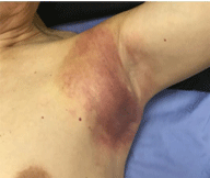
    Figure 1: Subcutaneous metastasis arising from primary gastric cancer at
the left axilla.
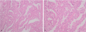
    Figure 2: Histopathological findings (hematoxylin and eosin staining). The
pathological reports after operation revealed moderately differentiated
adenocarcinoma: Tubular adenocarcinoma and papillary adenocarcinoma.
Lauren typing: intestinal type. (H&E, a*200, b*400).
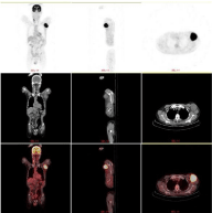
Figure 3: PET/CT suggests the lefty axillary subcutaneous metastases.
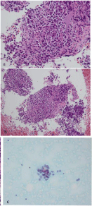
    Figure 4: The axillary mass biopsy, a gastric cancer cells were detected by
cells smear (H&E, *400), b&c gastric cancer cells were detected by paraffin
section (H&E, b*200, c*400).
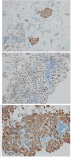
    Figure 5: Immunohistochemical findings. Magnification of all photographs
is 400*. a-c the tumor was positive for CK20 (d), PCK (e) and CDX-2 (f)
respectively.
Discussion
Epidemiological investigation on metastasis of gastric cancer is rare. At present, TNM staging is often used to staging malignant tumors, and cancer registries often only use “M0” and “M1” to indicate whether distant metastasis occurs. Therefore, there is a lack of information about specific distant metastasis sites [7].
The five-year cumulative risks of relapse (restricted to patients who underwent R0 resections and excluding in-hospital deaths) for patients with pathological stage T3 tumors, respectively, were 83 percent for D1 dissection and 72 percent for D2 dissection [8]. As a kind of so called “localized tumor”, gastric cancer, once with loco regional metastasis of, may be the most important signal of negative prognosis [9-11].
Metastases is mostly driven by the acquisition of genetic and/or epigenetic alterations within tumor cells and the formation of tumor microenvironment [12]. Metastasis of malignant tumors can occur as early as the stage of primary tumorigenesis [13]. If metastasis of cancer cells occurs before clinical detection, surgical resection may not prevent recurrence, invasion and metastasis of tumors. In our case, the patient was followed up regularly. Dynamic imaging monitoring showed no signs of recurrence. However, in the fifth year, he was diagnosed as distant subcutaneous metastasis. Because mild clinical manifestations and rare metastasis sites, the patient and physicians haven’t taken seriously, causing to missed the best surgical window.
Repeated changes in molecular phenotypes [14], immune evasion [15], drug resistance and so on [15], which make well cure well-confined tumor difficult. Through long-term observation by clinicians, some types of malignant tumors only metastasize to limited target organs [16]. Cutaneous or subcutaneous may be not a better growth microenvironment than liver or peritoneum. However, several cases of subcutaneous metastasis of gastric cancer, such as scalp metastasismandibular metastasis and so like, have been reported in succession [17,18]. We have sought here to report the rare case, which has provided clinical evidence for studying the special metastasis sites of gastric cancer.
Conclusion
We should pay enough attention to the local mass in patients with a history of gastric cancer. We continue to foresee more sensitive, specific and inexpensive monitoring methods will contribute to it, such as nanoparticles [19], liquid biopsy and etc. [20].
References
- Riihimäki M, Hemminki A, Sundquist K, Sundquist J, Hemminki K. Metastatic spread in patients with gastric cancer. Oncotarget. 2016; 7: 52307-3252316.
- Hu SC, Chen GS, Wu CS, Chai CY, Chen WT, Lan C. Rates of cutaneous metastases from different internal malignancies: Experience from a Taiwanese medical center. J Am Acad Dera told. 2009; 60: 379-387.
- Ferlay J, Shin HR, Bray F, Forman D, Mathers C, Parkin DM. Estimates of worldwide burden of cancer in 2008: GLOBOCAN 2008. Int J Cancer. 2010; 127: 2893-2917.
- Jemal A, Bray F, Center MM, Ferlay J, Ward E, Forman D. Global cancer statistics. CA Cancer J Clin. 2011; 61: 69-90.
- Forman D, Burley V. Gastric cancer: global pattern of the disease and an overview of environmental risk factors. Best Pract Res Clin Gastroenterol. 2006; 20: 633-449.
- Thomassen I, van Gestel YR, van Ramshorst B, Luyer MD, Bosscha K, Nienhuijs SW, et al. Peritoneal carcinomatosis of gastric origin: a populationbased study on incidence, survival and risk factors. Int J Cancer. 2014; 134: 622-628.
- Riihimäki M, Hemminki A, Sundquist K, Sundquist J, Hemminki K. Metastatic spread in patients with gastric cancer. Oncotarget. 2016; 7: 52307-52316.
- Bonenkamp JJ, Hermans J, Sasako M, van de Velde CJ, Welvaart K, Songun I, et al. Extended lymph-node dissection for gastric cancer. Dutch Gastric Cancer Group. N Engl J Med. 1999; 340: 908-914.
- Imano M, Yasuda A, Itoh T, Satou T, Peng YF, Kato H, et al. Phase II study of single intraperitoneal chemotherapy followed by systemic chemotherapy for gastric cancer with peritoneal metastasis. J Gastrointest. Surg. 2012; 16: 2190-2196.
- Ishigami H, Kitayama J, Kaisaki S, Hidemura A, Kato M, Otani K, et al. Phase II study of weekly intravenous and intraperitoneal paclitaxel combined with S-1 for advanced gastric cancer with peritoneal metastasis. Ann Oncol. 2010; 21: 67-70.
- Ishigami H, Kitayama J, Kaisaki S, Yamaguchi H, Yamashita H, Emoto S, et al. Phase I Study of Biweekly Intravenous Paclitaxel plus Intraperitoneal Cisplatin and Paclitaxel for Gastric Cancer with Peritoneal Metastasis. Oncology. 2010; 79: 269-272.
- Valastyan S, Weinberg RA. Tumor metastasis: molecular insights and evolving paradigms. Cell. 2011; 147: 275-292.
- Turajlic S, Swanton C. Metastasis as an evolutionary process. Science. 2016; 352: 169-175.
- Nakamura J, Okuyama K, Sato H, Yoda Y, Kai K, Noshiro H. Repeated changes of the molecular subtype in gastric metastasis from breast cancer: A case report. Mol Clin ONicole. 2016; 4: 695-698.
- Hanahan D, Weinberg RA. Hallmarks of cancer: the next generation. Cell. 2011; 144: 646-674.
- Filler IJ. The pathogenesis of cancer metastasis: the ‘seed and soil’ hypothesis revisited. Nat Rev Cancer. 2003 3: 453-458.
- Kawai S, Nishida T, Hayashi Y, Ezaki H, Yamada T, Shinzaki S, et al. World J Gastroenterol. 2013; 19: 1485-1498.
- Kirchberger MC. Unusual presentation of a cutaneous metastasis in the face arising from gastric cancer: a case report. SAGE Open Med Case Rep. 2018; 6: 2050313X18795080.
- Li R, Liu B. The application of nanoparticles in diagnosis and theranostics of gastric cancer. Gao J Cancer Lett. 2017; 386: 123-130.
- Pantel K, Alix-Panabieres C. Liquid biopsy and minimal residual disease - latest advances and implications for cure. Nat Rev Clin Oncol. 2019; 16: 409- 424.