Special Article - Surgical Case Reports
Austin J Surg. 2015;2(2):1053.
Surprise of an Acute Obstructive Nephrogram
Wani Bhushan N1* and Banode Pankaj2
Department of Surgery, MVPS DVP Medical College, India
*Corresponding author: Wani Bhushan N, Department of Surgery, MVPS’s Dr Vasantrao Pawar Medical College, Nashik, Department of Radiodiagnosis, JNMC Sawangi, India
Received: November 24, 2014; Accepted: March 18, 2015; Published: March 19, 2015
Abstract
The urographic nephrogram has an important role in the diagnosis of alterations to the renal parenchyma. The appearance of a unilateral, striated nephrogram becoming progressively denser during IV urography has been attributed to several processes. Here we are reporting a surprise finding of an acute obstructive nephrogram in a case on left ureteric colic and discussed a physicis behind the same.
Keywords: IV urography; Obstructive nephrogram; Acute obstructive uropathy; Striated nephrogram; Ureteric colic
Introduction
An obstructive nephrogram involving the entire kidney, owing to an acute ureteral obstruction is a well known phenomenon [1] and has been classically described. The urographic nephrogram plays an important role in the diagnosis of alterations to the renal parenchyma. The appearance of a unilateral, striated nephrogram becoming progressively denser during IV urography has been attributed to several processes; thus history, clinical examinations are of paramount.
Case
A 46 year old man presented with severe, colicky pain in left iliac fossa associated with nausea and vomiting, radiating to groin; with history of burning micturition of 3 months. He was admitted with provisional diagnosis of left ureteric calculus and worked up in that direction. All haematological investigations were with normal limits. Urine microscopy, it was found to be infected with presence of plenty pus cells and calcium oxalate crystals. Ultrasonography revealed left lower ureteric calculus without evidence of hydronephrosis and hydroureter.
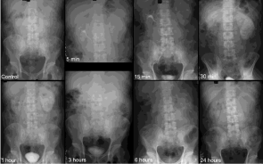
Intravenous Urography (IVU) was done; control film showed a soft tissue shadow in left renal area. A 5 minutes film after contrast injection, demonstrated diffuse nephrographic effects, with delay in opacification of the collecting system on the left with normal nephrogram on right side. In 15 minutes film, it was a dense nephrogram on the left side and a normal appearance of the collecting system on the right side. In 30 minutes film the density of left nephrogram had increased till one hour film and then gradually decreased. A 6, 12 and 24 hours films showed gradual disappearance of contrast on left side with radio-opaque impacted stone at lower ureter (Figure 1).

    Figure 1:  Intravenous urography (IVU), A Control film with soft tissue shadow
in left renal area; 5 minutes film after contrast material injection, demonstrates
diffuse nephrographic effects, with delay in opacification of the collecting
system on the left; 15 minutes film, shows the development of a dense
nephrogram on the left and a normal appearance of the collecting system on
the right; 30 minutes and 1 hour film, density of nephrogram has increased;
6, 12 and 24 hours film shows disappearance of contrast on left side with
radioopaque impacted stone at lower ureter.
Discussion
The IVU is traditional way of evaluating acute flank pain, sensitivity of which varies from 52-87% [1,2] as against newer modality like CT scan plan and contrast are more specific. The detection of calculi as a cause of obstructive uropathy depends on multiple factors; size and location of calculus, as well as the degree of obstruction. As a result, false-negative studies are not uncommon in the urographic evaluation of calculi. The urographic nephrogram plays an important role in the diagnosis of alterations to the renal parenchyma [3]. The presence of a standing column of contrast does not always indicate the site of obstruction, as it may be seen in normal individuals. The appearance of a unilateral, striated nephrogram becoming progressively denser during IV urography has been attributed to several processes; such as acute ureteral obstruction, acute pyelonephritis, infantile polycystic kidney, medullary sponge kidney, acute renal vein thrombosis, radiation nephritis, infection, and trauma [1-5].
Following the administration of the contrast agent, on the affected side, there occurs a characteristic sequence or ‘March of events’; after a delay, a nephrogram appeared which became denser; where it lasted for some hours, fine radial striations were seen. After a transitional phase showing both nephrogram and pyelogram, a dilated, dilute pyelogram (less dense than the normal side) appeared and eventually faded. If a stone was present, it did not move throughout the process. The pain was shown to be of ‘plateau’ type, increasing over half an hour to 3 hours, remaining very severe for half to 3, 6 or even 12 or 24 hours with only minor changes in its level, and then abating over halfan- hour to 2 hours. Comparing this with the radiological sequence, the nephrogram was present during the period of severe pain and changed to a pyelogram as the pain abated. Where a powerful analgesic was given, the pain was relieved but the radiological sequence appeared to continue independently [1,5-6].
As per Poiseuille’s law of laminar flow [6], under obstructive conditions, the pressure in the renal pelvis will rise until it is sufficient to oppose the pressure derived from glomerular filtration. Following the administration of the contrast agent, in an obstructed kidney, delayed accumulation of contrast is seen, resulting in a nephrogram that is initially of lower density. The pressure drop between glomerulus and renal pelvis will then be small and will principally occur at the ascending limb of the loop of Henle which will act as a pressure transmitter with minimal flow. This segment will thus serve to separate the filtration-resorption equilibrium taking place in the proximal convoluted tubule [6,7] (permitting accumulation of contrast medium there because it is not taken up by the tubular cells [8], so giving rise to the nephrogram) from the situation in the distal convoluted tubules and collecting tubules. Progressive concentration of contrast within the renal cortex and medulla occurs, producing the classically described “obstructive nephrogram” that may persist for some duration. Striations may occasionally be seen in the parenchyma, representing contrast material within dilated tubules. Dilatation proximal to the obstructing process may be seen as hydronephrosis and hydroureter, which may be minimal in the acute situation. A standing column of contrast may be observed proximal to the obstruction [9].
Then as all the renal tubules are semipermeable, and the distal convoluted tubule is particularly likely to lose water as part of the normal concentration process; although this process is grossly impaired in obstructive conditions, it is likely to be continuing in some tubules to some extent. Clearly this will permit retrograde flow up some tubules, while forward flow proceeds in others. Once such a system of complex flow is established, it can readily act as a gradual pressure reducing mechanism. Thus the nephrogram can gradually fade even in continuing obstruction and pyelosinus extravasations of contrast and mild clubbing of the calyces also may be seen [5,9].
References
- Dunnick NR, Sandler CM, Newhouse JH, Amis ES. Textbook of uroradiology, 3rd edn. Philadelphia, Pa: Lippincott Williams & Wilkins. 2001.
- Davies P, Price H. The urographic signs of acute on chronic obstruction of the kidney. Clin Radiol. 1980; 31: 205-213.
- Newhouse JH, Pfister RC. The nephrogram. Radiol Clin North Am. 1979; 17: 213-226.
- Hattery RR, Williamson B Jr, Hartman GW, LeRoy AJ, Witten DM. Intravenous urographic technique. Radiology. 1988; 167: 593-599.
- Dyer RB, Munitz HA, Bechtold R, Choplin RH. The abnormal nephrogram. Radiographics. 1986; 6: 1039-1063.
- Bretland P. Acute ureteric obstruction. London: Butterworth. 1972.
- Gottschalk Cw, Mylle M. Micropuncture study of pressures in proximal tubules and peritubular capillaries of the rat kidney and their relation to ureteral and renal venous pressures. Am J Physiol. 1956; 185: 430-439.
- Saxton HM. Review article: urography. Br J Radiol. 1969; 42: 321-346.
- Pollack HM. Some limitations and pitfalls of excretory urography. J Urol. 1976; 116: 537-543.