Clinical Image
Austin J Surg. 2025; 12(2): 1352.
Imaging Features of Annular Pancreas: A Rare Congenital Anomaly
Soukayna J*, Nadia B, Khadija ElJ and Leila J
Radiology Department, National Institute of Oncology, Rabat, Morocco
*Corresponding author: Jabour Soukayna, Radiology Department, National Institute of Oncology, Rabat, Morocco Email: dr.jaboursoukayna@gmail.com
Received: July 14, 2025 Accepted: July 24, 2025 Published: July 25, 2025
Clinical Image
The annular pancreas is a rare, silent congenital anomaly that leads to late onset of symptoms and difficult diagnosis. It is due to a failure of the ventral and dorsal pancreatic buds to fuse between weeks 4 and 8 of embryonic development, with the ventral bud encircling the first part of the duodenum.
The annular pancreas may be complete, with an annular duct completely surrounding the second part of the duodenum, or incomplete: the ring does not completely surround the duodenum, giving a “crocodile jaw” appearance.
Approximately 25-33% of adult cases of annular pancreas are asymptomatic, an incidental finding on imaging. However, it can cause pancreatitis, duodenal obstruction and, rarely, biliary obstruction. The most common symptoms in adults are abdominal pain, postprandial fullness, vomiting and gastrointestinal bleeding due to peptic ulcer disease.
Pancreatic tissue completely or incompletely surrounds the second part of the duodenum. Duodenal stricture and dilatation of the proximal duodenum may also be observed. In adults, it is frequently associated with pancreatitis.
MRI In addition to the characteristics of the annular pancreas, the anatomy of the pancreatic duct can be thoroughly assessed by magnetic resonance imaging. The annular duct generally connects to the main pancreatic duct or to the accessory duct (Santorini duct).
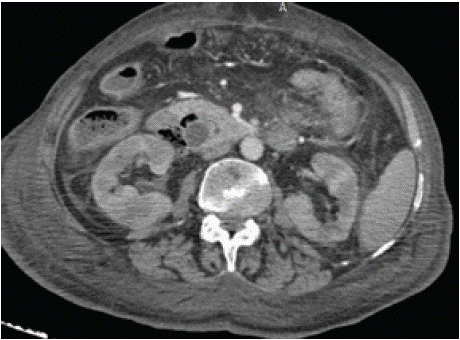
In symptomatic cases of annular pancreas, surgical management is the treatment of choice. Bypasses such as duodeno-jejunostomy or gastro-jejunostomy are the mainstay of surgical management 3 (Figure 1).

    Figure 1: Axial section of an injected CT scan showing the pancreatic
tissue (yellow arrow) with the wirsung duct (arrowhead) surrounding the
duodenum (red dot).
References
- Sandrasegaran K, Patel A, Fogel EL, et al. Annular pancreas in adults. AJR Am J Roentgenol. 2009; 193: 455-460.
- Zyromski NJ, Sandoval JA, Pitt HA, et al. Annular pancreas: dramatic differences between children and adults. J. Am. Coll. Surg. 2008; 206: 1019- 1025.
- Rondelli F, Bugiantella W, Stella P, et al. Symptomatic Annular Pancreas in Adult: Report of Two Different Presentations and Treatments and Review of the Literature. Int J Surg Case Rep. 2016; 20: 21-24.