
Case Series
Austin J Surg. 2025; 12(2): 1351.
Mouse in the Axilla – A Case Series of Fibroadenoma in the Axillary Ectopic Breast Tissue – A Rare Entity
Gnanaprakash S* and Manish Dewangan
Department of General Surgery, J.L.N. Hospital and Research Centre, Bhilai, Durg, Chhattisgarh, India
*Corresponding author: Dr. Gnanaprakash S, Department of General Surgery, J.L.N. Hospital and Research Centre, Bhilai, Durg, Chhattisgarh, India Tel: +91 9360967810; Email: gna1212prakash@gmail.com
Received: July 08, 2025 Accepted: July 24, 2025 Published: July 25, 2025
Abstract
Background: Axillary polymastia is a common variant of Ectopic Breast Tissue (EBT) with reported incidence of 2% to 6% in women. EBT can undergo the same physiological and pathological processes as the normally located breast. Though fibroadenoma commonly occurs in the breast, it is rare in the ectopic breast tissue. We report a series of four cases of fibroadenoma in axillary breast tissue over past 10 years in our hospital including a giant axillary fibroadenoma which we claim to be the largest till date in the English literature.
Keywords: Ectopic Breast Tissue; Fibroadenoma; Axilla; Supernumerary Breast
Introduction
Ectopic breast tissue occurs in 2 to 6% of the general population and is classically distributed along the embryonic milk line which extends from the axilla to the pubic region [1]. The axilla is the most common site, accounting for approximately 60 to 70% of accessory breast tissue [2]. The diagnosis of ectopic breast tissue is important because this tissue is also subjected to the same alterations and diseases, whether benign or malignant, which affect naturally positioned breasts [3]. Although carcinoma of the axillary accessory breast is rare, accounting for 0.3% of all breast cancers, the most frequent condition in the accessory breast is breast cancer followed by mastopathy and fibroadenoma [4]. Since publications describing this anomaly are rare in English literature, we decided to present a case series of fibroadenoma arising from axillary ectopic breast tissue over the past 10 years in our hospital. To the best of our knowledge, this is the first case series reporting four cases of fibroadenoma of the axillary breast tissue including one which fits into Giant axillary fibroadenoma.
Case Presentation
A Series of Four Cases Complied in Form of Table (Table 1):
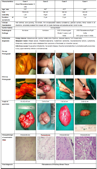
Table 1: A Series of Four Cases Complied.
Discussion
During the early weeks of embryonic development, the mammary milk lines, which represent two ectodermal thickenings along the sides of the embryo, extend from the axillary region to the groin. In normal development, most of the embryologic mammary ridges resolve, except for two segments in the pectoral region, which later become the breast, failure to involute may lead to ectopic breast tissue with (polythelia) or without (polymastia) a nipple/areolar complex [3]. Polythelia, in particular, has been associated with cardiac and urinary anomalies, which can be explained in part by the parallel embryologic development of mammary structures and the genitourinary system [3,4]. In our patients, we have excluded urological or cardiac abnormalities. Two hypotheses have been proposed on the embryogenesis of the supernumerary breast. One attributes the anomaly to the failure of regression and displacement of the milk line, while the other believes it develops from the modified apocrine sweat glands [5].
As compared to pectoral breast tissue, EBT demonstrates the same hormonal effects and is at risk of developing breast diseases. The possibilities of malignant masses include breast cancer, rhabdomyosarcoma, epidermoid carcinoma, neuroendocrine tumors, lymphoma, and melanoma. Axillary lymph node metastases from carcinoma of the breast (or of another cancer) are the single most important abnormality to exclude, as they are an important prognostic factor in breast cancer. Sometimes it could cause psychological disturbances in adolescence and it may give pain and discomfort
In 1915, Kajava published a classification system for supernumerary breast tissue that remains in use today. Class I consists of a complete breast with nipple, areola, and glandular tissue. Class II consists of nipple and glandular tissue but no areola. Class III consists of areola and glandular tissue but no nipple. Class IV consists of glandular tissue only. Class V consists of nipple and areola but no glandular tissue (pseudomamma). Class VI consists of a nipple only (polythelia). Class VII consists of an areola only (polythelia areolaris). Class VIII consists of a patch of hair only (polythelia pilosa) [7]. Our cases belongs to class IV. However, presently fibroadenoma is considered as hyperplasia of a single lobule of the breast, classified under ANDI (aberrations of normal development and involution).
Ectopic breast tissue without the presence of a nipple can cause delay in the diagnosis of malignancies. So earlier and more frequent metastasis, poorer prognosis are seen in ectopic breast tissue malignancies. Vigilant self examination of accessory breasts similar to that of normal breasts is encouraged, for earliest detection of lesions. Complete clinical examination of swellings of accessory breasts supplemented with FNAC is basic tool for management. Follow up after excision biopsy of Fibroadenoma in axilla is essential to detect at the earliest any further pathological changes.
Fibroadenoma is a very common benign condition in young females. However, they are very rare in axillary EBT with only few sporadic case reports in English literature. After extensively reviewing the English literature, found about 53 case reports since 2000. The mean size was 3.5cm and largest was 11cm. Our case is unique due to its size (15cm). We have not found any case in the literature having such a large sized fibroadenoma.
Conclusion
Fibroadenoma of accessory breast in axilla is a very rare occurrence with only 53 cases reported in the English literature (after 2000). The knowledge of this entity is essential for the clinicians because it poses diagnostic dilemma. High index of suspicion is mandatory for early diagnosis and management. Fine-needle aspiration cytology plays pivotal role for diagnosis.
References
- Coras, Brigitte Md, Landthaler, Michael Md, Hofstaedter, Ferdinand Md, Meisel, Claus Md, Hohenleutner, Ulrich Md. Fibroadenoma of the Axilla. Dermatologic Surgery. 2005; 31: p 1152-1154.
- Burdick AE, Thomas KA, Welsh E, Powell J, Elgart GW. Axillary polymastia. J Am Acad Dermatol. 2003; 49: 1154-1156.
- Mukhopadhyay M, Saha AK & Sarkar A. Fibroadenoma of the ectopic breast of the axilla. Indian J Surg. 2010; 72: 143-145.
- Sawa M, Kawai N, Sato M, Takeuchi T, Tamaki T, Oura S. Fibroadenoma of the axillary accessory breast: diagnostic value of dynamic magnetic resonance imaging. Jpn J Radiol. 2010; 28: 613-617.
- Aughsteen AA, Almasad JK, Al-Muhtaseb MH. Fibroadenoma of the supernumerary breast of the axilla. Saudi Med J. 2000; 21: 587-589.
- de Andrade JM, Marana HR, SarmentoFilho JM, Murta EF, Velludo MA, Bighetti S. Differential diagnosis of axillary masses. Tumori. 1996; 82: 596- 599.
- Surcel E, Koljonen V. Kajava Classification: The Person and the Research. Aesthetic Plast Surg. 2023; 47: 2177-2178.