
Case Report
Austin J Surg. 2025; 12(1): 1347.
Distorted Ileal Bladder: A Case Report
Tang F1,3#, Liao Y1#, Zhen Z2,3, Liu M1,3, Aers M2,3, Liu T2,3 and Peng J2,3*
¹Department of Urology, Jingzhou Central Hospital, Jingzhou Hospital Affiliated to Yangtze University, Jingzhou, China
²Department of Urology, Zhongnan Hospital of Wuhan University, Wuhan, China
³Medical Science Research Center, Zhongnan Hospital of Wuhan University, Wuhan, China
#These Authors Contributed Equally to this Work*Corresponding author: Jianping Peng, Department of Urology, Zhongnan Hospital of Wuhan University, Wuhan, China, Postal Code: 430071 Address: No.169, Donghu Road, Wuchang District, Wuhan, Hubei, China Tel: 17371290209; Fax: 027-67812892; Email: pengjianping@whu.edu.cn
Received: February 11, 2025; Accepted: March 04, 2025; Published: March 07, 2025;
Abstract
Background: The bricker bladder is a common treatment for muscleinvasive bladder cancer. This paper presents a rare case of a patient with bladder malignancy who underwent further total cystectomy and ileal replacement of the bladder due to tumor recurrence, after which the patient developed bladder stenosis and even bowel obstruction.
Methods: Our therapeutic approach was evaluated and reflected upon by reviewing the patient’s consecutive course of treatment.
Results: At the patient’s first medical visit, we found a torsion of the bricker bladder, which led to stenosis of the reconstructed bladder and upper urinary tract obstruction. After discussion, we performed open surgery to relieve the obstruction. However, we found that there were serious adhesions between the bricker bladder and the pubic bone, which made the surgery difficult to perform. After consultation with the gastroenterologists and in light of the patient’s condition, we finally underwent conservative treatment (ureteral stent placement). Six months later, the patient developed intestinal obstruction.
Conclusion: Multiple complications may occur after bricker bladder replacement, Regular follow-up is important for early detection and treatment. Our experience is that if a patient develops an ileocecal substitution obstruction, it is important to perform surgery on the patient’s condition as early as possible.
Keywords: Urethral reconstruction; Bladder cancer; Ileal bladder; Hydronephrosis
Introduction
Bladder cancer is one of the most common carcinomas of the urinary tract. It is classified into muscle-infiltrating bladder cancer and non-muscle-invasive bladder cancer according to whether the tumor invades the muscles. The gold standard treatment for muscleinvasive cancer is radical cystectomy coupled with urinary diversion [1]. Ileal substitution of the bladder is one of the more common methods. However, the postoperative period is associated with different complications. We report a case of ileal bladder stenosis with late intestinal obstruction. We hope that this case, together with our treatment measures, will provide ideas for managing related cases in the future.
Case Presentation
A 71-year-old female patient was admitted for bilateral hydronephrosis after radical cystectomy and ileal substitution of the bladder for bladder cancer. In June 2020, the patient was found to have bladder occupancy due to a physical examination, followed by a cystectomy of the bladder tumor. The postoperative pathology showed high-grade invasive uroepithelial carcinoma with tumor invasion into the submucosal layer and no intrinsic muscular infiltration. Immunohistochemical tests showed CK7 (+), CK20 (+), CD44 (+), Ki-67 (LI:60%), GATA-3 (+), P40 (+). On review cystoscopy in October 2020, bladder neoplasm was detected, so further cyst neoplasm electrosurgery was performed, and postoperative pathology results were returned: (Bladder neoplasm) High-grade invasive uroepithelial carcinoma with tumor invasion into the mucosal lamina propria. Further laparoscopic radical cystectomy and ileocystostomy were performed. Postoperative pathological return (bladder tumor resection + radical specimen) (Bladder) invasive uroepithelial carcinoma (high grade), two tumors, sizes 1.1*1.0*1.0cm and 1.3*0.6*0.4cm, respectively; both tumors invaded the subepithelial fibrous connective tissues; the closest distance to the superficial muscular layer was < 0.1cm; and there was no intravesical cancer embolus or nerve invasion. No tumor was seen in any of the ureteral stumps (left and right) sent for examination. No tumor was seen in the tissue of the cut edge of the ureter. No tumor metastasis was seen in 10 lymph nodes of the left pelvis and 6 lymph nodes of the right pelvis. The clinicopathological staging is pT1N0MX. After surgery, the patient had undergone bilateral nephrectomies two months earlier due to severe hydronephrosis and postoperative ureteroscopy revealed ileostomy stenosis. The patient had no other specific medical history. The patient had been admitted several times in the past for hydronephrosis with ureteral stenting. Physical examination: positive percussion pain in both kidney areas, bilateral renal fistulae were patent, and urine was slightly turbid and normal in color. The stoma was erythematous and drained a little urine. The patient was admitted to the hospital, and CT was perfected. The right renal pelvis and ureter were significantly dilated and watery and merged with the ileum at the distal end (Figure 1). Meanwhile, further X-ray urography showed: localized ileal segment stenosis (Figure 2). Abnormal kidney function results: BUN 13.30 mmol/L, Cr 185.90 umol/L, white blood cells elevated (15.88×10⁹g/L), and a positive urine culture. Calcitoninogen (PCT) was 17.89 ng/mL.
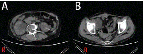
    Figure 1: CT images. (A) Hydronephrosis with marked dilatation of the right
renal pelvis and right ureter; (B) Ileal substitution of the bladder with the
right pubic.
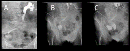
    Figure 2: X-ray urethrography images. (A) The left nephrostomy tube
was injected with an appropriate amount of contrast, and the left renal
pelvis-ureter was clearly shown throughout with visible dilatation, and the
contrast did not flow out of the right lower abdominal ileostomy; (B) The
right lower abdominal ileostomy was injected with contrast, and some of
the distal ilea were visible, and there was a narrow intestinal canal between
the proximal ileum that was visualized; (C) The ileum was separated from
the bladder, and the two sides were connected through the middle narrow
intestinal canal.
Considering the patient's history and examination findings, we speculate that the postoperative adhesions caused the bricker bladder to twist, thus triggering the stenosis. During surgery, the ileocecal substitute bladder entered the right iliac fossa downward, and the tube wall was tightly adherent to the pubic bone with a hard texture, which was difficult to loosen. Given this situation, we decided to temporarily place an ileal substitution bladder catheter for drainage and adjusted the nephrostomy tube. The patient had the nephrostomy tube removed in January postoperatively, and the follow-up patient had been replacing the ureteral stent but the overall result was still poor. After 6 months, the patient developed intestinal obstruction, and the examination revealed a soft tissue density shadow in the abdominal wall of the right lower abdomen. Dilated effusion of the adjacent intestinal canal and intestinal obstruction were considered, along with obvious dilated effusion of the right renal pelvis ureter (Figure 3). After a hospital-wide consultation, the patient was recommended for surgery to treat the intestinal obstruction. The surgery went well, and the patient's intestinal obstruction symptoms were resolved after the operation. Unfortunately, the patient was lost to follow-up.
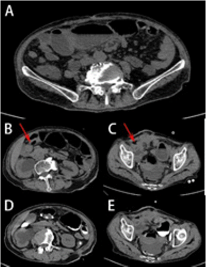
    Figure 3: Second CT images. (A) Air-fluid plane visible on emergency CT;
(B) and (C) Admission CT showed: right lower abdomen abdominal wall
soft tissue density shadow (Marked with a red arrow), incomplete intestinal
obstruction, right renal pelvis ureter significantly dilated fluid accumulation;
(D) and (E) CT-enhanced images.
Discussion
Bladder cancer (BC) is common worldwide and poses a significant public health challenge, with over 430,000 men and women diagnosed worldwide every year [1]. In general, bladder cancer is categorized into muscle-invasive bladder cancer (MIBC) and non-muscle-invasive bladder cancer (NMIBC) [2]. For MIBC, the guidelines recommend a combination of treatment modalities such as transurethral resection of bladder tumor (TURBT), and drug instillation in conjunction with the grading of the tumor [3-4]. Radical cystectomy (RC) is the standard surgical treatment for muscle-invasive bladder cancer and nonmuscle- invasive bladder cancer with a high risk of progression [5-7] After removing the tumor-bearing bladder, urinary diversion (UD) is mandatory. From a functional standpoint, the UD can be divided into continent reservoirs (Continent Pouches - Kock, Miami, and Indiana, and Orthotopic Neobladder) and non-continent reservoirs (Ileal Conduit - IC, and Cutaneous Ureterostomy - CU). These complex procedures involving bowel manipulation and multiple anastomoses might be responsible for most of the complications [8,9]. In contrast, ileal substitution cystoplasty is prone to complications such as stones, stoma stricture, and ureteral anastomotic stricture [10].
We report a complex case of ileal replacement bladder stenosis; few similar cases have been reported. The patient eventually underwent total bladder resection and ileal replacement bladder surgery due to the recurrence of bladder malignancy. Postoperatively, the patient began to experience symptoms of upper urinary tract fluid retention, which did not improve with multiple visits. Eventually, he developed a twisted stenosis of the ileocecal substitute bladder. Due to the difficulty of the surgery, we were not able to completely resolve the patient's twisted ileal replacement bladder for the first time and had to use a catheter to relieve the fluid accumulation. Eventually, the patient was seen again in the gastrointestinal surgery department for recurrent bowel obstruction. Ileal replacement of the bladder was proposed by Bricker in 1950, has been continuously popularized, and has been the most respected surgical procedure in the treatment of bladder cancer [7]. Ileal replacement of the bladder does not require a permanent ureteral stent, but complications associated with bowel-collar manipulation have been reported to be common [11]. Common complications of the bricker bladder are urinary leakage, bowel leakage, bowel obstruction, Parastomal hernia, and anastomotic stenosis. Ileostomy stenosis [12- 14]. The risk of stenosis is relatively high for these complications, and one report examined a significantly higher rate of stenosis in patients with Bricker (separation and regurgitation) anastomosis than with Wallace (conjoined and regurgitation) anastomosis [15].
However, twisting stenosis of the ileal bladder itself has rarely been reported. How to prevent complications after ileal replacement bladder surgery is a major postoperative concern. In this case, we clarified the cause of fluid retention through a series of examinations at the time of the patient's consultation, but unfortunately, we were unable to clarify the cause of the deformation of the replacement bladder through follow-up. We speculate that tumor recurrence may have caused adhesion of the replacement bladder to the surrounding tissues, or simple postoperative scar adhesion may have caused deformation of the replacement bladder by pulling.
Conclusion
Our case provides a lesson for future work. Early intervention is quite important for such complications, and it is worth reflecting on how to prevent such postoperative secondary obstruction, with more delicate surgical maneuvers and closer postoperative review playing an important role in preventing the appearance of such associated symptoms.
Ethics Statement
The publication of this case report was approved by the patient's written informed consent form and the ethics project review of Wuhan University School of Medicine (Grant No. JC2019-025).
References
- Bray F, Ferlay J, Soerjomataram I, Siegel RL, Torre LA, Jemal A. Global cancer statistics 2018: GLOBOCAN estimates of incidence and mortality worldwide for 36 cancers in 185 countries. CA Cancer J Clin. 2018; 68: 394- 424.
- Magers MJ, Lopez-Beltran A, Montironi R, Williamson SR, Kaimakliotis HZ, Cheng L. Staging of bladder cancer. Histopathology. 2019; 74: 112-134.
- Jin YH, Zeng XT, Liu TZ, Bai ZM, Dou ZL, Ding DG, et al. Treatment and surveillance for non-muscle-invasive bladder cancer: a clinical practice guideline (2021 edition). Mil Med Res. 2022; 9: 44.
- Slovacek H, Zhuo J, Taylor JM. Approaches to Non-Muscle-Invasive Bladder Cancer. Curr Oncol Rep. 2021; 23: 105.
- Neuzillet Y, Pradère B, Xylinas E, Allory Y, Audenet F, Loriot Y, et al. French AFU Cancer Committee Guidelines - Update 2022-2024: Muscle-Invasive Bladder Cancer (MIBC). Prog Urol. 2022; 32: 1141-1163.
- Lenis AT, Lec PM, Chamie K, Mshs MD. Bladder Cancer: A Review. JAMA. 2020; 324: 1980-1991.
- Witjes JA, Bruins HM, Cathomas R, Compérat EM, Cowan NC, Gakis G, et al. European Association of Urology Guidelines on Muscle-invasive and Metastatic Bladder Cancer: Summary of the 2020 Guidelines. European Urology. 2021; 79: 82-104.
- Stein R, Hohenfellner M, Pahernik S, Roth S, Thüroff JW, Rübben H. Urinary diversion--approaches and consequences.Dtsch Arztebl Int. 2012; 109: 617- 622.
- Korkes F, Fernandes E, Gushiken FA, Glina FPA, Baccaglini W, Timóteo F, et al. Bricker ileal conduit vs. Cutaneous ureterostomy after radical cystectomy for bladder cancer: a systematic review. Int Braz J Urol. 2022; 48: 18-30.
- Adnan S, Abu Bakar M, Khalil MAI, Fiaz S, Ahmad Cheema Z, Ali A, et al. Outcomes of Uretero-ileal Anastomosis in Bladder Cancer Cystectomies: Bricker vs. Wallace 1. Cureus. 2022; 14: e22782.
- Li Z, Liu Z, Yao K, Qin Z, Han H, Li Y, et al. An improved ileal conduit surgery for bladder cancer with fewer complications. Cancer Commun (Lond). 2019; 39: 19.
- Krafft U, Mahmoud O, Hess J, Radtke JP, Panic A, Püllen L, et al. Comparative analysis of Bricker versus Wallace ureteroenteric anastomosis and identification of predictors for postoperative ureteroenteric stricture. Langenbecks Arch Surg. 2022; 407: 1233-1240.
- Wang Z, Qin Y, Yang C, Wei X, Qian J, Tu S, et al. Conservative treatment of urinary fistula: Case report. Exp Ther Med. 2022; 24: 491.
- Li Z, Zhang Z, Ma H, Yao K, Qin Z, Han H, et al. Extraperitonealization of ileal conduit reduces parastomal hernia after cystectomy and ileal conduit diversion. Urol Oncol. 2022; 40: 162.e17-162.e23.
- Christoph F, Herrmann F, Werthemann P, Janik T, Schostak M, Klopf C, et al. Ureteroenteric strictures: a single center experience comparing Bricker versus Wallace ureteroileal anastomosis in patients after urinary diversion for bladder cancer. BMC urology. 2019; 19: 100.