
Case Report
Austin J Surg. 2024; 11(4): 1336.
Lipoma of the Psoas Muscle: A Case Report
Zouhry Ibrahim1,2*; Bahi Achraf1; Ghannou Nizar1; Halim El Mustapha2
¹Department of Visceral Surgery, Morocco
²HMIMV, Rabat, Morocco
*Corresponding author: Zouhry Ibrahim, Department of Visceral Surgery, HMIMV, Rabat, Morocco. Email: ibrahim.zouhry@um5r.ac.ma
Received: September 20, 2024 Accepted: October 09, 2024 Published: October 16, 2024
Summary
The lipoma is the most common soft tissue tumor, characterized by a benign proliferation of adipocytes. The intramuscular lipoma is an uncommon variant that accounts for less than 1% of all lipomas and can occur in any anatomical site, typically affecting individuals between the ages of 40 and 70 years. We report the case of a 60-year-old female patient presenting with a lipoma of the psoas muscle. We discuss the epidemiological aspects of psoas lipomas as well as the specificities related to diagnosis and therapeutic management.
Keywords: Lipoma; Psoas muscle
Introduction
Lipoma is the most common soft tissue tumor, characterized by a benign proliferation of adipocytes. Intramuscular lipoma is a rare variant, accounting for less than 1% of all lipomas, and can occur at any anatomical site, predominantly in individuals aged 40 to 70 years [1]. This report discusses the epidemiological aspects of psoas lipomas and highlights the diagnostic and therapeutic management peculiarities through a specific case.
Clinical Case
The patient is a 60-year-old woman with no significant medical history who presented with chronic abdominal pain associated with constipation, all while maintaining her general health status.Clinical examination revealed slight abdominal distension. Abdominopelvic Computed Tomography (CT) suggested a well-defined rounded mass with thin walls, consistent with a lipoma. This was supplemented by Magnetic Resonance Imaging (MRI), which demonstrated a large mass measuring 160 x 125 x 120 mm, well-defined and originating from the left iliopsoas muscle, displacing the digestive structures without signs of invasion, indicative of a psoas muscle lipoma. Blood tests showed normal complete blood count.The patient underwent a median laparotomy, which revealed a large, well-circumscribed fatty mass adherent to the psoas muscle. The mass was completely resected. Postoperative recovery was uncomplicated, and the patient was discharged on day six. Histopathological examination confirmed the diagnosis of lipoma.
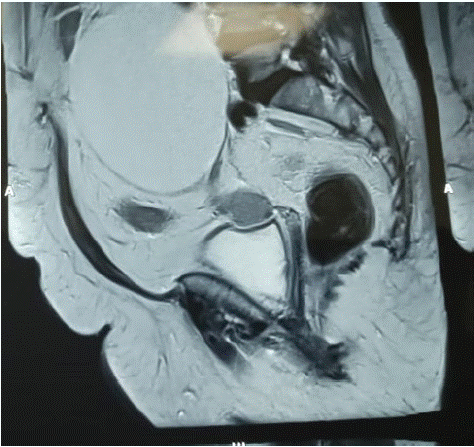
Figure 1: MRI showing a large mass related to an iliopsoas lipoma.
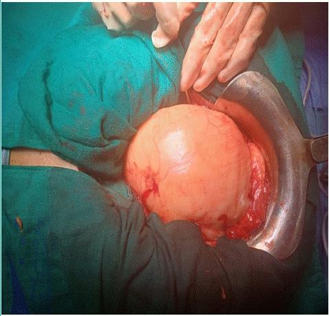
Figure 2: Intraoperative image of the lipoma.
Discussion
Lipoma is the most common soft tissue tumor, characterized as a benign proliferation of adipocytes. Intramuscular lipomas are an uncommon variant with poorly defined epidemiological characteristics. This rare condition accounts for 1.8% of all primary adipose tissue tumors and less than 1% of lipomas, predominantly affecting adults aged 40 to 70 years without a sex predominance [2]. Lipomas of the psoas muscle are particularly rare. Various risk factors have been suggested, including obesity, hypercholesterolemia, hormonal treatments, and genetic predisposition; however, our patient did not exhibit any of these factors.Most intramuscular lipomas remain asymptomatic, with pain being a rare and late symptom resulting from the compression of adjacent organs or peripheral nerves. Clinical examination may reveal a palpable soft mass, as seen in our patient. Computed Tomography (CT) and Magnetic Resonance Imaging (MRI) are the most commonly used complementary examinations for diagnosis, but they do not exclude the diagnosis of well-differentiated liposarcoma. On CT, lipomas exhibit fat density, while on MRI, the mass has the same signal as fat. The presence of a non-fatty component and/or enhancement after gadolinium injection should raise suspicion for liposarcoma. Intramuscular lipomas often lack a capsule and present with irregular margins. Rarely, areas of infarction or necrosis within the lipoma may suggest a liposarcoma. MRI also allows for assessment of relationships with neighboring organs [3]. Image-guided biopsies can be useful for diagnosis but are not always conclusive.The preferred treatment is complete surgical resection with the capsule. The recurrence rate is very low, often due to incomplete resections of lipomas. The risk of sarcomatous degeneration is considered negligible [4].
Conclusion
The psoas lipoma is a rare condition that primarily affects adults between the ages of 40 and 70, with no gender predominance. The diagnosis is based on imaging techniques and biopsy. The treatment involves complete surgical resection.
References
- Rastrelli M, Hoxhaj I, Di Maggio A, Sbaraglia M, Chiusole B, Tropea S, et al. Uncommon presentation of a giant psoas muscle lipoma: a case report and brief literature review. Ann Med Surg. 2023; 85: 488-91.
- Intramuscular lipoma: a review of the literature. Orthop Rev. 2024; 6.
- Nishida J, Morita T, Ogose A, Okada K, Kakizaki H, Tajino T, et al. Imaging characteristics of deep-seated lipomatous tumors: intramuscular lipoma, intermuscular lipoma, and lipoma-like liposarcoma. J Orthop Sci. 2007; 12: 533-41.
- Weniger M, D’Haese JG, Kunz W, Pratschke S, Guba M, Werner J, et al. En-bloc resection of a giant retroperitoneal lipoma: a case report and review of the literature. BMC Res Notes. 2024; 8: 75.