
1Department of Surgery, University of Texas Medical School at Houston, USA
2Michael E. DeBakey Institute for Comparative Cardiovascular Science and Biomedical Devices, Texas A&M University, USA
*Corresponding author: Shinil K Shah, Department of Surgery, University of Texas Medical School at Houston, USA
Received: November 15, 2014; Accepted: November 24, 2014; Published: November 25, 2014
Citation: Collom ML, Bajwa KS, Mehta SS, Walker PA and Shah SK. Endometriosis of the Appendix Masquerading as Appendicitis in a 34-Week Pregnant Woman: Case Report and Review of the Literature. Austin J Surg. 2014;1(8): 1040. ISSN: 2381-9030.
Endometriosis is most commonly found in women of childbearing age with the most common sites of disease being the ovaries, uterine ligaments, recto and vesicovaginal areas, pelvic peritoneum, cervix, labia and vagina. Endometriosis of the appendix is an extremely rare finding. In this report we describe a patient in her 3rd trimester of pregnancy that presented with acute abdominal pain with clinical examination, laboratory investigation and imaging consistent with acute appendicitis. She underwent laparoscopic appendectomy without complication but with the surprising pathological diagnosis of endometriosis of the appendix. We discuss the management and relevant literature of the diagnosis of endometriosis of the appendix in pregnancy.
Keywords: Endometriosis; Appendix; Appendicitis; Pregnancy
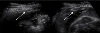
The patient presented is a 32-year-old woman who was 34 and 3/7th weeks pregnant. She presented to our facility with approximately 24 hours of right-sided abdominal pain associated with nausea and fevers. She did not report any similar previous episodes of pain. Her past medical and surgical history was unremarkable. She was febrile to 101F and her clinical examination demonstrated right sided abdominal tenderness with some localized guarding and rebound tenderness. Her laboratory studies were remarkable for a white blood cell count of 12.1 k/uL (reference range 4.5 – 11 k/uL) with left shift (86.1% neutrophils). The rest of her laboratory workup was essentially normal. An abdominal ultrasound demonstrated a dilated blind ending tubular structure in the right lower abdomen with tenderness noted over the structure consistent with a diagnosis of acute appendicitis (Figure 1).
Given suspicion for acute appendicitis, she was taken to the operating room for planned laparoscopic appendectomy. On examination, the appendix was noted to be inflamed with fibrinous exudates and no evidence of perforation, consistent with a clinical diagnosis of acute appendicitis. She was discharged on post-operative day 1 after an uneventful hospital stay.
Quite surprisingly, surgical pathology revealed no acute or chronic inflammation involving the appendiceal mucosa. Extensive decidual changes forming transmural nodules involving all layers of the appendiceal wall and periappendiceal soft tissues were noted with glandular structures noted in the area of decidual changes. Immunostaining for keratin (pancytokeratin), PAX8, vimentin, and calretinin on the epithelial structures surrounded by decidual tissue demonstrated PAX8 positivity for nuclei and keratin positivity for the cytoplasm. Stains for calretinin and vimentin were negative. Overall, the findings suggested transmural endometriosis of the appendix with extensive decidual changes.
At follow-up, the patient was doing well. She had no complications related to the pregnancy and delivered approximately 1 month after her operation.
Endometriosis of the appendix was first described by von Rokitansky in 1860 [1]. Although it is associated with pelvic pain, its clinical presentation can be varied. The development of endometriosis is still not entirely understood. There are several different etiologies that have been hypothesized, but the most widely accepted mechanism is retrograde menstruation from the fallopian tube into the peritoneum [2]. The most common location of an endometrial implant is the female pelvis, but it can occasionally be found in the umbilicus, kidney, lung, skin and diaphragm [3]. The gastrointestinal tract is affected very rarely and the percentage varies according to location. The recto-sigmoid (72%) and recto-vaginal septum (13%) are most commonly affected followed by the small intestine (7%), cecum (3.6%) and rarely the appendix (3%) [4]. In the gastrointestinal tract, endometriosis deposits usually only involve the serosa and subserosa. It is rare for the deposit to involve the muscular is propria, but if it does it typically causes symptoms and fibrosis [5]. The only gold standard for the diagnosis of endometriosis of the appendix is laparoscopy or laparotomy.
There are approximately twenty reported cases of endometriosis presenting as acute appendicitis during pregnancy [6-21]. The reported case is one of the more advanced gestational age presentations reported; there are three other cases in more advanced stages of pregnancy, (35 weeks gestation (2 cases), and at term (1 case) [14,15,20]. Additionally, most of the reported cases have occurred in women under the age of thirty, with two cases reported in individuals older than our reported patient (33 and 35 years old) [6,7].
Interestingly, in our patient, there was no pathologic evidence of acute appendicitis in the specimen. This is surprisingly a common finding. A large review of pathology specimens in patients treated for appendicitis demonstrated 14 cases of endometriosis in 4670 patients. In these specific cases, only 36% of specimens (5 cases) had evidence of acute inflammation in addition to findings of endometriosis [22].
It is interesting that our patient presented with signs and symptoms of acute appendicitis but without a pathological diagnosis of appendicitis. The presentation of patients with endometriosis of the appendix is varied. It is thought that bleeding and proliferation of ectopic endometrial tissue contributes to edema, inflammation, and luminal occlusion. Similar to endometriosis, cyclical pain may also be present [23]. The relatively high incidence of perforation noted during pregnancy is thought to be related to growth of endometrial tissue as a response to pregnancy related hormonal changes [21].
As with appendicitis in pregnancy, in the majority of the reported cases, open laparotomy either through a midline or transverse incision was required for treatment [6-20]. When the operative details were reported, there has been one reported case utilizing laparoscopy for the treatment of appendicitis secondary to endometriosis; in this case, an incidental carcinoid tumor was also discovered [11]. Pre term labor and delivery as well as fetal demise has been reported in certain cases, especially when perforation of the appendix is noted [7,19]. As with other cases of suspected appendicitis, timely intervention is key to preventing pregnancy related complications [24].
Endometriosis of the appendix presenting during pregnancy is an extremely rare presentation of appendicitis. It requires a high degree of suspicion amongst clinicians and most often, the diagnosis is made after definitive treatment. As with all cases of acute appendicitis in pregnancy, clinical suspicion should prompt quick intervention to prevent perforation and pregnancy related complications.
Abdominal ultrasound demonstrated a tubular structure (arrow) in the right lower quadrant. It measured 4.3 cm in length and 1.1 cm in diameter, consistent with a dilated appendix.
Austin Publishing Group is an emerging open access publisher specialising in Science, Technology and Medicine is dedicated to serve the biomedical community through its initiatives. Austin Publishing Group is an academic publisher with 100+ peer reviewed open access journals in various subjects such as biomedical, Pharma, Life Sciences, Environmental, Engineering and Management. Austin Publishing Group publishes Open Access eBooks providing free access to vast scientific literature.