
Case Report
Austin Surg Case Rep. 2025; 10(1): 1065.
Dual Pathology in Breast Carcinoma: Invasive Mucinous Carcinoma with Solid Papillary Ductal Carcinoma In Situ - A Rare Case Report
Binthaf PP¹*, Parag G² and Priya S³
1Final Year Junior Resident, Department of General Surgery, JLNH&RC, Bhilai, Chhattisgarh, India
2Unit Head and Chief consultant, DNB (General Surgery), MNAMS, FAIS, FALS (colorectal), FMAS, DipMas, Department of General Surgery, JLNH&RC, Bhilai, Chhattisgarh, India
3Senior Consultant, MBBS, DCP, DNB (Pathology) Department of Pathology, JLNH&RC, Bhilai,Chhattisgarh, India
*Corresponding author: Puthiya Purayil Binthaf, Final Year Junior Resident, Department of General Surgery, JLNH&RC, Bhilai, Chhattisgarh, India Tel: +919633695657; Email: binthaf967@gmail.com
Received: February 12, 2025; Accepted: March 10, 2025; Published: March 12, 2025;
Abstract
Background: Breast carcinoma is a heterogeneous disease, and the coexistence of multiple histological subtypes within the same tumor is rare. This case presents the unique coexistence of invasive mucinous carcinoma with solid papillary carcinoma in situ, presented at an advanced stage.
Case Presentation: A 78-year-old female presented with a palpable breast mass and axillary lymphadenopathy. Imaging revealed a large heterogeneous mass in the upper outer quadrant of the right breast extending to the periareolar region. Histopathological examination confirmed a dual pathology: invasive mucinous carcinoma and solid papillary carcinoma in situ. Immunohistochemistry revealed distinct patterns, aiding in precise subtype characterization. The late presentation posed challenges in surgical and oncological management.
Conclusion: The coexistence of these two rare subtypes is an unusual finding. While invasive mucinous carcinoma is generally associated with a favorable prognosis, the advanced stage and coexisting in situ component complicate treatment and prognosis. This case emphasizes the importance of a multidisciplinary approach for optimal management, furthermore highlights the importance of early detection and individualized therapeutic strategies to improve outcomes in such complex cases.
Keywords: Mucinous breast carcinoma; Solid papillary breast carcinoma; Case report
Abbreviations
BIRADS: Breast Imaging Reporting and Data System; DCIS: Ductal Carcinoma in Situ; MRM: Modified Radical Mastectomy; IHC: Immunohistochemistry; HRCT: High Resolution Computed Tomography.
Introduction
Breast cancer is the second most common cancer diagnosed in women. It is the leading cause of death from cancer in women worldwide. Amongst the different histological subtypes of breast cancer, Mucinous carcinoma and Solid papillary carcinoma are rare subtypes. Mucinous breast carcinoma accounts for approximately 4% cases [1] and Solid papillary carcinoma represents less than 1% cases [2] of all the diagnosed cases of breast cancer and both occurring in postmenopausal women. Both of these tumors display distinctive clinical, radiological and morphologic features and are believed to have good prognosis. Accurate diagnosis is therefore crucial for appropriate management. We intend to elaborate on one such rare case of co-existing mucinous breast carcinoma with solid papillary carcinoma in situ presented at an advanced stage.
Case Presentation
A 78-year-old woman presented with a mass in right breast for two months. On examination 8cm sized fungating mass was noted, extending into the upper outer quadrant and involving the skin (Figure 1A). Right side Mammogram showed BIRADS 4c lesion (Figure 2A, 2B). Tru-cut biopsy revealed invasive breast carcinoma with mucinous differentiation (Grade II) and DCIS component. HRCT thorax showed an ill-defined heterogeneous enhancing hypodense mass (Figure 2C). Subsequently, right modified radical mastectomy (MRM) with axillary dissection was done and specimen was sent for pathological evaluation. The gross analysis revealed an ill-defined grey-brown, gelatinous tumor in the central region, indurating the overlying skin (Figure 1B). Histopathological examination rendered a diagnosis of invasive mucinous carcinoma (Grade 2) (Figure 3A) with solid-papillary carcinoma in situ (Figure 3B). Total twenty-three axillary lymph nodes were retrieved from and one showed metastasis. Immunohistochemical markers (p63 and CK5/6) highlighted the intact myoepithelial cell layer around the solid papillary nodules and lost in the invasive areas. Neuroendocrine markers (chromogranin and synaptophysin) were positive in both the components (Figure 3C). The prognostic markers revealed strong estrogen receptor (ER), progesterone receptor (PR) positivity (Figure 3D, 3E) and negative for Her2neu (Figure 3F). Postoperative 3 cycles of chemotherapy (paclitaxel and carboplatin) were given, and the patient is on regular follow up for 3 months with an uneventful period.
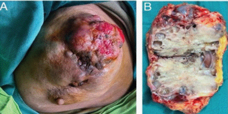
    Figure 1: Intraoperative images A) Fungating mass, Right breast B) Cut
surface of the specimen.
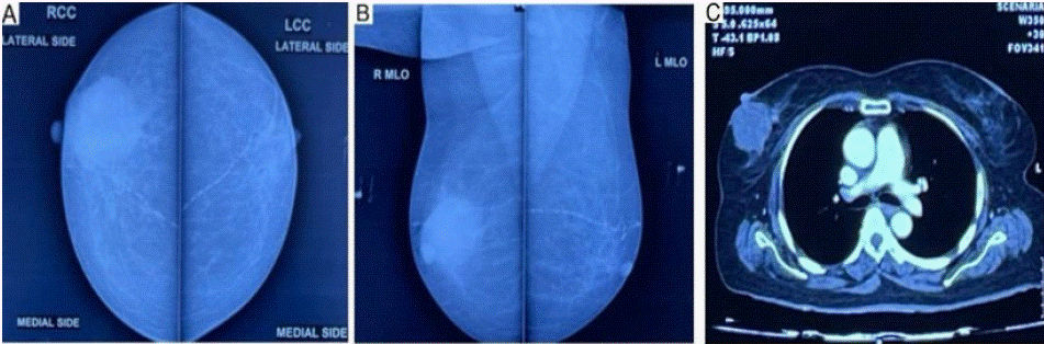
    Figure 2: Mammogram showing BIRADS 4C lesion, Right breast A) Craniocaudal view, B) Mediolateral oblique view, C) HRCT Thorax showing heterogeneous
enhancing hypodense mass lesion.
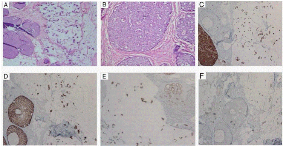
    Figure 3: A) Mucinous Breast Carcinoma, H&E, 4x view B) Solid papillary carcinoma in situ, H&E, 10x view, C) Chromogranin positive (IHC), D and E) ER, PR
positive (IHC), F) Her2neu negative.
Discussion
Mucinous and Solid papillary carcinoma of the breast are infrequent histological subtypes of breast cancer and occur in postmenopausal women. Solid papillary carcinoma shows low-grade cytological features with neuroendocrine differentiation [3]. These tumors can show intracellular and extracellular mucin, but if it is prominent then co-existence of mucinous breast carcinoma should be considered, as found in the present case. There are two histological subtypes of mucinous breast carcinoma: pure and mixed. Pure form shows more than 90% mucinous component and the mixed form shows 10-90% mucinous component with co-existing invasive ductal carcinoma [4]. However, the existence of invasive mucinous carcinoma with solid papillary carcinoma in situ is a rare occurrence.
Radioimaging varies from a well circumscribed mass (in case of pure mucinous) to an irregular, spiculated mass (likely to have mixed mucinous tumor or in solid papillary carcinoma) [5]. Ultrasonography may appear isoechoic in cases of pure mucinous cancer, which may tend to go under-reported as a benign lesion. However, mixed mucinous cancers demonstrate hypoechoic or heterogeneous mass [6]. In our case, a lobulated, heterogeneously hypoechoic lesion with multiple small cystic areas was noted.
Overall, mucinous and solid papillary carcinoma show lower frequency of axillary metastases and a better overall survival. However, mixed mucinous carcinoma or co-existing tumors show increased chances of nodal involvement [7]. In our case, one out of twenty-three lymph nodes demonstrated metastatic deposits.
The management of breast cancer requires a multi-disciplinary approach involving surgical resection, chemotherapy, radiation therapy, and targeted therapies based on the tumor stage and biomarkers. The treatment protocol of special types of breast cancer is still not well established. Surgery is the mainstay of treatment with or without adjuvant hormonal/ chemotherapy or radiotherapy, depending upon the tumor characteristics, margin status and stage. In our case, a MRM was performed, followed by adjuvant chemotherapy using a paclitaxel and carboplatin regimen.
Conclusions
This case contributes valuable insight into the diverse morphological spectrum of breast carcinomas, diagnostic and therapeutic challenges posed by the coexisting tumors, particularly in a late-stage presentation and reinforces the importance of tailored therapeutic strategies in managing such unique cases. The advanced stage at presentation underscores the need for increased awareness, early detection, and timely intervention in breast cancer cases.
Declarations
Funding
The authors received no financial support for the research, authorship, and/or publication of this manuscript.
Ethics Approval and Consent to Participate
This article is a case report which does not require an IRB approval. It is notable that the patient was not subjected to any experimental investigation or treatment at any point of time.
Consent for Publication
Written informed consent was taken from the patient. The identity of the patient is not revealed in the pictures or in the manuscript.
Authors Contribution
Dr Binthaf PP: Conducted the literature review, collected clinical data, drafted the manuscript, prepared visuals, and coordinated revisions with all co-authors.
Dr Parag Gupta: Conceptualized the study, Performed the surgical procedure, provided surgical insights, provided patient care, reviewed the manuscript to ensure clinical accuracy and relevance and coordinated revisions.
Dr Priya Sahu: Performed the histopathological examination, provided the definitive diagnosis, and contributed to manuscript preparation, coordinated revisions and interpretation of pathology findings.
All authors: Reviewed and approved the final manuscript.
References
- Tan PH, Ellis I, Allison K, Brogi E, Fox SB, Lakhani S, et al. The 2019 WHO classification of tumours of the breast. Histopathology. 2020; 77: 181-185.
- Guo S, Wang Y, Rohr J, Fan C, Li Q, Li X, et al. Solid papillary carcinoma of the breast: a special entity needs to be distinguished from conventional invasive carcinoma avoiding over-treatment. The breast. 2016; 26: 67-72.
- Maluf HM & Koerner FC. Solid papillary carcinoma of the breast. The American journal of surgical pathology. 1995; 19: 1237-1244.
- Di Saverio S, Gutierrez J & Avisar E. A retrospective review with long term follow up of 11,400 cases of pure mucinous breast carcinoma. Breast cancer research and treatment. 2008; 111: 541-547.
- Harvey JA. Unusual breast cancers: useful clues to expanding the differential diagnosis. Radiology. 2007; 242: 683-694.
- Memis A, Ozdemir N, Parildar M, Ustun EE, & Erhan Y. Mucinous (colloid) breast cancer: mammographic and US features with histologic correlation. European journal of radiology. 2000; 35: 39-43.
- Fein DA, Fowble BL, Hanlon AL, Hooks MA, Hoffman JP, Sigurdson ER, et al. Identification of women with T1-T2 breast cancer at low risk of positive axillary nodes. Journal of surgical oncology. 1997; 65: 34-39.