Clinical Image
Austin J Radiol. 2022; 9(4): 1199.
Skull Base Chordoma: The Thumb Sign
Khouchoua S*, Zahi H, Iraqi Houssaini Z, Jerguigue H, Latib R and Omor Y
Department of Radiology, Ibn Sina University Hospital Center, Morocco
*Corresponding author: Khouchoua S, Department of Radiology, National Institute of Oncology, Ibn Sina University Hospital Center, Avenue Allal El Fassi, 10000, Rabat, Morocco
Received: June 14, 2022; Accepted: July 07, 2022; Published: July 14, 2022
Clinical Image
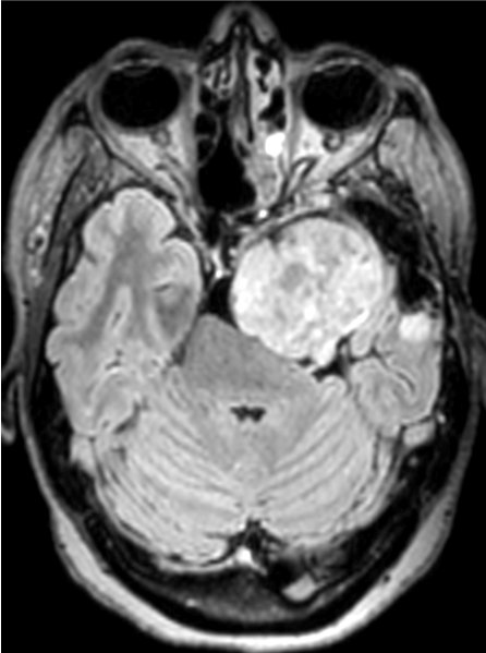
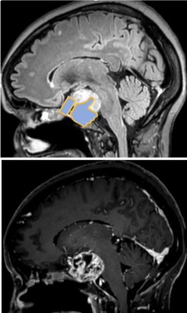
A 57 year old woman with no past medical history presented with headaches and progressive left vision loss. Subsequently, a contrast enhanced brain MRI was performed showing a large well defined skull base mass located laterally in the left petro clival fissure, exhibiting high T2 and FLAIR signal intensity (Figure A), with areas of low signal intensity on susceptibility weighted images consistent with calcifications and a heterogenous enhancement after injection. This tumor was responsible for mass effect on the orbital apex and optic nerve anteriorly and the mesio temporal lobe laterally. Most importantly, on the sagittal planes we can see the tumor projection indenting the pons which is displaced posteriorly. This finding is known as the thumb sign (Figure B and C) characteristic of chordomas. Given these MRI features and after a stereotactic biopsy the diagnosis of chordoma of the skull base was confirmed. Because of the major local extension, the patient underwent radiotherapy rather than surgical resection. Chordomas are rare skull base tumors arising from embryonal rest cells [1]. The major differential being chondrosarcomas with similar appearance but higher ADC values. The thumb sign is relatively specific of chordomas [2] although itcan be described in chondromas of the skull base and can also be seen in chondrosarcomas.

    Figure A: Contrast enhanced axial FLAIR image showing a large petro clival

mass with mass effect on both the orbital apex and the pons posteriorly.

    Figure B: Sagittal projections on FLAIR (1) and T1 gadolinium enhanced

after fat suppression (2) showing the heterogeneously enhanced mass with

extension posteriorly indenting the pons called the thumb sign.
References
- Kremenevski N, Schlaffer SM, Coras R, Kinfe TM, Graillon T, Buchfelder M. Skull Base Chordomas and Chondrosarcomas. Neuroendocrinology. 2020; 110: 836-847.
- Mark IT, Van Gompel JJ, Inwards CY, Ball MK, Morris JM, Carr CM. MRI enhancement patterns in 28 cases of clival chordomas. J Clin Neurosci. 2022; 99: 117-122.