
Clinical Image
Austin J Radiol. 2021; 8(3): 1131.
Ankle Localization of a Primary Bone Large B-Cell Lymphoma
Gérard B¹*, Brevet P¹, Demeyere M², Lequerré T¹, Vittecoq O¹ and Patenere C¹
¹Rheumatology Department, Rouen University Hospital, France
²Radiology Department, Rouen University Hospital, France
*Corresponding author: Gérard B, Rheumatology Department & CIC/CRB 1404, Rouen University Hospital, UNIROUEN, 76031 Rouen France
Received: March 30, 2021; Accepted: April 14, 2021; Published: April 21, 2021
Keywords
Primary bone lymphoma; Large B-cell lymphoma; Ankle bone tumor
Clinical Image
Plain X-Ray of a primary bone large B-cell lymphoma in an 84-year-old patient (Figure 1 and 2). She consulted for a swelling of the right ankle with functional impotence, in a context of altered general condition for many months. The radiographic appearance was typical, showing a shaft and metaphyseal “moth-eaten” osteolytic lesion, with periosteal reaction and thickening of the opposite soft tissues. The diagnosis of B-cell lymphoma was confirmed with histological examination of tissue samples from percutaneous computed tomography-guided biopsy of the lesion. Primary bone lymphoma is rare, accounting for 5% of malignant bone tumors, and mostly large B-cell lymphomas. Moreover, localization usually predominating at the roots of the axial skeleton, explained by the persistence of red bone marrow. Even if it presents in an unusual site, this case highlights the importance of considering typical imaging characteristics of a lesion.
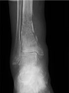
Figure 1:
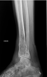
Figure 2: