Case Report
Austin J Radiol. 2025; 12(3): 1258.
Fissured Gallstone Mimicking Acute Cholecystitis: Value of the “Mercedes-Benz” Sign at CT Examination
Bourabaa Soukayna1,2* and Zain-Al-Abidine Khedid1,2
¹Emergency General Surgery Department, Ibn Sina University Hospital, Rabat, Morocco
²Mohammed V University of Rabat, Morocco
*Corresponding author: Bourabaa Soukayna, Emergency General Surgery Department, Ibn Sina University Hospital, Rabat, Morocco; Mohammed V University of Rabat, Morocco Email: soukayna.bourabaa@um5r.ac.ma
Received: June 22, 2025 Accepted: July 22, 2025 Published: July 24, 2025
Abstract
The “Mercedes-Benz sign” in CT imaging refers to the presence of partial radiolucencies within gallstones that resemble the star-like shape of the Mercedes-Benz symbol. This distinctive pattern is named after the iconic car logo due to its striking similarity. The sign is rare, with only a few dozen cases reported in the literature. We report the case of a 59-year-old woman who was admitted to the emergency department for gallstone mimicking acute cholecystitis.
Keywords: Case report; Cholecystitis; Mercedes-Benz sign
Introduction
The “Mercedes-Benz sign” is a radiological finding characterized by a triradiate pattern of nitrogen gas observed within gallstones. Initially noted on abdominal radiographs and later confirmed on CT scans in cases of calculus cholecystitis, this phenomenon is associated with acute abdominal pain. About half of gallstones show fluid-filled fissures, with less than half of these fissured stones containing gas. The gas-induced radiolucency typically appears in a triradiate shape, reminiscent of the Mercedes-Benz logo.
In 1966, Hay [1] wrote “Fissures within certain mixed gallstones have long been known”, Friedrich August Walter is said to have described gallstone fissures in vitro in 1796, and Bramson in 1846 discussed possible explanations for their occurrence. In 1931, Béla Breuer described their appearance on an abdominal radiograph [2].
Case Presentation
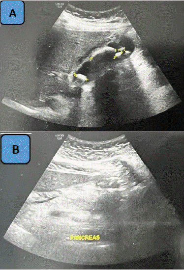
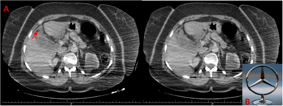
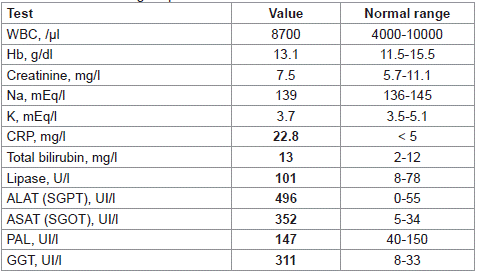
We present the case of a 59-year-old Caucasian female patient with a history of total thyroidectomy 10 years ago for multi-hetero-nodular goiter, after which she was placed on Levothyroxine. She was admitted to the emergency department with acute upper quadrant abdominal pain and fever persisting for the past 4 days. Upon examination, she was conscious with a Glasgow Coma Scale (GCS) score of 15, febrile, hemodynamically and respiratory stable, with normally colored conjunctivas. Abdominal examination revealed a positive Murphy's sign. The rest of the examination was unremarkable. Laboratory analysis showed a mild elevation in C-reactive protein (CRP) levels with evidence of hepatic cytolysis (Table 1), while the white blood cell (WBC) count was within normal limits. Emergency abdominal ultrasound (US) revealed a normal-sized, multi-lithiasic gallbladder with a thickened wall of 8 mm, compatible with acute lithiasic cholecystitis (Figure 1). Subsequent abdominal CT scan revealed a gallbladder with a thin wall containing an air-filled image at the fundus, consistent with a fractured stone, producing the Mercedes Benz sign (Figure 2). The patient was admitted for treatment and commenced on antibiotics, followed by laparoscopic cholecystectomy 48 hours later. She had an uneventful postoperative course and was discharged on postoperative day (POD) 1.

    Figure 1: (A) Abdominal US showing a normal-sized gallbladder with multiple
gallstones (plus), with a thickened wall measuring 8 mm. (B) The pancreas
is normal in size, with regular contours and homogeneous echostructure.

    Figure 2: (A) Axial abdominal CT scan showing a star-shaped pattern of air inside the gallbladder (red arrow) corresponding to the Mercedes-Benz sign, related
to the presence of gallstones; (B) Mercedes-Benz logo.

Table 1: Patients’ biological parameters.
Discussion
In the gallbladder, the “Mercedes-Benz sign” was first described by Kommerell and Wolpers [3] in 1938, depicting a star-shaped pattern of gas fissures within gallstones initially identified on abdominal radiographs [4]. Terms such as ‘crow's foot,’ ‘star-shaped,’ and ‘Jack stone’ were previously used to describe fissured gallstones but were later replaced by the term “Mercedes-Benz sign” [2]. This radiological finding is rare, with only a few cases reported in the literature, typically associated with conditions like biliary colic or cholecystitis.
Gallstones are categorized into two types: pigment stones, which appear hyperdense on CT due to calcification, and cholesterol stones, which are not visible on CT because they are embedded in cholesterolrich bile. Less than half of these stones contain gas [1,5], which may diffuse outside if the stone cracks, becoming the only detectable indicator of the gallstone. The gas accumulates within fissures, resulting in radiolucent lines within the calculus. The gas composition typically includes less than 1% oxygen, 6-8% carbon dioxide, and nitrogen [3]. The high carbon dioxide and low oxygen concentrations relative to those in normal air may be due to the diffusion of gas into the calculus that may originate from a gas-forming organism [6].
The Mercedes-Benz sign manifests as a triradiate radiolucent shadow in the right upper quadrant of the abdomen. The fissures are radial, often wider centrally or star-shaped, resembling the Mercedes- Benz logo, though they may show peripheral widening. These fissures, numbering from one to five and varying in length and distribution, typically run perpendicular to the middle point of each facet along the central two-thirds of the gallstone.
If the calculus is radiographed at right angles to its longitudinal axis, only the fissure tangential to the x-ray beam is shown.
Historically, Walter [7] first described star-shaped fissures in biliary calculi in vitro in 1796, with their radiographic appearance described much later in 1931 [8,9]. The presence of these fissures has been associated with the rapid formation and auto-fragmentation of calculi [4]. Numerous cases on record have documented gallstones complicated by infection. Akerlund [6] posited that the gas observed in these cases resulted from bacterial action or decomposition within the stones. It is known that B. coli can anaerobically generate gas, causing conditions like emphysematous cholecystitis, portal pylephlebitis, or in the urinary tracts, especially during diabetic coma (Olsson, 1962; Porter and Wright, 1961). However, in these situations, fermentation typically produces CO2, which dissolves readily in tissue fluids. Despite this understanding, various researchers, including Hay [1] and Hinkel (1950), have cultured the spaces within gas-containing stones and consistently found them sterile.
Gas-containing gallstones are a diagnostic challenge, often mistaken for gas or feces in bowel loops. They can be accurately identified using diagnostic imaging techniques such as CT and US, which can reveal specific attenuation values indicative of gas presence [10,11]. Hinkel [12,13] radiographed post-cholecystectomy gallbladders and found fissures in approximately 50% of gallstones. Most stones contained fluid, while around 46% also showed signs of gas. Another study using CT imaging on cases of cholecystolithiasis determined that gas was present in about 4% of calculi [14]. In a CT study of 21 calculi, central gas collections typically exhibited negative attenuation values as low as -290 HU [11]. Only densities below -100 HU are considered diagnostic, while values between -100 and 0 HU are deemed indeterminate, likely due to a mixture of gas and fluid within fissures [3,14].
References to further studies and detailed investigations have contributed to understanding the physical phenomenon of fissures within gallstones, suggesting potential links to crystalline composition and bile environment. Despite academic interest in gas formation mechanisms, the clinical relevance of the Mercedes-Benz sign lies in its indication of gallstones as a possible cause of abdominal pain, sometimes complicated by infection.
The interpretation of gas-containing gallstones on imaging studies like CT and US is critical, as their appearance can vary from highlevel echoes suggesting gas presence to isodense features resembling surrounding bile. Understanding these radiological nuances aids in accurate diagnosis and appropriate management of patients presenting with suspected gallstone-related symptoms.
Conclusion
The presence of gas inside gallstones, known as the Mercedes-Benz sign, occurs due to fissure development during crystallization. This can sometimes be mistaken for acute cholecystitis. Only abdominal CT can accurately diagnose this condition.
References
- Hay HRC. Gas in gal stones: a rare radiological sign in acute abdomen. Gut. 1966; 7: 387.
- Hao Xiang, Jason Han, William E Ridley, Lloyd J Ridley. Crow’s feet appearance: Fissured gallstones. Journal of Medical Imaging and Radiation Oncology. 2018; 62: 70.
- Kommerell B, Wolpers C. Gashaltige gallensteine. Fortschr Geb Rontgenstrahlen. 1938; 58: 156-174.
- Meyers MA, O’donohue N. The Mercedes-Benz sign: insight into the dynamics of formation and disappearance of gallstones. Am J Roentgenol Radium Ther Nucl Med. 1973; 119: 63-70.
- Nalda M, Zone J, Vela´n O. Signo del Mercedes Benz. Rev Chile Radiol. 2010; 16: 205–206.
- Akerlund A. Uber transparent gashaltige spaitbildungen in gallensteinen und ihre rontgendianostische bedeutung. Actaradiol. 1938; 19: 215-229.
- Walter FA. Anatomisches Museum, Berlin, 1796. Cited by Kommerell (12).
- Bauer KH. Uber Selbstzertrummerung von Gallensteinen und Neubildung von Steinen auf der Grundlage von Steintrummern. Arch klin Chir. 1931; 165: 53-80.
- Breuer B. Uber ein neues Rontgensymptom der Gallensteinkrankheit. Rontgenpraxis. 1931; 3: 879.
- Delabrousse E, Bartholomot B, Narboux Y, Barrali E, Chirouze C, Kastler B. Gas-containing gallstones: value of the \”Mercedes-Benz\” sign at CT examination. J Radiol. 2000; 81: 1639-1641.
- Becker CD, Vock P. Appearance of gas-containing gallstones on sonography and computed tomography. Gastrointest Radiol. 1984; 9: 323-328.
- Hinkel CL. Gas-containing biliary calculi. AJR. 1950; 64: 617-623.
- Hinkel CL. Fissures in biliary calculi further observations. AJR. 1954; 71: 979- 987.
- Fork FT, Nyman U, Sigurjonsson S. Recognition of gas in gallstones in routine computed tomograms of the abdomen. J Comput Assist Tomogr. 1983; 7: 805-809.