Clinical Image
Austin J Radiol. 2025; 12(2): 1256.
Isolated Sternal Involvement in Paget’s Disease of Bone
Boujida N, Jabour S, Elaitari K, El Bakkari A, Omor Y, Latib R and Amalik S
Radiology Department, National Institute of Oncology, Rabat, Morocco
*Corresponding author: Nadia Boujida, Radiology Department, National Institute of Oncology, Rabat, Morocco Email: nadiaboujida21@gmail.com
Received: April 10, 2025 Accepted: April 22, 2025 Published: April 28, 2025
Clinical Image
A 75-year-old man with a history of treated colonic adenocarcinoma presented for follow-up imaging. He was asymptomatic, with no clinical complaints. Laboratory findings were unremarkable, and previous imaging studies had shown stable disease. A follow-up contrast-enhanced CT scan of the chest, abdomen, and pelvis was performed as part of routine oncologic surveillance.
Description
Paget’s Disease of Bone is a chronic, localized disorder of bone remodeling, characterized by increased bone turnover without an inflammatory component. It typically affects large, non-contiguous areas of the skeleton [1]. It is the second most common metabolic bone disorder after osteoporosis. Despite this, it remains largely underdiagnosed and undertreated. The disease predominantly affects the elderly, being rare before the age of 40, and occurs more frequently in men than in women [2].
The etiology of Paget’s disease remains under investigation, with both genetic and environmental factors playing a role. Genetic evidence is substantial, with 15% to 40% of patients having a firstdegree relative affected by the disease.
In addition, intracellular inclusions resembling paramyxovirus nucleocapsids have been observed, and canine distemper virus has been detected in Pagetic osteoclasts.
Paget’s disease may be monostotic, involving a single bone, or polyostotic, affecting multiple bones.
It often presents with asymmetric involvement, such as unilateral femoral disease. Unlike other skeletal conditions, Paget’s disease rarely progresses to new skeletal sites over time [1].
The most commonly affected bones are the femur, spine, skull, sternum, and pelvis, although any bone can be involved.
Many patients are asymptomatic, with diagnosis made incidentally through elevated serum alkaline phosphatase levels or imaging performed for unrelated reasons.

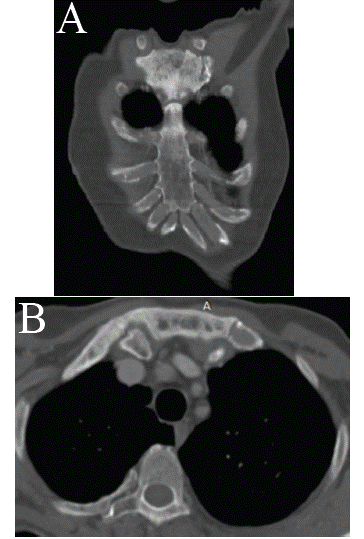
    Figure 1: Axial (B) and coronal (A) CT images in bone window: Welldemarcated
osteosclerosis of the sternal manubrium extending to the right
first rib, with preserved cortical margins, no periosteal reaction, and no
adjacent soft tissue involvement – findings consistent with Paget’s disease
of bone involving the sternum.
When symptoms are present, they may be due to the bone lesions themselves (e.g., bone pain, secondary osteoarthritis, or fractures), or due to compression of adjacent neural structures from bone expansion.
Diagnosis is primarily radiologic. In early disease, osteolytic lesions predominate. As the disease progresses, areas of sclerosis develop, resulting in the classic appearance of mixed lytic and sclerotic changes, with thickened trabeculae, cortical thickening, bone expansion, and deformity.
Although radiologic findings are usually characteristic, differential diagnoses such as sclerotic or lytic metastases may sometimes need to be considered.
Radionuclide bone scintigraphy is recommended during the initial evaluation to determine disease extent, especially in high-risk areas such as the skull base, spine, and long bones [1].
Symptomatic patients are treated with bisphosphonates to suppress bone turnover, promote healing of osteolytic lesions, and relieve pain. Analgesics and non-steroidal anti-inflammatory drugs (NSAIDs) may also be used for symptom control [3].
References