
Case Report
Austin J Radiol. 2025; 12(2): 1255.
Accessory Breast Tissue in Axilla Miming Lymphadenopathy in a Patient with Ovarian Tumor
Jaheddine Fadwa*
Department of Medical Imaging, Mohammed V University, 10100, Faculty of Medicine and Pharmacy, Rabat, Morocco
*Corresponding author: Jaheddine Fadwa, Department of Medical Imaging, Mohammed V University, 10100, Faculty of Medicine and Pharmacy, Rabat, Morocco Email: fadwa.jhd@gmail.com
Received: April 07, 2025 Accepted: April 21, 2025 Published: April 24, 2025
Abstract
Accessory breast tissue, is a rare anatomical variation that can present challenges in differential diagnosis, particularly when located in the axilla. Its appearance can be confusing, especially in the context of oncological conditions.
We present the case of a 42-year-old female with a known ovarian tumor who underwent a CT scan of the chest, abdomen, and pelvis. The CT scan revealed a mass in the right axilla, which was initially suggestive of lymphadenopathy. However, subsequent axillary ultrasound confirmed that the mass was accessory breast tissue rather than a lymph node.
Keywords: Accessory breast tissue; Lymphadenopathy; CT scan; Ultrasound
Case Presentation
We report the case of a 42-year-old female patient with an ovarian tumor who was referred to our radiology center for preoperative imaging, including chest, abdominal, and pelvic CT scans. The CT scans did not reveal any evident metastases, except for an abnormal right axillary lymph node.
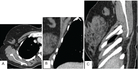
    Figure 1: CT finding of the right axilla: (A) Axial (B) Coronal and (C) Sagittal
Ct images depicting a right axillary lesion resembling a lymph node (arrow).
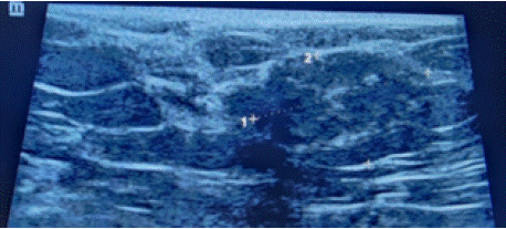
    Figure 2: Ultrasound of the right axilla showing a normal dense
fibroglandular tissue which is not connected to the pectoral breast tissue.
To further characterize the finding, an ultrasound of the axillary lymph node was performed. The ultrasound revealed normal dense fibroglandular tissue that was not connected to the pectoral breast tissue. No solid or cystic mass lesions were present. These findings were consistent with accessory breast tissue.
Discussion
Accessory breast tissue refers to normal breast tissue that is located apart from the primary breast area. It typically develops along the embryologic "milk line" and occurs in approximately 2 to 6% of women. The most frequent site for this tissue is in the axillary tail of the breast, though it can also appear along the inferior milk line beneath the breast. In some cases, supernumerary breast tissue may include a nipple and/or areolar complex [1].
Although lesions in the inferior part of the axilla can be visualized on the mediolateral oblique view of a mammogram, distinguishing them from abnormal axillary lymph nodes or other axillary abnormalities, such as tumors in accessory breast tissue, can be challenging. Sonography is the preferred imaging modality for assessing axillary masses and serves as an excellent tool for characterizing both palpable and non-palpable masses [2].
Accessory breast tissue is susceptible to the same conditions as normal breast tissue. It undergoes the usual hormonal fluctuations associated with the menstrual cycle, pregnancy, and postpartum changes. Like primary breast tissue, accessory breast tissue can develop cancer. Therefore, it is important to evaluate any palpable changes, suspicious calcifications, or masses in accessory breast tissue with the same diligence as for the primary breast tissue [1].
The potential for disease in accessory breast tissue should be taken into account, and axillary accessory breast tissue should be incorporated into screening examinations. Any abnormal findings within ectopic breast tissue should be assessed using the same diagnostic approach as for tissue located in the primary breast [2]. Accessory breast tissue can be surgically removed if it causes symptoms or complications, but such intervention is usually only required when the tissue becomes problematic [2].
Conclusion
Radiologists must be familiar with the normal appearance of accessory breast tissue across various imaging modalities, including CT scans, and need to consider accessory axillary breast tissue as a potential cause of an axillary mass.
References