
Clinical Image
Austin J Radiol. 2025; 12(2): 1253.
From Tendon to Bone: MR Features of Intraosseous Migration of Intratendinous Calcifications
Tlaite O*, Mohamed L and Jamal ELF
Radiology Department, Mohammed V Military Instruction Hospital, Mohammed V University, Morocco
*Corresponding author: Oubaddi Tlaite, Radiology Department, Mohammed V Military Instruction Hospital, Mohammed V University, Morocco Email: tlaite.oubaddi@gmail.com
Received: March 11, 2025 Accepted: March 28, 2025 Published: Aril 01, 2025
Clinical Image
Calcific tendinitis of the rotator cuff is a frequent cause of shoulder pain caused by the accumulation of hydroxyapatite crystals in the pre-insertional areas of the tendons. In rare instances, these calcifications may migrate to adjacent sites, including bone, resulting in notable bone marrow involvement. MRI is highly sensitive in the acute phase for detecting bone edema surrounding migrated intraosseous calcification and can also visualize adjacent soft tissue edema and bone erosion.
A 52-year-old man with no prior history of trauma or shoulder surgery, presented with acute severe pain in the left shoulder, associated with complete functional impairment. The patient underwent an MRI scan of the left shoulder, which revealed intraosseous calcification of the lesser tubercle (Figure 1A, White arrow) and bone marrow edema of the humeral head (Figure 1A, Yellow arrow), along with cortical erosion (Figure 1B, white arrow) and calcific tendinopathy of the subscapularis tendon (Figure 1C). Conservative treatment was initiated, including immobilization and nonsteroidal antiinflammatory drugs, resulting in favorable outcomes.
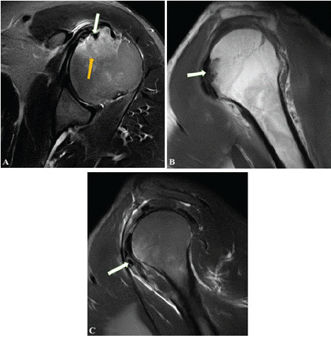
    Figure 1: (A) Axial T2 weighted fat saturated image showing intraosseous
calcification (White arrow) with bone marrow oedema of the lesser tubercle
(Yellow arrow), (B) Sagittal T1 weighted image showing cortical erosion
(White arrow), (C) Sagittal T2 weighted fat saturated image showing calcific
subscapularis tendinopathy.
References