
Case Report
Austin J Radiol. 2025; 12(1): 1252.
Aging and Aneurysms: Case Report of Moyamoya Disease in an Elderly Man
Dawadi K¹*, Tamang OY¹ and Poudyal B²
¹Consultant Radiologist, Department of Radiology and Imaging, Grande International Hospital, Kathmandu, Nepal
²Consultant Pediatrician, Department of Pediatrics, Grande International Hospital, Kathmandu, Nepal
*Corresponding author: Dr. Kapil Dawadi, Consultant Radiologist, Department of Radiology and Imaging, Grande International Hospital, Kathmandu, Nepal Email: kpldwd@gmail.com
Received: February 28, 2025; Accepted: March 17, 2025; Published: March 20, 2025;
Abstract
Moyamoya disease is a chronic progressive veno-occlusive disorder characterized by the stenosis of the intracranial internal carotid artery and its proximal branches, leading to the formation of fragile collaterals. It exhibits a bimodal distribution, with patients typically presenting in early childhood and middle age. Presentations include ischemia or intracranial hemorrhage, and it is rarely seen in the elderly population. We present a case of an elderly male who presented with intracranial hemorrhage.
Keywords: Moyamoya; Veno-occlusive; Elderly
Introduction
Moyamoya disease (MMD) characterized by progressive stenosis of the vascular branches of the internal carotid artery is a rare cerebrovascular disorder, leading to the development of abnormal, fragile collateral vessels. The condition was first described in 1957 in Japan, and the term "Moyamoya" was introduced by physicians Suzuki and Takaku in 1969. The word “Moyamoya” means "puff of smoke" in Japanese, referring to the hazy appearance of these collateral vessels on angiographic images in patients [1,2]. This disease shows a bimodal age distribution, with one peak occurring around 10 years of age and another peak between 30-40 years. It is rarely encountered in elderly patients, and females are found to be more affected than males [3]. In most cases, pediatric patients present with ischemia (approximately 75% of cases), whereas adults present with hemorrhages [4].
Case Presentation
A 69-year-old male was brought to our emergency department after experiencing sudden onset weakness in the left upper and lower limbs, along with altered sensorium. He had a medical history of Type II diabetes mellitus for the past 6 years, for which he was on regular medication and had well-controlled blood sugar levels. There was no other significant medical history.
Upon arrival at the hospital, the patient was drowsy. Clinical examination revealed a Glasgow Coma Scale (GCS) score of E3V4M5. Vital signs were stable. Neurological examination showed 5/5 power in the right upper and lower limbs, while power in the left upper and lower limbs was 3/5. Deep tendon reflexes were brisk on the left side. Based on the clinical findings, a provisional diagnosis of ischemic stroke was made, and the patient was taken for a CT scan of the head with CT angiography.
CT imaging of the head revealed a right basal ganglia hematoma extending into the bilateral lateral ventricles, third ventricle, and fourth ventricle, with no significant mass effect noted (Figure 1). CT angiography (CTA) showed non-visualization of the M1 segments of the bilateral middle cerebral arteries, with multiple collaterals present in the bilateral Sylvian fissures and basal ganglia. Distal reformation of the bilateral middle cerebral arteries was also observed (Figure 2). CT imaging of the head revealed a right basal ganglia hematoma extending into the bilateral lateral ventricles, third ventricle, and fourth ventricle, with no significant mass effect noted (Figure 1). CT angiography (CTA) showed non-visualization of the M1 segments of the bilateral middle cerebral arteries, with multiple collaterals present in the bilateral Sylvian fissures and basal ganglia. Distal reformation of the bilateral middle cerebral arteries was also observed (Figure 2).
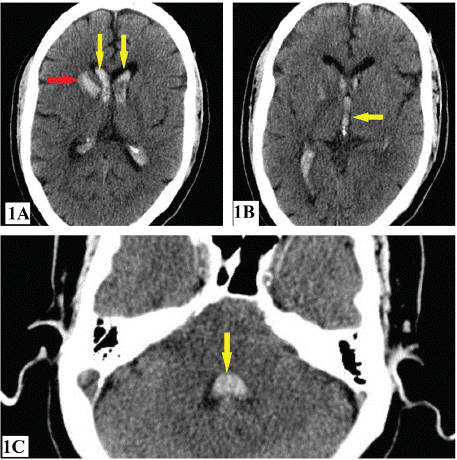
    Figure 1: Right basal ganglia bleed (red arrow in Figure 1A) with
intraventricular extension to bilateral lateral, 3rd and 4th ventricles (yellow
arrows in Figure 1A, 1B and 1C).
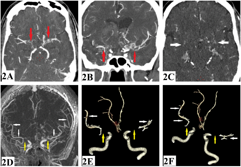
    Figure 2: Axial (2A) and Coronal (2B) MIP images showing non-visualization of the M1 segments of the bilateral middle cerebral arteries with multiple collaterals
(red arrows). Axial (2C) image showing collaterals in bilateral basal ganglia (white arrows). Coronal MIP (2D) and 3D VRT images (2E and 2F) showing
non-visualization of M1 segments of bilateral middle cerebral arteries (yellow arrows) with distal reformation (white arrows).
Additionally, a small focal contrast-filled outpouching was seen in the right basal ganglia adjacent to the hematoma (Figure 3). Based on these imaging findings, a diagnosis of Moyamoya disease with a pseudoaneurysm in the right basal ganglia was made. The patient was admitted for further management.
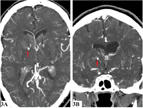
    Figure 3: Axial (3A) and Coronal (3B) images showing an aneurysm in right
basal ganglia (red arrows).
The patient’s condition deteriorated the following day, prompting a repeat CT scan of the head, which revealed an increase in the right basal ganglia and intraventricular bleeding, along with hydrocephalus (Figure 4). Extraventricular drainage was performed. However, the patient’s condition continued to deteriorate on the same day, leading to another CT scan, which showed a significant increase in the right basal ganglia and intraventricular bleeding, as well as worsening hydrocephalus. New findings compared to the previous imaging included midline shift to the left, subfalcine herniation, descending transtentorial herniation, and tonsillar herniation (Figure 5). Unfortunately, the patient expired the following day despite extensive management in the ICU.
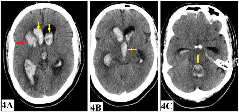
    Figure 4: Follow-up CT images showing increase in right basal ganglia bleed (red arrow in Figure 4A) and intraventricular hemorrhage with hydrocephalous
(yellow arrows in Figure 4A, 4B and 4C).
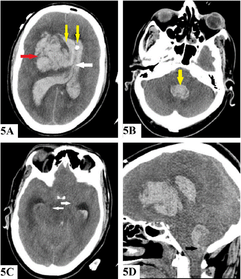
    Figure 5: Another follow-up CT showing further increase in right basal
ganglia bleed (red arrow in Figure 5A), and intraventricular hemorrhage and
hydrocephalous (yellow arrows in Figure 5A and 5B). Midline shift to the left
(white arrow in Figure 5A), descending transtentorial herniation (white arrows
in 5C) and cerebellar tonsillar herniation (black arrow in Figure 5D) were
new findings.
Discussion
Moyamoya disease is a rare progressive veno-occlusive vascular disorder characterized by chronic narrowing of the internal carotid arteries (ICAs), primarily at their terminal bifurcations leading to hypoperfusion of the brain parenchyma in the anterior circulation. The resulting ischemia stimulates the angiogenesis leading to proliferation of fragile collateral arteries which have an increased risk of rupture [5]. Patients with Moyamoya disease typically present with one of two phenotypes: (1) ischemic stroke or (2) hemorrhage [6]. Ischemic presentations due to hypoperfusion are more commonly seen in children, while hemorrhagic presentations are more prevalent in adults [7]. In pediatric patients with Moyamoya disease, hyperventilation conditions such as crying or other conditions such as fever or dehydration result in transient ischemic attack (TIA) episodes [8]. Adult patients more commonly present with cerebral hemorrhage owing to the fragile collateral vessels often leading to worse outcomes compared to those seen in children and adolescents [8]. This hemorrhage typically occurs in lobar areas, as most collateral vessels arise from the choroidal system. Aneurysm formation can also be seen in these collateral vessels [9].
The treatment modalities for Moyamoya disease (MMD) can be conservative or surgical. Conservative management primarily involves the use of antiplatelet drugs, anticonvulsants, and pain management. Treatment with antiplatelet drugs such as acetylsalicylic acid and/or clopidogrel is used to prevent new ischemic events. However, no single-drug regimen is accepted as a gold standard for managing ischemic or hemorrhagic complications. Additionally, rigorous control of risk factors such as dyslipidemia, hypertension, and diabetes is highly recommended [8].
The surgical treatment aims to minimize cerebral ischemia by enhancing cerebral blood flow (CBF) and decreasing the hemodynamic stress that can lead to cerebral hemorrhage [8]. Revascularization surgery prevents strokes and secondary hemorrhages in patients with MMD. The rate of recurrent ischemic attacks is lower with surgical treatment resulting in significantly better clinical outcomes compared to conservative treatment [8, 9].
The branches of the external carotid artery, such as the superficial temporal artery and occipital artery are used as donor vessels in surgical revascularization procedures. The surgical procedures can be divided into direct and indirect techniques. In direct bypass techniques, superficial temporal artery-middle cerebral artery anastomosis, superficial temporal artery-anterior cerebral artery anastomosis, and occipital artery-posterior cerebral artery (PCA) anastomosis can be done. Direct anastomosis procedures lead to an immediate increase in cerebral perfusion and is considered the firstline treatment [9].
Indirect methods are done when direct bypass is not feasible. Indirect methods are technically easier to perform, although they require more time to adequately restore blood flow [9]. The main indirect revascularization technique is synangiosis where collateral circulation toward the brain cortex is developed using connective tissues such as the scalp, muscle, and dura mater [9]. Several indirect techniques such as encephalo-duro-arteriosynangiosis, encephalomyo- synangiosis, and encephalo-duro-arterio-myo-synangiosis.
Conclusion
Moyamoya disease is a rare condition that leads to vascular occlusion and subsequent collateral formation in response to hypoperfusion. Patients may present with ischemic attacks or episodes of intracranial hemorrhage. The disease has a bimodal age distribution, typically occurring in infancy or early childhood and middle age, making it rare in the elderly population.
Consent
Written informed consent was obtained from the patient to publish this report in accordance with the journal's patient consent policy.
References
- Suzuki J, Takaku A. Cerebrovascular “Moyamoya” disease: disease showing abnormal net-like vessels in base of brain. Arch Neurol. 1969; 20: 288–299.
- Suzuki J, Kodama N. Moyamoya disease--a review. Stroke. 1983; 14: 104– 109.
- Kim JS. Moyamoya disease: epidemiology, clinical features, and diagnosis. J Stroke. 2016; 18: 2–11.
- Wakai K, Tamakoshi A, Ikezaki K, Fukui M, Kawamura T, Aoki R, et al. Epidemiological features of moyamoya disease in Japan: findings from a nationwide survey. Clin Neurol Neurosurg. 1997; 99: S1–S5.
- Pollak L. Moyamoya disease and moyamoya syndrome. N Engl J Med. 2009; 361: 98.
- Liu XJ, Zhang D, Wang S, Zhao YL, TeoM, Wang R, et al. Clinical features and long-termoutcomes of moyamoya disease: a single-center experience with 528 cases in China. J Neurosurg. 2015; 122: 392–399.
- Zhang H, Zheng L, Feng L. Epidemiology, diagnosis and treatment of Moyamoya disease. Exp Ther Med. 2019; 17: 1977–1984.
- Shang S, Zhou D, Ya J, et al. Progress in moyamoya disease. Neurosurg Rev. 2020; 43: 371–382.
- Lee SU, Oh CW, Kwon OK, et al. Surgical treatment of adult moyamoya disease. Curr Treat Options Neurol. 2018; 20: 22.