
Clinical Image
Austin J Radiol. 2025; 12(1): 1251.
Glomus Tumor of the Finger: MRI Diagnosis
Cherif A*, Imrani K, Jaheddine F, Moatassimbillah N and Nasser I
Department of Radiology, IBN Sina Hospital, Morocco
*Corresponding author: Asma CHERIF, Department of Radiology, IBN Sina Hospital, Rabat, Morocco Email: asmacherif27@gmail.com
Received: February 10, 2025; Accepted: February 28, 2025; Published: March 03, 2025
Clinical Image
Glomus tumors are rare benign mesenchymal tumors, accounting for 5% of all hand tumors, developed from the neuromyoarterial glomus, whose role is essentially thermoregulation and microcirculation. The pathogenesis remains unknown and the diagnosis may be difficult, as the pain may be considered neuropathic [1].
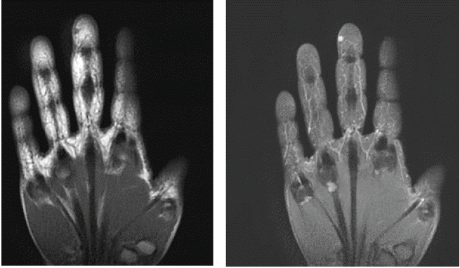
It is revealed by a sharp pulpal pain described as electrical, triggered by shocks, aggravated by pressure on the fingertips and by contact with cold. Associated signs should be sought: pain on precise pressure of a pencil tip, bluish subungual staining, nail dystrophy, and a possible bulge visible in case of a large tumor [2].
MRI sequences show a small (4x 5 mm) well-defined soft tissue mass on the ulnar aspect of the third distal finger respecting the cortex. It presents in T1 hyposignal, T2 hypersignal with vivid and homogeneous enhancement on post contrast sequences.
References
- H Chagraoui, M Alj, MEl Jazouli, S Chiheb. Les tumeurs glomiques: caractéristiques épidémiologiques et cliniques (série de 52 cas), Annales de Dermatologie et de Vénéréologie – FMC. 2022; 2: A289.
- Dominique Le Viet, Ombretta Spingardi. Tumeurs glomiques des doigts, Revue du Rhumatisme. 2023; 90: 205-210.