
Case Report
Austin J Radiol. 2025; 12(1): 1250.
Desmoid Tumor Mimicking Acute Appendicitis: Unusual Diagnostic of Right Iliac Fossa Pain
EL AZOUZI M, BENTAHAR A, SLIMANI A, ELGUAZZAR A and EL FIKRI A
1Radiology Department, Military Hospital Mohammed V, Rabat, Morocco
2Radiology Department, Military Hospital Moulay Ismail, Meknes, Morocco
3General surgery Department, Military Hospital Avicenna, Marrakech, Morocco
4Radiology Department, Military Hospital Avicenna, Marrakech, Morocco
*Corresponding author: EL AZOUZI Mehdi, Radiology Department, Military Hospital Mohammed V, Rabat, Morocco Tel: +212622216733; Email: Mehdi.med.maroc@gmail.com
Received: February 03, 2025; Accepted: February 17, 2025; Published: February 20, 2025
Abstract
Desmoid tumors are rare neoplasms that can occur at any age, with a predominance in women. Their exact aetiology remains poorly understood, but three factors appear to influence their development: genetic, traumatic and hormonal.
The main manifestation of these tumors is a painless mass that gradually increases in size, sometimes accompanied by symptoms associated with compression of neighbouring structures. The diagnosis is suspected by radiological examination and confirmed by pathological examination, which reveals a benign proliferation of fibroblasts.
The serious nature of these tumours lies in their tendency to recur and their potential for local invasiveness. Surgical treatment, consisting of carcinological excision, is preferred when local conditions allow, or other treatment options may be considered, such as radiotherapy.
Keywords: Desmoid tumor; Abdominal wall; Aggressive fibromatosis; Right iliac fossa pain
Introduction
Desmoid tumors are rare lesions with no metastatic potential. However, they have a strong tendency to invade locally and to recur. They account for 3% of all soft tissue tumours and 0.03% of all neoplasms [1].
We report the case of a young female patient with no history of trauma, surgery or pregnancy who presented to emergency with right iliac fossa pain and was found to have a desmoid tumor of the abdominal wall which progressed favourably after surgical treatment.
In this case report, we discuss the clinical and radiological aspects of desmoid tumors and the treatment modalities.
Case Presentation
A 30-year-old woman presented to the emergency department complaining of acute right iliac fossa pain, which had been present for two days. The pain started suddenly, remained constant and intensified over time. She also suffered loss of appetite and nausea. There were no signs of fever, vomiting, diarrhoea, constipation or urinary problems. The patient had no previous medical or surgical history and had never suffered from appendicitis or diverticulitis. On examination, she appeared healthy, with normal vital signs and a body mass index (BMI) of 23 kg/m2. Abdominal examination revealed a soft, undistended abdomen with normal bowel sounds and deep McBurney point tenderness. Rebound tenderness in the right iliac fossa, Rovsing's sign, psoas sign and obturator sign were all negative. Analysed blood parameters and urine cytobacteriological examination were within the normal range.
The patient was referred to the radiology department on suspicion of appendicitis. An abdominal ultrasound examination revealed a heterogeneous mass at right antero-lateral abdominal wall. Magnetic resonance imaging (MRI) was performed to better characterise the mass and revealed a well limited mass measuring 40x35mm, developed at the expense of the fascia of the right external oblique muscle, isointense to the muscles on T1-weighted-sequences (T1WI) and hyperintense on T2-weighted sequences (T2WI) and short tau inversion recovery (STIR) image with heterogeneous post-contrast enhancement. This mass was surrounded by a pseudo-capsule hypointense on T1WI and T2WI (Figure 1).
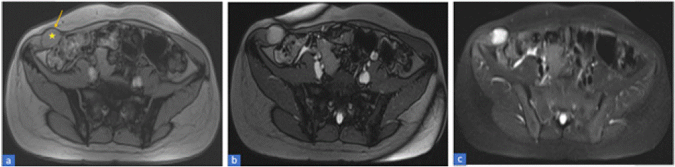
    Figure 1: Axial MRI images, in T1-weighted-sequences (a), T2-weighted-sequences (b) and STIR (c) showing a well-limited mass (star) developed at the
expense of the fascia of the right external oblique muscle, isointense to the muscles on T1WI and hyperintense on T2WI and STIR image. This mass is
surrounded by a pseudo-capsule (arrow) which is hypointense on T1WI and T2WI.
A wide tumor resection with safety margins and reconstitution of the parietal defect was performed (Figure 2).
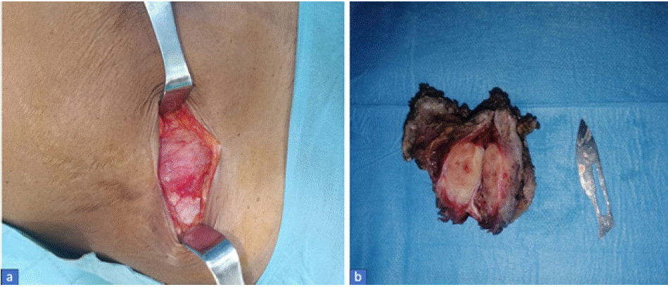
Figure 2: Macroscopic appearance of the tumour before (a) and after (b) excision.
The histological diagnosis was made in our training centre thanks to the anatomopathological study of the surgical specimen (Figure 3).
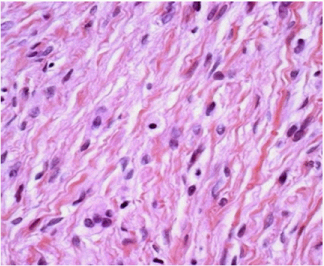
    Figure 3: Microscopic view of the excised desmoid tumor showing fascicles
of fibroblastic spindle cells with abundant intercellular collagen.
The patient subsequently progressed well.
Discussion
Desmoid tumors (DT) are defined by the World Health Organisation (WHO) as a clonal fibroblastic proliferation that occurs in deep soft tissue and is characterised by an inability to metastasise, although it may be multifocal in the same limb or part of the body [2].
It is also known as aggressive fibromatosis. It is a rare tumour. It is estimated that around 3.7 new cases occur each year in every million people [3].
Unlike fibrosarcomas, these tumours are distinguished by the fact that, although locally aggressive, they do not spread to other parts of the body [4].
They can occur at any age, and more frequently affect women in their reproductive years (20 to 40), with a sex ratio of 3:1 [5].
Desmoid tumours can develop in all the musculoaponeurotic structures of the body, but are mainly located in the abdominal region. tree forms are described:
• DT of the abdominal wall which develop in the musculoaponeurotic structures of the abdominal wall, essentially the rectus and obliquus muscles. They may rarely develop from the pelvic wall; in which case they mimic an ovarian tumor.
• Extra-abdominal DT, mainly involving the posterior part of the trunk, the chest wall and the lower limbs.
• Intra-abdominal DT invading the pelvis, mesentery and retroperitoneum, sometimes with vaginal, bladder, ureteral or rectal repercussions [6].
A clinical classification of DT proposed by Ferenc et al is based on three criteria: size (diameter), doubling time in months (expansion) and tumour location (site). This classification is equivalent to the TNM classification used for malignant tumours [7].
There is a well-known association with patients with a history of abdominal surgery, trauma, oestrogen therapy, Familial Adenomatous Polyposis (FAP) [8].
Medical imaging plays a vital role in the diagnosis, extension assessment and follow-up of desmoid tumours:
• Abdominal ultrasound: is the examination often performed as a first line of defence following the discovery of an abdominopelvic mass on clinical palpation. It does not provide specific signs for making the diagnosis, but it does help to localise the tumor, assess its size and relationship, and guide percutaneous biopsy; Desmoid tumours generally appear as a solid, heterogeneous, hypoechoic, welllimited mass. Ultrasound is particularly useful for early detection of tumor recurrence, and should be performed during follow-up consultations with patients who have already undergone surgery [9].
• Computed tomography (CT)-scan: is the examination of choice for the diagnosis of intra-abdominal desmoid tumors. The lesion may appear hypo-, iso- or hyperdense in relation to the muscle tissue, depending on its composition. The degree of enhancement after intravenous administration of contrast is variable. The advantage of a CT scan is that it enables the limits of the tumor and its deep extension to be better assessed, and the tumor's relationship with the major vessels, nerves and neighbouring organs to be assessed in the preoperative work-up [10].
• Magnetic resonance imaging (MRI): is the best examination for exploring desmoid tumors, which appear as heterogeneous images with hypo- or often iso-intense in relation to the muscle in T1WI, and variable signal in T2WI depending on the predominant component (cellular or fibrous). Enhancement is often intense. MRI can give a more accurate picture of the tumor's dimensions, pinpoint its origin and determine which structures it infiltrates, and thus rule out certain differential diagnoses [11].
The differential diagnosis arises above all with fibrosarcoma and fibroblastic proliferations, and nodular fasciitis [12].
The definitive diagnosis must be established by histopathological analysis. DT are composed of elongated fibroblasts and myofibroblasts, characterised by elongated, tapering cytoplasm, typical elongated, vesicular nuclei and multiple small nucleoli. The cells are arranged in a linear fashion and separated from each other by collagen. These tumors tend to evolve over time [13].
Wide local excision followed by reconstruction of the defect is the treatment of choice. Complete resection of the abdominal wall containing the tumor with negative margins is necessary when the lesion is close to or involves the peritoneum. Incomplete excision may lead to local recurrence [14].
Radiotherapy is used in patients with inoperable tumors, local recurrence or incompletely excised lesions. Chemotherapy and endocrine therapy have also been used to treat DT in patients in whom resection is technically impossible due to diffuse tumour infiltration [15].
Conclusion
Desmoid tumors, also known as aggressive fibromatoses, are rare, non-metastatic fibroblastic proliferations, but locally aggressive due to their infiltrative nature, resulting in frequent recurrences. Diagnosis of these tumors is relatively straightforward, but the difficulty lies in the high frequency of recurrence, which can be reduced by carcinological surgical resection with healthy margins. However, if the margins are invaded, additional external radiotherapy may be required.
Conflict of Interest Statement
The authors declare that there is no conflict of interest.
All authors confirm that they have obtained written consent from the patient for publication of the article.
References
- Teo HEL, Peh WCG, Shek TWH. Case 84: desmoid tumor of the abdominal wall. Radiology. 2005; 236: 81-84.
- Master SR, Mangla A, Shah C. Desmoid Tumor. In: StatPearls. Treasure Island (FL): StatPearls Publishing. 2024.
- The desmoid tumor I. Incidence, sex-, age- and anatomical distribution in the Finnish population Am J Clin Pathol. 1982; 77: 665-673.
- Kumar V, Khanna S, Khanna AK, Khanna R. Desmoid tumors: experience of 32 cases and review of the literature. Indian J Cancer. 2009; 46: 34-39.
- Lahlou MK, M Soufi, M Bensaid, R Messrouri. Fibromatose agressive de la paroi abdominale (tumeurs desmoïdes). J. Afr. Cancer. 2009; 1: 223-228.
- Montagliani L, Duverger V. Les tumeurs desmoïdes. J Chir. 2008; 145: 20-26.
- Hoos A, Lewis J, Antonescu CR, et al. Characterization of molecular abnormalities in human fibroblastic neoplasms: A model for genotypephenotype association in soft tissue tumors. Cancer Res. 2001; 61: 3171- 3175.
- Cotte E, Glehen O, Monneuse O, et al. Tumeurs desmoïdes associées à la polypose adénomateuse familiale. Gastroentérologie Clinique et Biologique. 2004; 28: 574-581.
- Wang Y, Tang J, Luo Y. Sonographic diagnosis of fibromatosis. J Clin Ultrasound. 2008; 36: 330-334.
- Clark SK, Phillips RKS. Desmoids in familial adenomatous polyposis. Br J Surg. 1996; 83: 1494-1504.
- MBH Amor, L Ploton, L Ceugnart, et al. Imagerie par résonance magnétique des tumeurs desmoïdes: critères d’évaluations actuels. Bulletin du Cancer. 2020; 107: 359-363.
- MC Kinnon JC, Neifeld JP, Kay S. Management of desmoid tumors. Surg Gynecol obstet. 1989; 169: 104-106.
- Sturt JNH, Clark SK. Current ideas in desmoid tumors. Familial Cancer. 2006; 5: 275–285.
- C Escobar, R munker, et al. Update on desmoid tumors. Annals of oncology. 2012; 23: 562-569.
- Schlemmer M. Desmoid tumors and deep fibromatoses. Hematol Oncol Clin North Am. 2005; 19: 565-571.