Review Article
Austin J Radiol. 2025; 12(1): 1248.
Exploring Chest Disease Classification Methods Using X-ray Image Analysis
Jyoti Gupta¹*; Manju Lata Joshi¹; Nand Kishor Gupta²
¹Department of Computer Science & Engineering, Poornima University, Jaipur, Rajasthan, India
²Department of Electrical Engineering, Poornima University, Jaipur, Rajasthan, India
*Corresponding author: Jyoti Gupta, Department of Computer Science & Engineering, Poornima University, Jaipur, Rajasthan, India. Email: 2023mtechjyoti16533@poornima.edu.in
Received: December 09, 2024; Accepted: December 30, 2024; Published: January 06, 2025
Abstract
The World Health Organization has suffered from the limited diagnosis support systems and limited physicians. Especially in rural areas, almost all cases are handled by a single physician that is time consuming and tiring. Computer added diagnostic systems are being developed to solve this problem. The automated computer added diagnostic tools are of great significance for patient screening. The computer-aided detection based on Chest X-Ray Radiographs (CXR) play an important role in the diagnosis and treatment planning of the patients having lung diseases such as COVID-19, pneumonia etc. This review article presents a brief overview of all the available computer-aided systems to classify chest diseases using X-ray images. This review emphases the most common chest diseases such as Covid-19 and pneumonia along with different deep learning and machine learning techniques as available in the literature. This review paper can be useful for the researchers who are working in these areas for further improvements and advancements in the current technologies.
Keywords: Deep Learning; Chest X-ray Images; Image Classification; Radiology; Pneumonia; COVID-19.
Introduction
Chest diseases including pneumonia and COVID-19, which kill millions of people because they are not detected in time. For the diagnosis of disorders of the chest, a chest X-ray is thought to be the most suitable imaging modality. It takes time to accurately diagnose chest conditions with a chest radiograph. For the purpose of manually diagnosing chest disorders such pneumonia and Covid-19, a radiologists' experience is crucial. Consequently, it is difficult to diagnose chest disorders only from a chest radiograph. As a result, it is believed that computer-aided diagnosis will progress to assist radiologists in identifying regions of interest as well as positive or negative cases of Covid-19 and pneumonia. Techniques for machine learning and deep learning have proven to be effective for medical aided diagnosis. All of these strategies are helpful, but only if they can achieve an accuracy level that is comparable to that of a human. As previously indicated, chest X-rays can be utilized to identify pneumonia. Because of COVID-19, pneumonia is severe. It has a huge, fast effect on the lungs. The primary distinction between COVID-19- caused pneumonia and conventional pneumonia is that the former damages only a portion of the lungs, whilst the latter affects the entire respiratory system.
By listing the available deep learning technologies, highlighting the challenges, and outlining the essential future research, the research contributions were investigated.
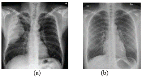
Chest X-ray radiography is used more frequently in clinical practice despite CT's superior sensitivity in detection. Its benefits are inexpensive, radiation-dose-minimum, easy to use, and widely accessible in community or general hospitals. Figure 1 provides examples of chest X-ray images of both COVID and non-COVID cases.

    Figure 1: Chest X-ray image of (a) COVID-19 patient and (b) Normal
patient.
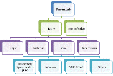
Manually diagnosing of this disease is time-taking, vulnerable to human error, and crucially necessitates the assistance of radiologists. It is imperative to consult a highly skilled radiologist because, as Figure 2 shows that anomalies identified in the early stages of COVID-19 may be similar to those observed in certain other pulmonary syndromes, including Viral Pneumonia (VP) and SARS-CoV-2.

Figure 2: Pneumonia types identified from CXR images [31].

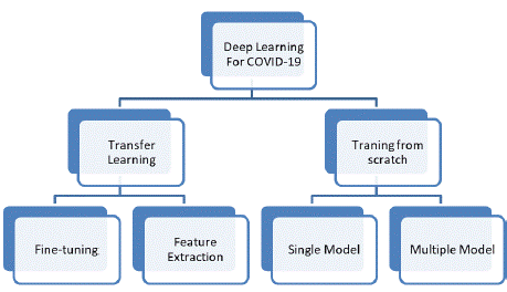
    Figure 3: Tasks of deep learning to COVID-19 detection on chest X-ray
images [32].
Correct interpretation of imaging results can be difficult due to the rapid onset of the disease and similarities to other respiratory conditions including pneumonia Any automated system aimed at diagnosing COVID-19 should consider other respiratory problems for a more comprehensive and accurate diagnosis. Pneumonia can arise from an infection of the lungs caused by the novel coronavirus-related disease COVID-19. Figure 2 illustrates several forms of pneumonia.
The Following are the Main Contributions of the Study:
• Table 1 provides an overview of the widely used COVID-19, pneumonia datasets that are openly accessible.
    Dataset
    Size
    Classes
    Ref.


    COVID Chest X-ray
    686    images
    Positive    COVID-19
    [35]


    COVID-19 Chest X-ray
    53    images
    Positive    COVID-19
    [36]


    COVID-19-Radiography database
    21,173    images
    Normal,    positive COVID-19, opacity, and viral pneumonia
    [34]


    Paediatric-CXR b
    5856    images
    Normal,    bacterial-pneumonia, viral- pneumonia
    [37]


    Pneumonia CXR
    2356    images
    viral-    pneumonia
    [38]
Table 1: Publicly available CXR datasets.
• It gives a summary of the various popular Deep Learning based approaches that have been applied in relevant studies.
• It describes in detail how well the various Deep Learning models perform.
This work aims to identify COVID-19, pneumonia from chest X-ray images with reduced computational power requirements, increased accuracy, and quick processing times.
Detection of Covid-19 using Deep Learning
Various methods for COVID-19 identification with deep learning and chest radiography images have been suggested. This section goes over the suggested techniques for detecting COVID-19 in chest X-ray images. Additionally, as seen in Figure 2, we offer a categorization of several deep learning approaches used to identify COVID-19 diseases in chest X-ray images. As per the reviewed articles, we discovered that training from scratch and transfer learning are the two primary methods used for COVID-19 detection utilizing chest X-ray images. While some methods used chest X-ray datasets to optimize the deep learning models for COVID-19 detection training, other methods applied classification techniques such as Support Vector Machine (SVM) for transfer learning using the features extracted from the taught models. In some respects, multiple deep learning models were developed to detect COVID-19, while others were trained with one CNN model from scratch.
Transfer Learning-based Methods: There are two types of transfer learning approaches: 1) Methods that rely on fine-tuning; 2) Features extraction techniques followed by classification techniques. This section reviews methods that employed X-ray images of chest and used transfer learning to localize COVID-19. We discuss feature extraction-based classification techniques after reviewing Strategies based on positive change.
A) Fine-Tuning Based Methods: The optimization method involves importing knowledge from another domain and loading into a deep learning model for a particular task, this method uses learning models to optimize small registered data sets [12]. A few methods have adjusted the training models on chest X-ray images using commercially available neural networks. For the purpose of leveraging chest X-ray images for COVID-19 early detection, a transfer learning strategy utilizing a pre-trained deep learning model on ImageNet was proposed in [13].
B) Features Extraction: This section reviews studies using classification methods to diagnose COPD by extracting deep features or common symptoms from chest X-ray images. The deep features that were extracted were from both conventional features like cooccurrence matrix features and ImageNet-based pretrained deep learning models. The suggested techniques to identify COVID-19 utilizing characteristics taken from chest X-ray images are described in the ensuing paragraphs.
Training from scratch: Reviews studies that described deep learning methods employing datasets of chest X-ray images that were trained from scratch for COVID-19 identification. As explained in the next subsections, single-model-based methods and multi-modelbased methods are two groups into which these methods can be separated.
A) Single model-based methods: In this approach, a single model-based Convolutional Neural Network was trained on chest X-ray images in order to detect COVID-19. In [14], a straightforward architecture with two convolution layers was created, trained, and displayed. Three public datasets provided data for this neural network's training: 1) The Joeseph Paul dataset, which includes 262 patients' 542 chest X-ray images [15]. 2) COVID-19 dataset developed at Qatar University by researchers [16,18]. 3) The Kermany et al. dataset [17].
B) Multiple model-based methods: The following paragraph provides more information on the several model-based techniques that are discussed in this chapter. These techniques integrate and train multiple deep learning models to identify COVID-19 from chest X-ray images. In [19], an autoencoder and a convolutional neural network were trained, with the autoencoder's latent space serving as the convolutional neural network's input. Using a dataset of chest X-ray images that included 400 positive cases of COVID-19 and normal cases is 400, this technique outperforms a VGG16 model by 2% [15]. A different approach is presented in [20] that uses chest X-ray images datasets to train ResNet50v2 and Xception networks for COVID detection and feature learning.
Literature Survey
We analyze CXR images showing signs of Viral infections with SARS-CoV-2 using DL techniques. Key research papers to implement transfer learning using data sets of CNN architectures trained on ImageNet. The sections below provide an overview of the CNN method for COPD detection and the data analyze for this study.
A CNN-based Model for COVID-19 Forecasting
A model was created for preprocessing and classifying a given chest X-ray picture using CNN architectures that have already been trained. Selecting the best preprocessing enhancement processes for optimal performance improvement requires a thorough understanding of the issue, data collection, and production environment COVID-19 diagnosis system based on Deep Learning, with defined steps to go including the later below.
Preprocessing: Preprocessing is required to improve the image quality and, in turn, the performance of the model. It needs to be resized, normalized, and sometimes converted to grayscale. Image preprocessing is the process of formatting images before using them for inference and model training. This preprocessing stage would be applied to images used in testing and training. In order to prepare image data for model input, preprocessing is required. In CNN systems, normalization and picture scaling are essential steps that maintain image stability and enhance model performance. A Convolutional Neural Network learns faster and the gradient descent is more stable when normalization is applied. Histogram equalization is an alternative image processing technique that uses image intensity histograms to enhance the global contrast of images, such as those found in Heidari et al. [10]. Shear is a type of image distortion that can be used to improve photos for computer vision applications, such as those found in Khan et al. [11], or to create or correct perspective angles.
The reason of resizing images is to maintain consistency, and resizing ensures that all input images are of the same size, which is essential for stable model training and inference. By resizing, strike a balance between computational cost and preserving the spatial features that a CNN can effectively learn.
This section provides a description of the preprocessing methods used in our studies. The various preprocessing methods utilized in our studies are listed in Table 2.
Names of Author
Year
Methods of image preprocessing
Heidari et al. (2020) [22]
2020
Histogram equalization technique, bilateral low-pass filter, and image scaling of 224 by 224 pixels
Jain et al. (2020) [23]
2020
640 by 640 pixels is the new size of the image.
Gaussian blur and rotation as augmentation techniquesKhan et al. (2022) [24]
2022
Techniques for augmentation: rotation, shear, reflections in both horizontal and vertical planes
The resolution of each image was lowered to 224 * 224 * 3.Islam et al. (2020) [25]
2020
The image has been resized to 224 by 224 pixels.
Sousa et al. (2022) [26]
2022
Image augmentation involving zoom, breadth, height, flipping horizontally and vertically, range rotation, and resizing the input from 200 * 200 to 300 * 300
Abbas et al. (2021) [27]
2021
Select five additional random angles for translation, rotation, rotation up, rotation down, and right/left. Histogram transformation
Gupta et al. (2022) [28]
2022
Resize the image to 128*128*3 pixels
Asnaoui and Chawki (2021) [29]
2021
Normalization of energy levels
Finite difference adaptive histogram equalization
Using geometric transformations (such as rescaling, rotating, shifting, shearing, zooming, and flipping) as methods for enhancement
Image scaled to 299 * 299 pixels for Inception Resnet V2.Jia et al. (2021) [30]
2021
Reduced image size to 299 x 299 x 3 for mobileNet adaptation
Images resized with the Modified ResNet to 256 x 256 x 3.
Table 2: Several methods for preparing Chest-X-ray images.
Traditional CNN Architecture: These days, CNN architectures can perform at an expert level on par with humans in a number of challenging visual tasks, including pathology identification and medical picture processing. There have been several different CNN designs put forth in the literature since the first effective CNN was created in 1998. It was referred to as LeNet and was extensively utilized for the handwritten digit recognition application. The developer of it was Yann LeCun. LeNet is a shallow design compared to existing models, with three convolutional, two averages pooling, and two fully connected layers. For feature extraction, CNN is utilized. an outline of a standard Convolutional Neural Network for COVID-19 prediction, which is explained in more detail below.
1. Input Layer: The input layer represents the starting point of the model. It takes the raw data (images, text, etc.) and passes it on to the next layers. For image data, this layer usually takes input in the form of a 3D array: height × width × channels (e.g., 256x256x3 for a colour image).
2. Batch Normalization: Normalizes the input to a layer by scaling and shifting, which helps improve training stability and speed.
3. Convolutional Layer: The convolutional layer is created by convolving learnable filters, frequently referred to as kernels, with the input images. The method is an element-wise point product and a sum to generate a number as a feature map element.
4. Pooling Layer: Pooling layer Convolutional networks can have both the basic convolutional layers and local and/or global pooling layers.
5. Flatten Layer: Converts multidimensional data into a 1D array. This is necessary because Dense layers expect input as a onedimensional vector rather than a multi-dimensional matrix.
6. Fully Connected Layer (Dense Layer): It is a fully integrated layer for distribution services and is widely used. Otherwise, they are called dense layers, and these layers receive higher properties. The map generated from the previous layer is converted into a probability vector indicating the probability of the input falling into each category.
7. Dropout Layer: Used to prevent overfitting by randomly setting a fraction of input units to zero during training.
A Summary of the Review Articles
Researchers have written a number of papers in recent years that concentrate on the automatic identification of respiratory illnesses including1 pneumonia and Covid-19. The dataset Chest X-ray14, made available by Wang et al. in 2017 [4], was used in the research conducted by Varshni et al. [3]. A deep learning-based method for the categorization of COVID-19 and pneumonia infection was presented by Mahmud et al. [5]. The Kaggle database provided the dataset for these investigations [2]. Li et al.'s method [1] for COVID-19 infection detection was proposed. The suggested method successfully distinguishes between Community-Acquired Pneumonia (CAP) and COVID-19 pneumonia. The COVNet deep learning model, which is a three-dimensional CNN architecture, is used for training. A publicly accessible dataset containing COVID-19 and Community- Acquired Pneumonia (CAP) CT scan samples is used. A sensitivity and specificity rate of 90% and 96%, respectively, were attained by the COVNet model. A deep learning-based model was proposed by Xu et al. [6] as a method for COVID-19 infection identification. Panwar et al. [7] presents a method based on convolutional neural networks, in which COVID-19 and normal images are classified using a 24-layer CNN model. In another work Hasan et al. [33] proposed weighted models of InceptionV3, MobileNetV2, VGG16 and DenseNet169 and Xception were proposed to detect pneumonia and normal cases by applying the transfer tilt concept to obtain a great performance of 92% accuracy. In Table 3. the various dataset and deep learning-based methods used by some Authors are listed.
Sr. No.
Authors
Objective
Dataset
Deep Learning Based Method
Result
1
Li et al.'s [1]
COVID-19 infection detection
dataset consisted of 4356 chest CT exams from 3,322 patients
COVNet deep learning model
sensitivity and specificity rate 90% and 96%
2
Kermany D [2]
Chest X-Ray Images for Classification
OCT dataset,
dataset includes 84,484 imagesCapsule network
accuracy of 99.6%,
3
Varshni et al. [3]
Diagnosis of chest conditions
dataset Chest X-ray14 consists of 112,120 frontal chest X-ray images
DenseNet-169 and Support Vector Machine
more than 90% accurate
4
Wang et al. [4]
Classification and Localization of Common Thorax Diseases
dataset Chest X-ray14 consists of 108,948 frontal-view X-ray images
DenseNet-169
accuracy of 80%,
5
Xu et al. [6]
COVID-19 infection identification
618 transverse-section CT samples
deep learning-based, Computed tomography
accuracy rate was 86.7%
6
Chowdhury [9]
screening viral and COVID-19 pneumonia
dataset comprises 3616 images of COVID-19-positive cases
Arti?cial intelligence, machine learning
accuracy, precision, sensitivity 99.7%
7
Heidari et al. [10]
images global contrast
dataset involving 8474 chest X-ray images
CNN model and Histogram equalization
accuracy of 94.5 %, 98.0% specificity
8
Khan et al. [11]
Enhance Images
dataset consisted of 3224 images from both COVID-19 infected and Healthy
Shear is a type of image distortion
Accuracy of 96.53%
9
N. Tajbakhsh. [12]
medical image analysis
database consisting of 121 CTPA datasets with a total of 326 Pes
CNN model fine-tuning
Accuracy of 95%
10
E. Irmark. [14]
COVID-19 disease detection
Contains 542 frontal chest x-ray images
deep convolutional neural network
accuracy of 99.20%
11
T. Rahman, A. Khandakar [18]
COVID-19 detection using chest X-ray images
dataset consisting of 18,479 CXR images
Image Enhancement technique
accuracy, precision, sensitivity, F1-score, and specificity were 95.11%, 94.55%, 94.56%, 94.53%, and 95.59% respectively
12
H. Hanafi, A. Pranolo [19]
Automatic COVID-19 disease detection based on X-ray images
45 COVID-19, 931 Bacteria
Pneumonia, 660 Virus
Pneumonia, 1203 Normalautoencoder and a convolutional neural network
83.5% Accuracy
13
Trivedi et al. [21]
automatic detection of pneumonia using chest X-ray images
dataset of 5856 chest X-ray images
Deep learning, CNN based architectures, Mobile Net
97.34% of Accuracy
Table 3: Some Authors list with their work.
Classifying images takes an extremely long time. Training a model with greater accuracy requires a larger collection of labelled data. It is costly and time-consuming to gather these datasets, though. Transfer learning strategies using already trained deep learning models could be one way to overcome these challenges. Transfer learning involves using large and diverse datasets such as ImageNet to pre-train a deep neural network model. The model must then be exported to another data set in the same area. Two possible approaches in transfer learning are feature extraction and fine-tuning.
Conclusions
This article examines Several deep learning (DL) methods for identifying COVID-19 from chest X-ray pictures are discussed in this article along with the current status of the field's research. This publication explains the various datasets and pre-trained CNN models from earlier studies. It has been determined that methods like transfer learning, data augmentation, and cross-validation can improve the generalization and adaptability of deep learning models. The article describes several datasets from earlier research. The automatic diagnosis of COVID-19 by DL techniques holds great potential; nevertheless, greater collaboration between computer scientists and medical professionals is required to develop more dependable and efficient DL models.
The deep learning methods for COVID-19 identification utilizing chest X-ray images are compiled in this review paper. Additionally, the datasets of chest X-ray images that were utilized in the methodologies that were assessed are summarized in this article. The article also discusses the tactics that have been assessed and identifies areas that could want improvement.
References
- Li L, Qin L, Xu Z, Yin Y, Wang X, Kong B, et al. Artificial intelligence distinguishes COVID-19 from community acquired pne2umonia on chest CT. Radiology. 2020; 296: 200905.
- Kermany D, Zhang K, Goldbaum M. Labeled Optical Coherence Tomography (OCT) and Chest X-Ray Images for Classifcation. Mendeley Data. 2018: V2.
- Varshni D, Thakral K, Agarwal L, Nijhawan R, Mittal A. Pneumonia Detection Using CNN based Feature Extraction. in 2019 IEEE International Conference on Electrical, Computer and Communication Technologies (ICECCT). 2019: 1–7.
- Wang X, Peng Y, Lu L, Lu Z, Bagheri M, Summers R. ChestX-ray14: Hospitalscale Chest X-ray Database and Benchmarks on Weakly-Supervised Classifcation and Localization of Common Thorax Diseases. 2017.
- Mahmud T, Rahman MA, Fattah SA. CovXNet: A multi-dilation convolutional neural network for automatic COVID-19 and other pneumonia detection from chest X-ray images with transferable multi-receptive feature optimization. Comput Biol Med. 2020; 122: 103869.
- Xu X, Jiang X, Ma C, Du P, Li X, Lv S, et al. A deep learning system to screen novel coronavirus disease 2019 pneumonia. Engineering. 2020; 6: 1122–1129.
- Panwar H, Gupta PK, Siddiqui MK, Morales-Menendez R, Singh V. Application of deep learning for fast detection of COVID-19 in X-rays using nCOVnet. Chaos Solitons Fractals. 2020; 138: 109944.
- COVID-19 Radiography Database-Kaggle.
- Chowdhury ME, Rahman T, Khandakar A, Mazhar R, Kadir MA, Mahbub ZB, et al. Can AI help in screening viral and COVID-19 pneumonia? IEEE Access. 2020; 8: 132665–132676.
- Heidari M, Mirniaharikandehei S, Khuzani AZ, Danala G, Qiu Y, Zheng B. Improving the performance of CNN to predict the likelihood of COVID-19 using chest X-ray images with preprocessing algorithms. Int J Med Inform. 2020; 144: 104284.
- Khan SH, Sohail A, Khan A, Lee YS. COVID-19 detection in chest X-ray images using a new channel boosted CNN. Diagnostics. 2022; 12: 267.
- Tajbakhsh N, Shin JY, Gurudu SR, Hurst RT, Kendall CB, Gotway MB, et al. Convolutional neural networks for medical image analysis: Full training or fine tuning?. IEEE Trans Med Imag. 2016; 35: 1299–1312.
- Qjidaa M, Mechbal Y, Ben-fares A, Amakdouf H, Maaroufi M, Alami B, et al. ‘Early detection of COVID19 by deep learning transfer model for populations in isolated rural areas. Proc Int Conf Intell. Syst Comput Vis. (ISCV). 2020: 1–5.
- Irmak E. ‘A novel deep convolutional neural network model for COVID-19 disease detection. Proc Med Technol Congr. (TIPTEKNO). 2020: 1–4.
- Cohen JP, Morrison P, Dao L, Roth K, Duong TQ, Ghassemi M. COVID-19 image data collection: Prospective predictions are the future. 2020: arXiv: 2006.11988.
- Chowdhury MEH, Rahman T, Khandakar A, Mazhar R, Kadir MA, Mahbub ZB, et al. ‘Can AI help in screening viral and COVID-19 pneumonia?. IEEE Access. 2020; 8: 132665–132676.
- Kermany DS, Goldbaum M, Cai W, Valentim CCS, Liang H, Baxter SL et al. Identifying medical diagnoses and treatable diseases by image-based deep learning. 2018; 172: 1122–1131.
- Rahman T, Khandakar A, Qiblawey Y, Tahir A, Kiranyaz S, Kashem SBA, et al. Chowdhury, Exploring the effect of image enhancement techniques on COVID-19 detection using chest X-ray images. Comput Biol Med. 2021; 132: 104319.
- Hanafi H, Pranolo A, Mao Y. CAE-COVIDX: Automatic COVID-19 disease detection based on X-ray images using enhanced deep convolutional and autoencoder. Int J Adv Intell Inform. 2021; 7: 49–62.
- Rahimzadeh M, Attar A. A modified deep convolutional neural network for detecting COVID-19 and pneumonia from chest X-ray images based on the concatenation of xception and ResNet50 V2. Informat Med Unlocked. 2020; 19: 100360.
- Trivedi M, Gupta A. A lightweight deep learning architecture for the automatic detection of pneumonia using chest X-ray images. Multimedia Tools and Applications. 2022; 81: 5515-5536.
- Heidari M, Mirniaharikandehei S, Khuzani AZ, Danala G, Qiu Y, Zheng B. Improving the performance of CNN to predict the likelihood of COVID-19 using chest X-ray images with preprocessing algorithms. Int J Med Inform. 2020; 144: 104284.
- Jain G, Mittal D, Thakur D, Mittal MK. A deep learning approach to detect Covid-19 coronavirus with X-ray images. Biocybern Biomed Eng. 2020; 40:1391–1405.
- Khan SH, Sohail A, Khan A, Lee YS. COVID-19 detection in chest X-ray images using a new channel boosted CNN. Diagnostics. 2022; 12: 267.
- Islam MZ, Islam MM, Asraf A. A combined deep CNN-LSTM network for the detection of novel coronavirus (COVID-19) using X-ray images. Inform Med Unlocked. 2020; 20: 100412.
- De Sousa PM, Carneiro PC, Oliveira MM, Pereira GM, da Costa Junior CA, de Moura LV, et al. COVID-19 classification in X-ray chest images using a new convolutional neural network: CNN-COVID. Res Biomed Eng. 2022; 38: 87–97.
- Abbas A, Abdelsamea MM, Gaber MM. Classification of COVID-19 in chest X-ray images using DeTraC deep convolutional neural network. Appl Intell. 2021; 51: 854–864.
- Gupta A, Gupta S, Katarya R, Ajum. InstaCovNet-19: A deep learning classification model for the detection of COVID-19 patients using Chest X-ray. Appl Soft Comput. 2021a; 99: 106859.
- El Asnaoui K, Chawki Y. Using X-ray images and deep learning for automated detection of coronavirus disease. J Biomol Struct Dyn. 2021; 39: 3615–3626.
- Jia G, Lam HK, Xu Y. Classification of COVID-19 chest X-Ray and CT images using a type of dynamic CNN modification method. Comput Biol Med. 2021; 134: 104425.
- Subramaniam K, Palanisamy N, Sinnaswamy RA, Muthusamy S, Mishra OP, Loganathan AK, et al. A comprehensive review of analyzing the chest X-ray images to detect COVID-19 infections using deep learning techniques. Soft Comput. 2023; 27: 14219–14240.
- Alahmari SS, Altazi B, Hwang J, Hawkins S, Salem T. A Comprehensive Review of Deep Learning-Based Methods for COVID-19 Detection Using Chest X-Ray Images. IEEE Access. 2022; 10: 100763-100785.
- Rabiul Hasan MD, Ullah SMA. Enhancing Pneumonia Diagnosis: An Ensemble of Deep CNN Architectures for Accurate Chest X-Ray Image Analysis. Lecture Notes in Networks and Systems. LNNS. 2024; 867: 255– 269.
- Tawsifur R. COVID-19 Radiography Database.
- Cohen JP, Morrison P, Dao L, Roth K, Duong T, Ghassemi M. COVID-19 Image Data Collection: Prospective Predictions Are the Future. arXiv. 20202006.11988.
- Chung A, Wang L, Wong A, Lin ZQ, McInnis P, Gunraj H. Figure 1 COVID-19 Chest X-ray.
- Mooney P. Chest X-ray Images (Pneumonia) [(accessed on 1 November 2022)].
- Kermany DS, Goldbaum M, Cai W, Valentim CCS, Liang H, Baxter SL, et al. Identifying Medical Diagnoses and Treatable Diseases by Image-Based Deep Learning. Cell. 2018; 172: 1122–1131.