
Research Article
Austin J Radiol. 2024; 11(5): 1247.
First Varicocele Embolizations in West Africa, in Dakar (Senegal) and Fair-Embo Concept
Diallo M¹*; Diallo A²; Ndaw MDB¹; Diack A¹; Amar NI¹; Diop AD²; Mbengue A¹; Diop AN³
¹Dakar Main Hospital, Senegal
²Fann University Hospital, Dakar, Senegal
³UFR Health Gaston Berger University, Saint-Louis, Senegal
*Corresponding author: Moustapha DIALLO, Specialist in military hospitals, Main Hospital of Dakar, medical imaging department, Dakar, Senegal. Tel: 00 221 77 65 72 700 Email: daddykr@hotmail.fr
Received: November 25, 2024; Accepted: December 13, 2024 Published: December 20, 2024
Summary
Objectives: Evaluation of our first experience of varicocele embolization with the FAIR-Embo concept.
Materials and Methods: This is a retrospective, descriptive and analytical study of fourteen (14) varicocele embolization procedures performed in 13 months between January 2022 and February 2023. The embolizations were performed with a sclerosing agent, surgical sutures and a coil after a right femoral venous vascular access followed by catheterization of the left testicular veins.
Results: The mean age of the patients revolved around 33 +/- 6 years with extremes of 21 and 47 years. The dominant indication was a varicocele in a context of primary infertility and concerned half of our patients (50%). All our patients had a varicocele on ultrasound which was bilateral in 9 patients or 64.28% and unilateral left in 5 patients or 35.71%. The overall spermiological profile before embolization was oligo-astheno-terato-necro zoospermia with an average sperm density of 29.35 million/ml. The success rate was 85.71% with 2 cases of failure due to anatomical variants. We had noted a clear improvement in sperm parameters after embolization with the occurrence of three (3) cases of pregnancies within an average time of 9 months.
Conclusion: The varicocele embolization is a good alternative to surgical treatment. It is, indeed, an effective method, performed on an outpatient basis and now available in West Africa thanks to the FAIR-Embo concept.
Keywords: Varicocele; Embolization; Fair-embo; Dakar; Senegal
Introduction
The varicocele is defined as a dilatation and reflux of the veins of the scrotal pampiniform plexus. It is usually primary, due to valvular incontinence of the testicular vein (or internal spermatic vein), [1] or secondary to obstruction of the renal vein or extrinsic compression of the spermatic vein.
Varicocele was historically described by Celsius, Hippocrates, and mentioned by Ambroise Pare in 1541 who drew the connection between the symptoms of varicocele and reflux in the spermatic vein [2].
It is found in 35% of men with primary infertility and in 71% to 81% of men with secondary infertility [3]. The existence of a varicocele is associated with a high risk of alteration of sperm parameters and abnormally high levels of free radicals, exposing spermatozoa to DNA damage [4-7]. Furthermore, two recent meta-analyses conclude that after varicocelectomy, there is an improvement in spermogram parameters [8-9] and a reduction in nucleotide damage to spermatozoa [10].
The diagnosis of varicocele is first clinical and the Doppler ultrasound of the scrotum serves as the paraclinical benchmarking reference examination allowing to confirm the diagnosis and to evaluate its impact. The spermogram is the essential examination to qualitatively and quantitatively assess the sperm.
Varicoceles generally fall into clinical and sonographic stages/ categories [8-11-12] but there is still no universally accepted classification of the severity of varicoceles, leading to great heterogeneity in the literature [2-13-15].
Some inconsistencies also exist in terms of recommendation of therapeutic strategy and indication. In this particular context, the European Association of Urology (EAU) guidelines on reproductive health suggest treatment of varicoceles in adults with abnormal semen parameters and otherwise unexplained infertility [11].
The basis of varicocele treatment is occlusion of the spermatic vein. Surgical treatment is the gold standard treatment, meaning the treatment set as a reference, but embolization by interventional radiology technique is a good alternative.
The embolization by interventional radiology technique is an equally effective method, with fewer complications and can be performed on an outpatient basis [16].
It has been widely practiced for several years in developed countries but unfortunately, its accessibility is very limited in West Africa because embolization agents are very rarely available and their cost is also high.
Fortunately, with the Fair-Embo Concept [17], this barrier linked to the inaccessibility of embolization agents is removed with the use of suture threads which are available, at a very low cost and with an easy-to-use technique.
In Senegal, for instance, the embolization of varicoceles is made possible thanks to this Fair-Embo Concept, and the main objective of this study is to describe our technique and give the first results.
Materials and Methods
This was a retrospective, descriptive and analytical study of fourteen varicocele embolization procedures performed from January 2022 to February 2023 at the main hospital in Dakar in a multipurpose angiography room equipped with a General Electric brand device and the OPTIMA IG 5 330 model.
We included all patients who underwent varicocele embolization during this period and who had a complete file, namely a testicular Doppler ultrasound and a spermogram before and after embolization. Four files with incomplete information were excluded.
Data entry and processing were performed using Kobocollect software and analyzed with MICROSOFT EXCEL 2016 software. The parameters studied were the age of the patients, the indication for embolization, the ultrasound data before and pre-embolization (grade of reflux according to the Hirshen classification and testicular volumes), the spermogram data before and after embolization (density, mobility, vitality, morphology), the embolization agent, the technical success of the embolization and the occurrence of pregnancy. Our technique consisted of right femoral venous vascular access with a 05 French (Fr) diameter and 10 cm or 25 cm long valve introducer. Catheterization of the left renal vein, most often with a 04 Fr Cobra probe mounted in a TerumoR hydrophilic guide, allowed venography to be performed in order to visualize the ostium of the left testicular vein. The latter was then catheterized with the same probe and the same guide, then an angiography confirmed spontaneous reflux or after Valsalva maneuver, up to the pampiniform plexus and to look for possible collaterals. The Cobra probe was brought as distal as possible to the foot of the sacroiliac joint or upstream of the most proximal collateral. Subsequent embolization was most often performed with first a sclerosing agent (an ampoule of aetoxisclerolR 2%) injected in Valsalva in the form of foam obtained with a mixture of air and sclerosing product. This injection was done under digital compression of the spermatic cord by the patient held for 10 minutes to allow satisfactory contact with the wall of the vessels. This was followed by the release of absorbable suture threads 2.0 VicrylR with a length of approximately 02 cm and on average between 06 and 10, flushed with physiological serum (figure 2). The guide was introduced into the probe and pushed until it exited the catheter, objectifying the release of the threads into the vein. In the case where coils were used, their release was done under control and a coil of a length ranging from 20 cm and a diameter of 14 mm was sufficient to obtain embolization. Technical success at the end of the procedure was objectified by an occlusion of the testicular vein downstream of the embolization site without permeability of the collaterals. Figures 1 and 2 show the equipment used and Figures 3 and 4 illustrate embolization in two patients with sclerosing agent, sutures and coil. At the end of the procedure, the silet was removed and then manual compression was performed for about 5 minutes followed by a compressive dressing that will be left in place for 2 hours. The patients were kept under observation and then executed after approximately 3 to 4 hours with a prescription for painkillers to be taken as needed. They were advised to stop physical exertion and lifting heavy loads for a week and to stop sexual intercourse for 48 hours. The total cost of the procedure is 400,000 FCFA (four hundred thousand CFA francs).
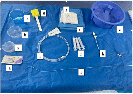
    Figure 1: Embolization materials.
Note: a, b and c: cups; d: tampon; e: sterile compress; f: sterile field; g:
water tray; h: suture thread; i: hydrophilic guide; j: syringes; k: desilet; l:
cobra probe.
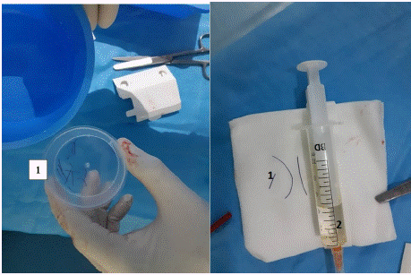
    Figure 2: Preparation of the suture threads.
Note: 1: Pieces of suture threads. 2: Syringe with physiological serum for
flushing.
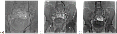
    Figure 3: Non-subtraction frontal angiography image showing varicocele
embolization with sutures and sclerosing agent.
Note: (a): Diagnostic stage with the left testicular vein flowing back to the
pampiniform plexus. (b): Therapeutic stage and (c) post-treatment control.
1 Cobra catheter; 2 Incompetent left gonadal vein; 3 Digital compression of
the left spermatic cord; 4 Embolization of the gonadal vein at the end of the
procedure.
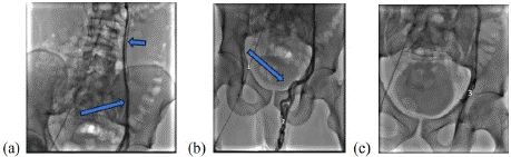
    Figure 4: Non-subtraction frontal angiography image showing coil
embolization of varicocele.
Note: Diagnostic step in (a and b) with the testicular vein flowing back to the
pampiniform plexus (single arrow) and a collateral flowing below the plane
of the iliac crest (short arrow). In (c), Coils in place performing embolization
downstream of the collateral.
1 Cobra probe; 2 Veins of the left pampiniform plexus; 3 coils.
Results
The mean age of the patients was 33 years with extremes of 21 years and 47 years.
The indication for embolization was a varicocele in a context of infertility with disturbance of the spermogram in 7 patients, i.e. half (50%) and symptomatic varicoceles with scrotal pain and heaviness for the other 7 patients.
On Doppler ultrasound, the varicocele was bilateral in 9 patients (64.28%) and unilateral on the left in 5 patients (35.71%). Ultrasound grade III was the most common, noted in 9 patients (56.25%) and grade II in 7 patients (43.75%). Testicular hypotrophy was noted bilaterally in 7 patients and unilaterally on the left in 1 patient.
Table 1 summarizes the data from the patients' ultrasound scans.
![]()
    Features
    Attendance
    Percentage


    Affected side


    right side
    0
    0%


    left side
    5
    35,1%


    bilateral form
    9
    64,25%


    Ultrasound grade of varicocele


    Left side


    grade I
    0
    0%


    grade II
    5
    35,1%


    grade III
    9
    64,25%


    Right side


    grade I
    1
    7,14%


    grade II
    4
    28,57%


    grade III
    4
    28,57


    Testicular volume


    Left


    = 10 cc
    6
    42,85%


    < 10 cc
    8
    57,14%


    Right


    = 10 cc
    7
    50%


    < 10 cc
    7
    50%
Table 1: Distribution of patients according to ultrasound data.
In patients with altered spermograms, the overall abnormality was oligo-astheno-teratozoospermia with a mean sperm density of 29.35 million/ml, mean motility of 18%, mean vitality of 36%, and 70% abnormal shape (Table 2).
![]()
    Sperm parameters
    General averages


    Density (Million/ml)
    29,35


    Mobility (%)
    18,09


    Vitality (%)
    36,64


    Abnormal form
    70,64
Table 2: Overall averages of spermogram parameters before embolization for
all patients.
The embolization agents used were sutures associated with a sclerosing agent in 7 patients (50%) and used alone in 4 patients (28.5%). One patient had been embolized with a coil (7.14%).
Our technical success was 85.71% with 2 failures (14.8%) of testicular vein catheterization due to anatomical variants. The procedures lasted between 30 and 60 minutes depending on the ease of catheterization and the embolizing agent used.
The postoperative course was simple. We had no complications during or after embolization.
All patients had a control spermogram at 6 months after embolization and a clear improvement was noted concerning the mobility, vitality and concentration of spermatozoa. The patient who had azoospermia had a timid induction of spermatogenesis with a sperm concentration at 0.67 million. (Table 3) summarizes the comparison of spermograms before and 06 months after embolization.
![]()
Sperm parameters
Pre-embolization spermogram
Spermogram at
6 months post embolizationDensity (Million/ml)
29,35
49,80
Mobility (%)
18,09
36,6443,10
54,40Vitality (%)
70,64
66,30
Abnormal morphology (%)
Table 3: Mean spermogram parameters before and after embolization.
On the follow-up ultrasound performed 3 months later, testicular volumes had increased in 11 patients, including two on the left side and 9 on the right side. A slight reflux (grade 1) persisted in two patients. (Table 4)
![]()
    Ultrasound Characteristics
    Before Embolization
    After Embolization


    Ultrasound grade of varicocele


    Left side


    grade I
    0
    1


    -grade II
    5
    0


    -grade III
    9
    0


    Right side


    -grade I
    1
    1


    -grade II
    4
    0


    -grade III
    4
    0


    Testicular volume


    Left


    = 10 cc
    6
    7


    -< 10 cc
    8
    7


    Right


    -= 10 cc
    7
    11


    -< 10 cc
    7
    3
Table 4: Reflux grade and testicular volume before and after embolization.
In patients who were in a context of primary infertility, 3 cases of pregnancy (21.42%) had occurred within an average of 9 months after embolization.
Discussion
Varicocele is a cause of male infertility and usually a pathology of young subjects. The mean age of our patients is 33 years and half of our patients were consulted for primary infertility. Our study is comparable to that of Zini L et al [18], Bonnet Q et al [19] and Giuseppe F. et al [20] where the mean age of their patients is 31 and 33 years. Varicoceles are found in approximately 15% of adult men and 35% of those who complain of primary infertility. The existence of a varicocele is associated with a high risk of alteration of sperm parameters and abnormally high levels of free radicals, exposing spermatozoa to DNA damage [4-7].
Varicocele can cause scrotal pain with heaviness, outside the context of infertility. Scrotal pain represented the second reason for consultation in our study. This was also the case in the study conducted by Oumar Y et al [21].
Varicoceles are mostly located on the left, sometimes bilateral and very rarely isolated on the right as shown in our study and several other studies. We had 9 patients (64.28%) with bilateral varicoceles and 5 patients (35.71%) with unilateral left varicoceles. Bilateral damage was the most common in the Tunisian study of Jallouli H et al. [22] as well as that of Yigal Gat et al. [23]. In the series of Binhazzaa M et al. [24] the left damage was the most common (83.6%), as well as in the series of Diallo A.B et al [25] (84.9%) and Benazzouz M et al [26] (89.74%). One explanation is the primary valvular incompetence of the spermatic veins which affects the left side in 40% and the right side in 23%. Also, the opening of the left spermatic vein at a right angle into the left renal vein and its long vertical course are factors favoring reflux.
Another explanation for this left predominance is the Nutcracker Syndrome (NS) or the Nutcracker phenomenon. This involves compression of the left renal vein between the superior mesenteric artery and the aorta, leading to a decrease in blood flow from the left renal vein to the inferior vena cava and its diversion to the spermatic vein [27]. In the presence of an isolated right varicocele of recent onset, it is recommended to look for a right renal neoplastic process.
In our study, varicocele was associated with bilateral testicular hypotrophy in 7 patients, i.e. 50%, and unilateral left testicular hypotrophy in 1 patient. On the other hand, Traoré M [28] found hypotrophy in 1 patient (3%), which was more marked on the left than on the right. The explanation for testicular hypotrophy would be that the ischemic lesions generated by reflux affect the seminiferous tubules, Sertoli cells, germ cells and later Leydig cells [29]. The suffering of these different components of the testicular parenchyma will have repercussions on trophicity, therefore on the volume which in turn will decrease depending on the severity of the reflux. After embolization, an increase in testicular volumes can be noted. This was not the case in our patients. This could be explained by the short delay of the ultrasound control, carried out 3 months after embolization. Benazzouz M et al [26] report that two prospective randomized studies in adolescents showed an increase in homo and contralateral testicular size after treatment of varicocele compared to those who had not been treated.
The other impact of varicoceles is the alteration of spermogram parameters in a global or segmental manner. The overall abnormality on the spermograms of our patients was oligo-asthenoteratozoospermia, as found in most studies, notably that of Traoré M [28].
These results, except for necrozoospermia, correspond to the "stress pattern" that was described by Mac Leod. This is a nonpathognomonic association of spermogram abnormalities noted in cases of varicocele: a decrease in mobility, sperm density and a number of abnormally shaped spermatozoa greater than 15%. The mechanism of alteration of sperm parameters during varicocele has been described by Clavijo R et al [30] as the association of 3 elements: Oxidative stress, testicular hypoxia and hyperthermia. Our embolization technique is almost akin to that of other teams, particularly in terms of vascular access (most often right femoral vein), the catheterization probe (Cobra type) and the bilaterality of the treatment. We can cite the studies of Zini L et al [18], Bilreiro C et al [31] and Keoghane S et al [32]. In patients with bilateral varicocele, unilateral left embolization allowed correction of the right varicocele in 57.14%. Right varicoceles are most often supplied by the left side and treatment of the latter is sufficient to make the varicocele disappear on the right. However, some teams systematically treat both sides in the case of bilateral varicocele. The most commonly used embolization agents are coils, biological glue and more rarely sclerosing agents. Coils and biological glue are currently not very accessible in West Africa because of their high cost and low availability.
Fortunately, sutures have recently been proposed as an embolization agent by Vidal V. of the Fair-Embo project "Fair and equitable embolization" which demonstrated that existing embolization agents (coils, calibrated particles and liquid agents) can be easily and effectively replaced by simple fragments of surgical suture threads [17]. They act as foreign bodies inside the vessels and cause the formation of an aggregate that ends up obstructing the vessel. These sutures are available everywhere in the world, at any time and they are very affordable.
The technique of delivering suture fragments through the catheter is less delicate, it is identical to the injection of gelatin sponge torpedoes or uncontrolled delivery coils. The use of suture threads of appropriate diameter according to the internal diameter of the catheter is essential for the suture fragments to progress correctly in the catheter. The disadvantage is that they are not radiopaque and the only way to ensure their release is to introduce a guide into the catheter that will be pushed to the exit, guaranteeing the release of the thread. Also, their use lengthens the procedure times because embolization takes longer to be done compared to conventional agents. We had effectively used coils in a patient and in comparison, with suture threads, embolization was obtained in a shorter time.
We are the first to have used suture threads for varicocele embolization. Moreover, suture threads were effectively used by Diop AD et al [33] in 2020 in Dakar (SENEGAL) to treat a renal arterial pseudoaneurysm.
The effectiveness of coils and biological glue is no longer to be demonstrated. They have been used successfully in the studies of Bilreiro C et al [31] and Keoghane et al [32]. The biological glue must be mixed with Lipiodol (radiopaque oil) and this adds an additional cost.
Sclerosing agents are often used in association in the treatment of varicoceles. They are effective with rare complications. They are more accessible in our region and with a low cost. We had associated them most often with suture threads. Zini L et al [18] had used them in association with an occlusive endovascular metal stent. Varicocele embolization is effective with a technical success rate of over 90%, proven by several studies comparing it to Subinguinal Microsurgery (SIM), which is the reference surgical technique for surgical treatment. Taking into account our overall activity outside the study period, the failure rate of our center is estimated at 5.8% due to anatomical variants of the testicular vein making its catheterization impossible.
A meta-analysis by Liu et al [34] comparing endovascular and surgical treatments of varicocele in 2138 patients concluded that endovascular treatment of varicocele is associated with similar rates of recurrence and subsequent pregnancy outcomes compared with surgical treatment, but with lower rates of adverse events. The efficacy of varicocele embolization is also reflected in the improvement of spermogram parameters and this was the case in all our patients.
Giuseppe Fallara et al, in their meta-analysis of prospective trials comparing any treatment for varicocele (surgical or radiological) [19], reported that treatment for varicocele results in a significant improvement compared to baseline in terms of sperm concentration (mean difference 14.22 million/ml), progressive motility (mean difference 9.76%), and normal morphology (mean difference 6.14%). Çayan et al [35], demonstrated that induction of spermatogenesis is possible after embolization in men with non-obstructive azoospermia and without associated genetic abnormalities. Similarly, in a study by Esteves [36] involving peripheral azoospermic patients, varicocele cure allowed induction of spermatogenesis in 43.9% of patients.
The contribution of varicocele treatment on natural fertility is reported by Abdel-Meguid et al [37] in their randomized controlled study with better fertility in the treated patient group (n = 75) compared to the untreated group (n = 75) and the spontaneous pregnancy rate was 32.9% in the treated group against 13.9% in the untreated group. In our study we had an induction of pregnancy in the wives of 3 patients (14.28%) after varicocele embolization.
Conclusion
Varicocele embolization is an effective technique already proven by several studies comparing it with the standard surgical treatment, meaning the benchmarking reference surgical treatment. It is still not widely available in West Africa due to difficult access to embolization materials and limited human resources in interventional radiology. Thanks to the Fair-Embo Concept, embolization is made accessible by suture threads, which are widely available and at very low cost. Our team is the first to have used suture threads in the embolization of varicoceles. Our initial results are consistent with those in the literature and, therefore, a technical success with suture threads associated with a sclerosing agent or used alone.
The limitation of this study is the small number of scopes of its patients, and a broader study will need to be carried out to confirm the effectiveness of suture threads in the embolization of varicoceles.
References
- Bigot JM, Tassart M, Le Blanche A. Traitement endovasculaire des varicocèles Encycl Méd Chir. Radiodiagnostic-Urologie-Gynécologie. 2003; 10: 34–450.
- Bertolotto M, Freeman S, Richenberg J, Belfield J, Dogra V, Huang DY, et al. Ultrasound evaluation of varicoceles: systematic literature review and rationale of the ESUR-SPIWG Guidelines and Recommendations. J Ultrasound. 2020; 23: 487–507.
- Gorelick JI, Goldstein M. Loss of fertility in men with varicocele. Fertil Steril. 1993; 59: 613–6.
- Kass EJ, Reitelman C. Adolescent varicocele. Urol Clin North Am. 1995; 22: 151–9.
- Zini A, Azhar R, Baazeem A, Gabriel MS. Effect of microsurgical varicocelectomy on human sperm chromatin and DNA integrity: a prospective trial. Int J Androl. 2011; 34: 14–9.
- Mostafa T, Anis TH, El-Nashar A, Imam H, Othman IA. Varicocelectomy reduces reactive oxygen species levels and increases antioxidant activity of seminal plasma from infertile men with varicocele. Int J Androl. 2001; 24: 261–5.
- Practice Committee of the American Society for Reproductive Medicine, Society for Male Reproduction and Urology. Report on varicocele and infertility: a committee opinion. Fertil Steril. 2014; 102: 1556–60.
- Baazeem A, Belzile E, Ciampi A, Dohle G, Jarvi K, Salonia A, et al. Varicocele and male factor infertility treatment: a new meta-analysis and review of the role of varicocele repair. Eur Urol. 2011; 60: 796–808.
- Agarwal A, Deepinder F, Cocuzza M, Agarwal R, Short RA, Sabanegh E, et al. Efficacy of varicocelectomy in improving semen parameters: new metaanalytical approach. Urology. 2007; 70: 532–8.
- Zini A, Dohle G. Are varicoceles associated with increased deoxyribonucleic acid fragmentation? Fertil Steril. 2011; 96: 1283–7.
- Minhas S, Bettocchi C, Boeri L, Capogrosso P, Carvalho J, Cilesiz NC, et al. European Association of Urology Guidelines on Male Sexual and Reproductive Health: 2021 Update on Male Infertility. Eur Urol. 2021; 80: 603–20.
- Baazeem A, Belzile E, Ciampi A, Dohle G, Jarvi K, Salonia A, et al. Varicocele and male factor infertility treatment: a new meta-analysis and review of the role of varicocele repair. Eur Urol. 2011; 60: 796–808.
- Bertolotto M, Cantisani V, Drudi FM, Lotti F. Varicocoele. Classification and pitfalls. Andrology. 2021; 9: 1322–30.
- Dubin L, Amelar RD. Varicocele size and results of varicocelectomy in selected subfertile men with varicocele. Fertil Steril. 1970; 21: 606–9.
- Lotti F, Frizza F, Balercia G, Barbonetti A, Behre HM, Calogero AE, et al. The European Academy of Andrology (EAA) ultrasound study on healthy, fertile men: Scrotal ultrasound reference ranges and associations with clinical, seminal, and biochemical characteristics. Andrology. 2021; 9: 559–76.
- Lima SS, Castro MP, Costa OF. A new method for the treatment of varicocele. Andrologia. 1978; 10: 103–6.
- Vidal V, Hak JF, Brige P, Chopinet S, Tradi F, Bobot M, et al. In Vivo Feasibility of Arterial Embolization with Permanent and Absorbable Suture: The FAIREmbo Concept. Cardiovasc Intervent Radiol. 2019; 42: 1175–82.
- Zini L, Rigot JM, Ballereau C, Dehaene JL, Lemaitre L, Mazeman E. Apport de l’embolisation de la varicocèle chez 51 patients infertiles. Andrologie. 2001; 11: 56–60.
- Bonnet Q, Coppens L, Delvigne A, Waltregny D. [Favorable impact of left antegrade sclerotherapy of clinical left varicocele on spermogram]. Prog Urol. 2020; 30: 281–7.
- Fallara G, Capogrosso P, Pozzi E, Belladelli F, Corsini C, Boeri L, et al. The Effect of Varicocele Treatment on Fertility in Adults: A Systematic Review and Meta-analysis of Published Prospective Trials. Eur Urol Focus. 2023; 9: 154–61.
- Diarra Y. Aspect de l’échographie doppler dans le diagnostic de la varicocèle au CHU Gabriel Toure de Bamako–mali: Mémoire en ligne. Bamako. Université de Bamako. 2023.
- Jallouli H, Hadj Slimen M, Sahnoun A, Kechou S, Ben Amar S, Bahloul A, et al. [Surgical treatment of varicocele improves fertility and facilitates medically assisted procreation]. Prog Urol. 2008; 18: 543–9.
- Gat Y, Bachar GN, Zukerman Z, Belenky A, Gornish M. Varicocele: a bilateral disease. Fertil Steril. 2004; 81: 424–9.
- Binhazzaa M, Bounasr E, Perez G, Almont T, Soulie M, Faruch M, et al. Comparison of subinguinal microsurgical varicocelectomy vs percutaneous embolization in infertile men. Prog Urol. 2016; 26: 1178–84.
- Diallo AB, Bah I, Barry M, Diallo TMO, Bah MD, Kanté D, et al. La varicocèle de l’adulte: aspects anatomo-cliniques et resultats therapeutiques au service d’urologie-andrologie du CHU de Conakry, Guinee. African Journal of Urology. 2015; 21: 137–41.
- Benazzouz MH, Essatara Y, El Sayegh H, Iken A, Benslimane L, Nouini Y. [Impact of varicocele on testicular volume and sperm parameters]. Pan Afr Med J. 2014; 19: 334.
- Dong W, Yao Y, Huang H, Han J, Zhao X, Huang J. Surgical management of nutcracker phenomenon presenting as left varicocele in adolescents: a novel approach. J Pediatr Urol. 2014; 10: 424–9.
- Traoré O. varicocèle chez l’adulte au service d’Urologie du CHU Gabriel Touré. PhD Thesis. USTTB. 2021.
- Boyer L, Hermabessière J, Boyer-Medeville C, Boissier A, Viallet JF. Role of transrectal echography in the evaluation of male infertility. Apropos of 91 studies. J Radiol. 1994; 75: 321–6.
- Clavijo RI, Carrasquillo R, Ramasamy R. Varicoceles: prevalence and pathogenesis in adult men. Fertil Steril. 2017; 108: 364–9.
- Bilreiro C, Donato P, Costa JF, Agostinho A, Carvalheiro V, Caseiro-Alves F. Varicocele embolization with glue and coils: A single center experience. Diagn Interv Imaging. 2017; 98: 529–34.
- Keoghane SR, Jones L, Wright MP, Kabala J. Percutaneous retrograde varicocele embolisation using tungsten embolisation coils: a five year audit. Int Urol Nephrol. 2001; 33: 517–20.
- Diop AD, Diop AN, Hak J-F, Di Bisceglie M, Bartoli J-M, Guillet B, et al. Hemostatic embolization of renal artery pseudoaneurysm using absorbable surgical suture (FairEmbo concept). Diagn Interv Imaging. 2020; 101: 757–8.
- Liu Q, Zhang X, Zhou F, Xi X, Lian S, Lian Q. Comparing Endovascular and Surgical Treatments for Varicocele: A Systematic Review and Meta-Analysis. Journal of Vascular and Interventional Radiology. 2022; 33: 834-840.e2.
- Cayan S, Kadioglu A, Orhan I, Kandirali E, Tefekli A, Tellaloglu S. The effect of microsurgical varicocelectomy on serum follicle stimulating hormone, testosterone and free testosterone levels in infertile men with varicocele. BJU Int. 1999; 84: 1046–9
- Esteves SC, Miyaoka R, Roque M, Agarwal A. Outcome of varicocele repair in men with nonobstructive azoospermia: systematic review and meta-analysis. Asian J Androl. 2016; 18: 246–53.
- Abdel-Meguid TA, Al-Sayyad A, Tayib A, Farsi HM. Does varicocele repair improve male infertility? An evidence-based perspective from a randomized, controlled trial. Eur Urol. 2011; 59: 455–61.