
Case Report
Austin J Radiol. 2024; 11(1): 1227.
Neurogenic Heterotopic Ossification in the Non-paretic Limb: A Case Report
Zahra SM Husain*; Abdullah Saad Al Driweesh
Department of Medical Imaging, Dammam Medical Complex, Dammam, Saudi Arabia
*Corresponding author: Zahra SM Husain Department of Medical Imaging, Dammam Medical Complex, Dammam, Saudi Arabia. Email: Zahra.4@live.com
Received: January 31, 2024 Accepted: February 19, 2024 Published: February 26, 2024
Abstract
Heterotopic Ossification (HO) is a pathological condition characterized by the formation of bone outside the skeletal tissues, which is usually caused by either traumatic or neurogenic factors. Neurogenic HO is a rare complication that can occur after a cerebral or spinal injury. The condition can manifest with a range of symptoms and is often challenging to diagnose clinically. Although various imaging techniques have been employed to diagnose HO, clinicians and radiologists may occasionally encounter radiological features of HO that can resemble other disease conditions. In this report, we present a unique case of neurogenic HO that developed in the non-paretic limb of a patient with Traumatic Brain Injury (TBI).
Keywords: Heterotopic Ossification; Hemiplegia; CT; MRI
Introduction
Heterotopic Ossification (HO) is a pathological condition characterized by the formation of lamellar bone in the soft tissue adjacent to a joint. It is commonly associated with Spinal Cord Injury (SCI), Traumatic Brain Injury (TBI), burn injury, and orthopedic trauma [1]. HO can also occur in patients with post-stroke hemiplegia, although the incidence is low (0.5–1.2%) as reported by Varghese [2].
The pathogenesis of HO is not fully understood. It is postulated that the major pathogenic factors are immobilization and aggressive joint mobilization to preserve joint range of motion. Other factors that may predispose to HO are spasticity, fracture, infection, deep vein thrombosis and pressure ulceration. HO can be categorized into traumatic and neurogenic types. Traumatic HO may develop after direct injury or surgery to the muscles (mainly following total joint arthroplasty). Neurogenic HO may develop after neurological insults, such as brain injury or SCI [3].
HO in the non-paretic limb is rare; there are only a few case reports in the literature. We present here the case of an elderly man who had right-sided post-stroke hemiplegia with HO in the non-paretic limb.
Case Report
A 75-year-old male known case of hypertension, type II diabetes and ischemic heart disease. He presented to the emergency department with five hours history of right sided weakness. Neurological examination revealed motor power of 1/5 in the right upper and lower limb with no sensory deficit. Glasgow Coma Scale (GCS) was 13/15. Non-enhanced CT scan of the brain was performed and showed left basal ganglia acute intra-parenchymal hemorrhage (Figure 1). The diagnosis of acute hemorrhagic stroke was established. The patient was admitted under the neurology team and was managed conservatively. Following discharge, he underwent two sessions of physiotherapy after which he lost the follow-up.
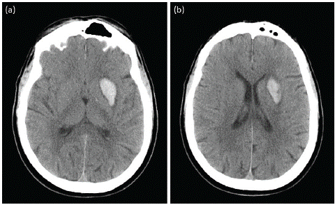
Figure 1: Non-enhanced CT scan of the brain at the level of basal ganglia (a) and lateral ventricles (b). There is intra-parenchymal hematoma in the left basal ganglia and extending to the ventricular level. Surrounding brain tissue edema causing mass effect and partial effacement of the ipsilateral frontal horn of the lateral ventricle.
Ten months after his initial visit, the patient presented to the emergency department with two weeks history of left hip pain with decreased range of motion. No history of trauma was reported. Pelvis radiograph was done and demonstrated peri-articular calcification involving the medial and lateral aspect of the left hip joint (Figure 2). Some of the calcifications are corticated. No fractures or soft tissue swelling. The patient received analgesia and was given an appointment with the orthopedist.
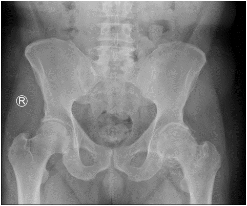
Figure 2: Frontal pelvis radiograph. Left hip joint peri-articular corticated calcification involving the medial joint space to a greater extent. No right hip joint peri-articular calcification.
The orthopedist requested pelvis CT (Figure 3) and MRI (Figure 4) for the patient. CT scan clearly demonstrated the presence of peri-articular corticated fragments with no pseudo-arthrosis. In MRI, the peri-articular fragments follow the signal intensity of the bone marrow in all sequencessuggestive of mature heterotopic ossification. Axial T2* sequence (Figure 5) showed fluid collection in the posterior aspect of the femoral neck denoting adventitious bursa secondary to the pressure effect.
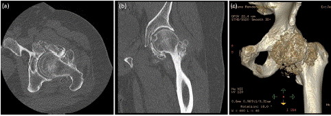
Figure 3: CT scan of the pelvis in bone window axial (a), coronal (b) and 3D bone reconstruction (c). All peri-articular calcifications are corticated and have variable sizes and shapes.
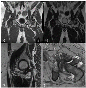
Figure 4: Pelvis MRI coronal T1 weighted image (a), coronal (b) and sagittal (C) T2 weighted images and axial gradient (d) sequence. All peri-articular calcifications follow the bone marrow signal intensity in all sequences.
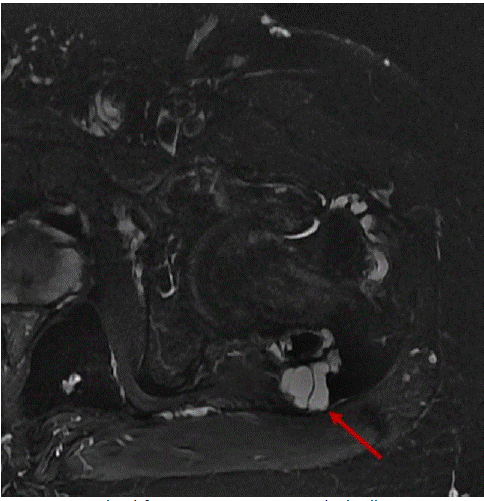
Figure 5: Axial T2* fat-saturation sequence. Fluid collection adjacent to the posterior aspect of the femoral neck suggestive of adventitious bursa (arrow).
Based on the combined imaging features, the diagnosis of mature heterotopic ossification was established. The patient was not a candidate for surgery due to his multiple comorbidities and therefore he was managed conservatively.
Discussion
The occurrence of HO in post-stroke hemiplegia is extremely uncommon, with only a handful of cases documented in the literature. The exact cause of HO is still unknown. Some researchers have suggested that neuronal control mechanisms are involved in the development of HO. Others have argued that a specific neurotransmitter may regulate this metabolic process. HO, osteoporosis, Charcot joint, obesity- related increased bone density, and other conditions are believed to be associated with this control mechanism [4-5]. There are some factors that may initiate or stimulate the neurogenic HO. These include: long-lasting coma, mechanical ventilation, increased muscle tone, and restricted movement in the affected limb. The precise mechanisms of how these factors influence HO formation are unclear [6].
In addition to these neurogenic factors, some researchers have claimed that immobilization, passive ROM exercises, transfer activities and repeated micro-traumas may be the main causes. Recent studies have supported the idea that HO may result from trauma. Michelsson et al. suggested that immobilization and forceful mobilization are the key factors that induce heterotopic bone formation [7]. A histological study in rabbits has demonstrated that HO develops within 2–5 weeks after passive exercise of immobilized limbs. This model reflects traumatic HO rather than alteration of neurogenic HO pathways [8]. Ackerman et al. have confirmed the same findings in their experimental study [9]. Some researchers propose that ongoing ROM exercises aggravate the inflammation and thus increase the HO mass. On the other hand, other researchers have indicated that ROM exercise preserves or enhances joint mobility, but does not stimulate HO formation [10].
The histological features of early HO lesions include a uniform number of proliferative cells that resemble a sarcoma. Mature HO lesions are distinguished by the specific peripheral ossification pattern. These lesions are enclosed in a thick fibrous pseudocapsule [11]. The radiological findings correlate with the histological stage. Early non-ossified lesions are hardly detectable, while mature HO lesions are easily identifiable by the peripheral ossification pattern, also termed “eggshell ossification”. CT is more accurate in detecting the zonal pattern. MRI, however, may be confusing due to the heterogeneous appearance, the peripheral oedema and the potential core contrast enhancement that may raise diagnostic dilemmas [12]. Clinical manifestations are often related to local inflammation, nerve compression or stiffness. Furthermore, it may be challenging to distinguish the early stage of HO from Deep Venous Thrombosis (DVT), cellulitis, osteomyelitis or from bone-forming tumours based on clinical examination alone [13].
Conservative management consists of physical therapy, bisphosphonates or NSAIDs. The clinical evidence for the efficacy of bisphosphonates is scarce. Prophylactic radiation and indomethacin have been shown to decrease the occurrence of postoperative HO, particularly in the context of hip arthroplasty [14]. Surgical removal is often required for the treatment of severe HO that impairs mobility or causes pain or neurological symptoms. The optimal timing for surgery is still debated, but there is a general consensus on prioritizing the clinical situation over the stage of the lesion. In fact, early removal of HO is not linked to higher recurrence rates [15].
Conclusion
In the context of stroke patients presenting with spontaneous joint pain or limitation, it is recommended that clinicians consider HO as a possible diagnosis. The clinical significance of HO development on the unaffected side in post-stroke hemiplegia is highlighted, as it may exacerbate the patient's functional status. It is important to note that HO can occur in either hemiplegic or non-hemiplegic extremities in patients with traumatic brain injury, where the extent of neurologic damage cannot be definitively established.
References
- Lal S, Hamilton BB, Heinemann A, Betts HB. Risk factors for heterotopic ossification in spinal cord injury. Arch Phys Med Rehabil. 1989; 70: 387–390.
- Varghese G. Heterotopic ossification. Phys Med RehabilClin North Am. 1992; 3: 407–415.
- Johns JS, Cifu DX, Keyser-Marcus L, Jolles PR, Fratkin MJ. Impact of clinically significant heterotopic ossification on functional outcome after traumatic brain injury. J Head Trauma Rehabil. 1999; 14: 269–276.
- Garland DE. Heterotopic ossification. In: Nickell VL, Botte MJ, editors. Orthopedic rehabilitation. New York: Livingstone. 1992; 453–469.
- Jones KB, Mollano AV, Morcuende JA, Cooper RR, Saltzman CL. Bone and brain: a review of neural, hormonal, and musculoskeletal connections. Iowa Orthop J. 2004; 24: 123–132.
- Zeilig G, Weingarden HP, Levy R, Peer I, Ohry A, Blumen N. Heterotopic ossification in Guillain-Barre syndrome: incidence and effects on functional outcome with long-term follow-up. Arch Phys Med Rehabil 2006; 87: 92–95.
- Michelsson JE, Rauschning W. Pathogenesis of experimental heterotopic bone formation following temporary forcible exercising of immobilized limbs. ClinOrtop. 1983; 176: 265–272.
- Michelsson JE, Ganroth G, Andersson LC. Myositis ossificans following forcible manipulation of the leg. A rabbit model for the study of heterotopic bone formation. J Bone Joint Surg Am. 1980; 62: 811–815.
- Ackerman LV. Extraosseous localized non-neoplastic bone and cartilage formation (so-called myositis ossificans): clinical and pathological confusion with malignant neoplasms. J Bone Joint Surg Am. 1958; 40A: 279–298.
- Garland DE, Shimoyama ST, Lugo C, Barras D, Gilgoff I. Spinal cord insults and heterotopic ossification in the pediatric population. ClinOrthop. 1989; 245: 303–310.
- Hoda SA. Enzinger and Weiss’s soft tissue tumors, 6th edition. Adv Anat Pathol. 2014; 21: 216.
- Kransdorf MJ, Meis JM. From the archives of the AFIP. Extraskeletal osseous and cartilaginous tumors of the extremities. Radiographics. 1993; 13: 853–84.
- Kransdorf MJ, Meis JM, Jelinek JS. Myositis ossificans: MR appearance with radiologic-pathologic correlation. Am J Roentgenol. 1991; 157: 1243–8.
- Popovic M, Agarwal A, Zhang L, Yip C, Kreder HJ, Nousiainen MT, et al. Radiotherapy for the prophylaxis of heterotopic ossification: a systematic review and meta-analysis of published data. Radiother Oncol. 2014; 113: 10–7.
- Chalidis B, Stengel D, Giannoudis PV. Early excision and late excision of heterotopic ossification after traumatic brain injury are equivalent: a systematic review of the literature. J Neurotrauma. 2007; 24: 1675–86.