
Case Presentation
Austin J Pulm Respir Med 2018; 5(1): 1059.
Endobronchial Mucosa-Associated Lymphoid Tissue (MALT) Lymphoma with Atelectasis after Remission of Gastric MALT Lymphoma
Yagi T, Kanazawa H*, Sunanaga Y, Sakaki Y and Soejima M
Department of Respiratory Medicine, Kagoshima Kouseiren Hospital, Japan
*Corresponding author: Kanazawa H, Department of Respiratory Medicine, Kagoshima Kouseiren Hospital, 22-25 Tenpozan, Kagoshima, 890-0061, Japan
Received: February 21, 2018; Accepted: April 23, 2018; Published: April 30, 2018
Abstract
A 90-year-old woman with the chief complaint of dyspnea was referred to our hospital. Seven years earlier, she had received radiation treatment for a gastric Mucosa-Associated Lymphoid Tissue (MALT) lymphoma. Chest computed tomography revealed right lower lobe atelectasis and narrowing and stenosis of the bronchi. Bronchoscopy was performed; histological and immunohistochemical examination of the bronchial biopsy specimen yielded a diagnosis of MALT lymphoma. Her clinical course and the absence of Helicobacter pylori (H. pylori) suggested that the bronchial lesions were metastases of the previous gastric MALT lymphoma. The spread of gastric MALT lymphoma to endobronchial lesions is rare, and bronchoscopic examination is useful to detect the macroscopic and microscopic characteristics of endobronchial MALT lymphoma.
Keywords: MALT; Lymphoma; Atelectasis; Endobronchial involvement; Bronchoscopy; Gastric lesion
Abbreviations
COPD: Chronic Obstructive Pulmonary Disease; CT: Computed Tomography; H. pylori: Helicobacter Pylori; LEL: Lymphoepithelial Lesions; MALT: Mucosa-associated Lymphoid Tissue; NHL: Non-Hodgkin’s Lymphoma; PS: Performance Status; s-IL2R: soluble Interleukin-2 Receptor; SpO2: Percutaneous Oxygen Saturation
Introduction
Mucosa-associated Lymphoid Tissue (MALT) lymphoma comprised of monoclonal B cells is a type of non-Hodgkin’s lymphoma (NHL) that commonly occurs in the stomach [1]. These lesions rarely metastasize from the stomach to other sites, especially to the airways. This report describes a patient with multiple bronchial MALT lymphoma lesions that developed after the remission of gastric MALT lymphoma. Furthermore, we discuss bronchoscopy was useful for both diagnosis and determining the extent of lesion expansion.
Case Report
A 90-year-old woman who had received radiation treatment for gastric MALT lymphoma seven years earlier was referred to our hospital after experiencing exertional dyspnea for three weeks. Physical examination demonstrated tachypnea; peripheral SpO2 was 82%; and breath sounds of the right lower lung field were absent. Laboratory examinations were within the normal range.
Chest CT showed severe and extensive area of stenosis, thickened bronchial walls, and polyp-like lesions from the trachea to the bilateral main bronchus, atelectasis of the right lower lobe, resulting in complete interruption of the right lower bronchus and right pleural fluid (Figures 1A-D).
Flexible bronchoscopy, performed with the consent of the patient, revealed edematous mucosa mimicking multiple nodules and a thickened bronchial wall irregularly extending from the carina to the bilateral main bronchus with mild stenosis. The right middle bronchus was completely obstructed (Figures 2A-D).
Histological examination of the bronchial biopsy specimen showed diffuse infiltrates of lymphoid cells that were centrocyte-like; however, there was no evidence of nuclear divisions and lymphoid follicles within the bronchial wall. Lymphocytes had formed Lymphoepithelial Lesions (LEL) in the epithelium. Immunohistochemical examination revealed the lymphocytes were strongly positive for CD20 and CD79a (indicating B-cell origin) and were also positive for Ki-67, but negative for κ and λ light chain restrictions. These findings suggested MALT lymphoma. Her serum concentration of s-IL2R was normal, and H. pylori were not observed. The patient did not receive chemotherapy because of her older age and poor Performance Status (PS) and she was transferred to a palliative care hospital.
Discussion
MALT lymphoma is characterized by neoplastic marginal cells that vary in the colonization of reactive germinal centers, plasma differentiation, and destructive epithelial infiltration, forming lymphoepithelial lesions in various organs [1]. These lymphomas are generally low-grade, progress slowly, and occur most frequently in the stomach, but can occur in the lungs, thyroid, and salivary glands [2]. Invasion or metastasis to other sites is relatively rare [1].
Lesions in multiple organs may be a relapse of the primary clone or metachronous lesions. These can be differentiated by comparing the rearrangement patterns of their immunoglobulin heavy chain genes [3].
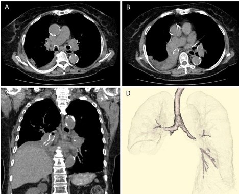
    Figure 1: Chest CT showed aggressive stenosis from carina to bilateral main bronchus, complete obstruction of the right lower bronchus, atelectasis of the right
lower lobe, and right pleural fluid (A-C). Three-dimensional CT showed irregular bilateral main bronchial walls, and stenosis and obstructive lesions at left main
bronchus and right middle bronchus (D).
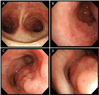
    Figure 2: Bronchoscopic findings showed stenosis for an edematous mucosa and an irregular bronchial wall from the carina to the bilateral main bronchus (A:
carina, B: left main bronchus, C: right middle bronchus), and the obstruction of the right basal bronchus (D).
However, our patient was negative for H. pylori, a major pathogenic agent in gastric MALT lymphoma, and genetic screening was not performed. The airway lesions were most likely metastatic rather than primary, because of their multiple occurrences and because the patient had been in complete remission from primary gastric MALT lymphoma [3].
Widespread tracheobronchial stenosis cause by MALT lymphoma is extremely rare and should be differentially diagnosed from other airway obstruction-related diseases. Tumors in this patient presented with atelectasis of the right lower lobe, which owing to her age and poor PS was thought to be caused by aspiration or a foreign body in the airway. However, bronchoscopy supported the diagnosis of MALT lymphoma. A biopsy is necessary for the diagnosis of MALT lymphoma, because macroscopic findings are non-specific and few bronchial NHLs are of the MALT type [4]. Moreover, tumors in patients with MALT lymphoma and atelectasis have been previously reported to be of bronchial origin rather than metastases from gastric tumors [4,5].
Conclusion
In conclusion, we report a rare case of bronchial MALT lymphoma associated with atelectasis presenting seven years after gastric MALT lymphoma. Bronchoscopy is necessary to diagnose patients with atelectasis and airway lesions.
References
- Swerdlow SH, Campo E, Harris NL, Jaffe ES, Pileri SA. WHO Classification of tumors of Hematopoietic and Lymphoid Tissues. IARC Lyon. 2008: 214–219.
- Isaacson P, Wright DH. Malignant lymphoma of mucosa-associated lymphoid tissue: a distinctive type of B-cell lymphoma. Cancer. 1983; 52: 1410–1416.
- Kawamata N, Miki T, Fukuda T, Suzuki K, Sumi Y, Ohdama S, et al. Determination of a common clonal origin of gastric and pulmonary mucosa-associated lymphoid tissue lymphomas presenting five years apart. Intern Med. 1995; 34: 220-223.
- Solomonov A, Zuckerman T, Goralnik L, Ben-Arieh Y, Rowe JM, Yigla M. Non-Hodgkin's lymphoma presenting as an endobronchial tumor: report of eight cases and literature review. Am J Hematol. 2008; 83: 416–419.
- Hardy K, Nicholson DP, Schaefer RF, Hsu, SM. Bilateral endobronchial non-Hodgkin’s lymphoma. South Med J. 1995; 88: 367–370.