
Case Series
Austin J Pulm Respir Med. 2023; 10(2): 1099.
Exploring Diverse Presentations of Cryptogenic Organizing Pneumonia: A Case Series
Ramana Prasad VV¹*; Chirali Shah²; Kranthi Kumar²
¹Department of Pulmonology, KIMS Krishna Institute of Medical Sciences, India
²Senior Resident, Department of Pulmonology, Krishna Institute of Medical Sciences and Research, Minister Road, Secunderabad 500003
*Corresponding author: Ramana Prasad VV Department of Pulmonology, KIMS Krishna Institute of Medical Sciences, India. Email: ramanaprasadpulmo@gmail.com
Received: June 26, 2023 Accepted: August 02, 2023 Published: August 09, 2023
Abstract
Cryptogenic Organizing Pneumonia (COP), the idiopathic form of Organizing Pneumonia (OP) previously known as “bronchiolitis obliterans organizing pneumonia,” is a well-documented medical condition with distinct clinicoradiological features and diagnostic criteria. COP can exhibit a diverse range of radiological presentations, including multiple patchy alveolar opacities (typical pattern), a solitary focal lesion, or diffuse bilateral infiltration. Corticosteroid therapy is typically effective in achieving rapid clinical and imaging improvement, but caution must be exercised in cases of active infections where corticosteroids are contraindicated, posing a challenge for OP diagnosis. This case series emphasizes the need for healthcare providers to be knowledgeable about the various atypical clinical manifestations of COP, recognize the diverse and uncommon radiological presentations that may mimic other pulmonary pathologies, and consider COP in the differential diagnosis to ensure appropriate management and enhance patient outcomes.
Keywords: Cryptogenic organising pneumonia; Solitary pulmonary nodule; Focal pneumonia
Introduction
Organizing Pneumonia (OP), previously known as Bronchiolitis Obliterans Organizing Pneumonia (BOOP), is a specific type of diffuse interstitial lung disease that primarily affects the small airways within the alveolar wall [1,2]. OP typically presents with characteristic radiographic features, including ground-glass opacification and/or consolidation distributed along the bronchovascular bundles. These findings tend to be more prominent in the peripheral or subpleural regions when observed on a Chest X-Ray (CXR) [2,3]. On chest Computed Tomography (CT), bilateral migratory patchy alveolar opacities are often seen, and they have a tendency to resolve spontaneously.
OP can manifest with uncommon radiographic patterns such as focal pneumonia, perilobular consolidations, curved bands of consolidation, single or multiple nodules, and a Diffuse Micronodular Pattern (DMP) [1,4,5]. Diagnosis of OP is challenging due to the nonspecific respiratory symptoms commonly observed, including cough, fever, and shortness of breath, which are also typical of infectious pneumonias. Consequently, OP is frequently misdiagnosed as an infectious etiology, leading to initial treatments involving antimicrobial agents. Ultimately, patients may undergo invasive procedures to obtain tissue samples for diagnosis. Histopathological examination of OP typically reveals the presence of granulation tissue characterized by budding fibrin exudates transitioning into collagen-containing fibroblasts and myofibroblast proliferation, interspersed with loose connective tissue within the distal air spaces [1,6]. OP has associations with various conditions, including connective tissue diseases, infections, medication side effects, acid reflux disorder, organ transplant reactions, and adjacent malignancies [1,6-8]. When no identifiable causes are found, it is referred to as Cryptogenic Organizing Pneumonia (COP).
Case Series
We present a case series of four middle-aged patients who presented with acute-onset dry cough, low-grade fever, shortness of breath, and chest pain. Upon examination, their vital signs were stable, and general and systemic assessments were unremarkable. Initial routine blood and sputum analyses provided inconclusive results. These patients were initially treated with antibiotics for suspected community-acquired pneumonia but were later referred to our center for further diagnostic workup.
The diagnostic workup encompassed a thorough evaluation to rule out other potential causes of their atypical presentations. Chest X-ray in the posterior-anterior view revealed well-defined rounded lesions in the right mid-zone for two patients and consolidation in the right lower lobe for another patient (Figure 1). Imaging studies, including High-Resolution Computed Tomography (HRCT), played a pivotal role in identifying characteristic features of Cryptogenic Organizing Pneumonia (COP) in different anatomical locations. CT-guided biopsies were performed on these lesions, confirming histopathological findings consistent with COP (Figure 1).
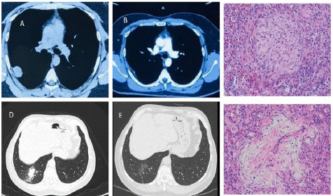
Figure 1: Radiology and Histopathology Images. A – Solitary Pulmonary Nodule in right middle lobe; B – Resolution of nodule post 1 month of corticosteroid therapy; C - Typical Masson body (intraluminal plug of mucopolysaccharide-rich fibroblast proliferation without evidence of collagenous fibrosis);D – right lower lobe consolidation; E – complete resolution of consolidation post corticosteroid therapy; F - Histopathology of organizing pneumonia (or BOOP), characterized by intraluminal plugs of proliferating fibroblasts that fill distal airways and peribonchiolar air spaces.
The absence of infectious, neoplastic, or other inflammatory etiologies, along with the histopathological findings, supported the diagnosis of COP. Consequently, the patients were initiated on therapeutic doses of oral corticosteroids, specifically prednisone. Subsequent follow-up visits demonstrated significant clinical improvement, and serial imaging studies confirmed the regression of radiological opacities related to COP and a reduction in the size of the solitary pulmonary nodule.
Discussion
Organizing Pneumonia (OP) is a lung injury response characterized by the presence of inflammatory cells and connective tissue matrix in the distal airspaces of the lungs. It presents with various imaging patterns observed on High-Resolution Computed Tomography (HRCT) of the chest. OP can be associated with different conditions such as connective tissue disorders, infections, drug reactions, hypersensitivity pneumonitis, and aspiration. When OP cannot be attributed to a specific underlying cause, it is termed cryptogenic organizing pneumonia. The initial descriptions of OP can be traced back to the late 1800s and early 1900s [9].
Cryptogenic Organizing Pneumonia (COP), which was previously known as "bronchiolitis obliterans organizing pneumonia," is a well-documented form of OP. It presents with distinct clinical, radiological, and pathological features. Radiologically, COP can manifest in three main patterns: multiple patchy alveolar opacities (typical pattern), a solitary focal lesion, or diffuse bilateral infiltration [2].
The diagnosis of COP relies on the identification of typical pathological and clinical-radiological features, as well as the exclusion of other possible causes or associated disorders. On routine chest radiographs and HRCT imaging, patients with COP typically exhibit bilateral and diffuse alveolar opacities with preserved lung volumes. These opacities are often peripherally distributed and may migrate and regress spontaneously [9]. Radiological phenotypes reported by Cordier et al, in association with COP include airspace consolidation in subsegmental/subpleural areas of one or both lungs, changes consistent with fibrosis, and diffuse, rapidly progressive fulminant disease [10]. HRCT scans reveal the presence of multifocal areas of airspace consolidation that are primarily located in peripheral or peribronchial regions, preferentially affecting the lung bases, and exhibiting changes over a few weeks [11]. Consolidated areas may show air bronchograms, and ground-glass attenuation is frequently observed alongside consolidation. However, honeycombing is typically absent. Less common patterns, such as band-like, reversed halo, atoll, or crazy paving, have also been described but lack specificity (Table 1) [11]. The presence of multiple areas of consolidation that vary over time or appear in different lung regions should raise suspicion of an immunologic/inflammatory process, potentially indicating OP. However, the differential diagnosis should include eosinophilic pneumonia or vasculitis.
![]()
    Radiographic imaging patterns    of organising pneumonia


    Consolidation


    Subpleural and/or peribronchial


    Mid to lower lung zone predominance


    Can be perilobular


    Opacities may migrate, wax, wane or disappear


    Spontaneous regression of consolidated areas may occur


    Combination of bilateral subpleural consolidation and    mid to lower zone predominance observed in majority of patients


    Other patterns


    Focal with single nodule or mass


    Nodular (variable size, can be solitary or multiple)


    Reversed halo sign (ground-glass opacity, surrounded by    a crescent or ring of consolidated parenchyma)


    Ground-glass opacities (usually bilateral, patchy, seen    in up to 90% of patients with cryptic organising pneumonia)


    Parenchymal bands (often associated with multifocal    consolidations)


    Perilobular (arcade-like or polygonal opacities that    are poorly defined and border secondary pulmonary lobules)


    Fibrotic


    Reticular opacities with basilar predominance, architectural    distortion and superimposed alveolar opacities


    Honeycomb change, traction bronchiectasis


    Rare changes


    Diffuse micronodules (centrilobular or peribronchial)


    Mediastinal lymph node enlargement


    Pleural effusion
Table 1: Radiological Presentation of Cryptogenic Organising Pneumonia [9].
Focal Organizing Pneumonia (FOP) is a rare subtype of OP characterized by an isolated focal lesion on chest imaging, which can resemble lung cancer [8]. FOP accounts for approximately 10-15% of all OP cases, with the majority being of unknown cause [12,13]. Patients with FOP are typically asymptomatic or exhibit mild symptoms, and it is more prevalent among middle-aged male smokers [12-14]. CT features of FOP show considerable variation, including solitary or multiple nodules or masses with irregular margins, typically located in the peripheral areas of the lungs [15,16]. Mediastinal lymphadenopathy involvement is uncommon in OP, but some studies have reported enlarged mediastinal nodes in a subset of COP patients. A retrospective study by Niimi et al. [17] showed that enlarged mediastinal nodes were present in 36% (eight out of 22) of the patients with COP.
The diagnosis of FOP requires histopathological identification of a predominant pattern of OP, characterized by the presence of polypoid intraluminal plugs consisting of proliferating fibroblasts and myofibroblasts within alveolar ducts and airspaces, with varying degrees of bronchiolar involvement [2-4,18]. Surgical resection of the lung lesion is often performed in suspected cases of lung cancer [13,14].
Treatment for OP typically involves corticosteroid therapy, which leads to rapid clinical and imaging improvement. Corticosteroids have shown efficacy in managing symptomatic and progressive COP and are recommended as the initial treatment option [19]. Most patients recover completely with oral corticosteroids, although relapses are common.
Conclusion
This series of cases highlights the significance of recognizing the varied and uncommon presentations of COP, which can imitate other pulmonary conditions such as infectious pneumonias, malignancies, lung diseases related to connective tissue disorders, hemorrhage, sarcoidosis, interstitial lung diseases, aspiration or chemical pneumonitis, drug or radiation-induced lung diseases, or sequestrations. A timely and accurate diagnosis necessitates a comprehensive assessment that includes imaging studies and histopathological examination. Since corticosteroid therapy is the mainstay treatment but contraindicated in active infections, the diagnosis of OP can be challenging. Therefore, it is crucial to emphasize that healthcare providers should remain mindful of the possibility of atypical manifestations of COP and consider it in the differential diagnosis to ensure appropriate management and enhance patient outcomes.
Author Statements
Conflict of Interest
The authors declare that there is no conflict of interest.
References
- Cordier JF. Organising pneumonia. Thorax. 2000; 55: 318-28.
- Epler GR. Bronchiolitis obliterans organizing pneumonia. Arch Intern Med. 2001; 161: 158-64.
- Montesinos JJ, Laguna MA. Case 1: cryptogenic organizing pneumonia. AJR Am J Roentgenol. 1998; 171: 835.
- Huo Z, Feng R, Tian X, Zhang H, Huo L, Liu H. Clinicopathological findings of focal organizing pneumonia: a retrospective study of 37 cases. Int J Clin Exp Pathol. 2015; 8: 511-6.
- Baque-Juston M, Pellegrin A, Leroy S, Marquette CH, Padovani B. Organizing pneumonia: what is it? A conceptual approach and pictorial review. Diagn Interv Imaging. 2014; 95: 771-7.
- Cottin V, Cordier JF. Cryptogenic organizing pneumonia. Semin Respir Crit Care Med. 2012; 33: 462-75.
- Lebargy F, Picard D, Hagenburg J, Toubas O, Perotin JM, Sandu S, et al. Micronodular pattern of organizing pneumonia: case report and systematic literature review. Medicine. 2017; 96: e5788.
- Lohr RH, Boland BJ, Douglas WW, Dockrell DH, Colby TV, Swensen SJ, et al. Organizing pneumonia. Features and prognosis of cryptogenic, secondary, and focal variants. Arch Intern Med. 1997; 157: 1323-9.
- Raghu G, Meyer KC. Cryptogenic organising pneumonia: current understanding of an enigmatic lung disease. Eur Respir Rev. 2021; 30: 210094.
- Cordier JF. Cryptogenic organising pneumonia. Eur Respir J. 2006; 28: 422-46.
- Roberton BJ, Hansell DM. Organizing pneumonia: a kaleidoscope of concepts and morphologies. Eur Radiol. 2011; 21: 2244-54.
- Cordier JF, Loire R, Brune J. Idiopathic bronchiolitis obliterans organizing pneumonia: definition of characteristic clinical profiles in a series of 16 patients. Chest. 1989; 96: 999-1004.
- Watanabe K, Harada T, Yoshida M, Shirakusa T, Iwasaki A, Yoneda S, et al. Organizing pneumonia presenting as a solitary nodular shadow on a chest radiograph. Respiration. 2003; 70: 507-14.
- Melloni G, Cremona G, Bandiera A, Arrigoni G, Rizzo N, Varagona R, et al. Localized organizing pneumonia: report of 21 cases. Ann Thorac Surg. 2007; 83: 1946-51.
- Maldonado F, Daniels CE, Hoffman EA, Yi ES, Ryu JH. Focal organizing pneumonia on surgical lung biopsy: causes, clinicoradiologic features, and outcomes. Chest. 2007; 132: 1579-83.
- Kohno N, Ikezoe J, Johkoh T, Takeuchi N, Tomiyama N, Kido S, et al. Focal organizing pneumonia: CT appearance. Radiology. 1993; 189: 119-23.
- Niimi H, Kang EY, Kwong JS, Carignan S, Müller NL. CT of chronic infiltrative lung disease: prevalence of mediastinal lymphadenopathy. J Comput Assist Tomogr. 1996; 20: 305-8.
- Zhao F, Yan SX, Wang GF, Wang J, Lu PX, Chen B, et al. CT features of focal organizing pneumonia: an analysis of consecutive histopathologically confirmed 45 cases. Eur J Radiol. 2014; 83: 73-8.
- Bradley B, Branley HM, Egan JJ, Greaves MS, Hansell DM, Harrison NK, et al. Interstitial lung disease guideline: the British Thoracic Society in collaboration with the Thoracic Society of Australia and New Zealand and the Irish Thoracic Society. Thorax. 2008; 63: v1-58.