
Case Report
Austin Plastic Surg Open Access. 2025; 3(1): 1005.
Aesthetically Driven Management of Frontal Sinus Fractures: A Case Series
Singh S1*, Shah K2, Landge J3 and Gavali P4
1Post Graduate Student, Department of Oral and Maxillofacial Surgery, Government Dental College and Hospital Chhatrapati Sambhajinagar Maharashtra India
2Professor & Head of Department, Department of Oral and Maxillofacial Surgery, Government Dental College and Hospital Chhatrapati Sambhajinagar Maharashtra India
3Associate Professor, Department of Oral and Maxillofacial Surgery, Government Dental College and Hospital Chhatrapati Sambhajinagar Maharashtra India
4Assistant Professor, Department of Oral and Maxillofacial Surgery, Government Dental College and Hospital Chhatrapati Sambhajinagar Maharashtra India
*Corresponding author: Dr. Sunny Singh, Department of Oral and Maxillofacial Surgery, Government Dental College and Hospital Chhatrapati, Sambhajinagar, Maharashtra, India Tel: 9991993866; Email: dr.sunnysingh1994@gmail.com
Received: June 27, 2025 Accepted: July 22, 2025 Published: July 24, 2025
Introduction
Frontal sinus fractures, although relatively uncommon among facial injuries, pose unique diagnostic and therapeutic challenges due to their anatomical complexity and critical aesthetic implications. Comprising part of the anterior skull base and overlying the frontal lobe, the frontal sinus is divided into anterior and posterior tables. While posterior table fractures may compromise the central nervous system and often warrant aggressive management, anterior table fractures, when isolated, offer the possibility of a more conservative, aesthetically oriented repair.
Traditionally, the surgical approach to frontal sinus trauma has been influenced by the perceived risk of these complications. Surgeons frequently opted for sinus obliteration or cranialization, particularly in the presence of posterior table fractures or frontonasal duct injuries. While effective in preventing intracranial complications, these interventions often resulted in significant morbidity, prolonged recovery times, and suboptimal cosmetic outcomes. Over time, this has led to a reassessment of indications for such procedures, especially in cases limited to the anterior table.
Modern management strategies emphasize the importance of individualized treatment planning. With improvements in diagnostic imaging—particularly high-resolution computed tomography (CT)— surgeons can now more accurately assess fracture patterns, sinus patency, and associated injuries. These advancements, combined with innovations in surgical tools, endoscopic access, and low-profile fixation systems, have paved the way for techniques that prioritize both functional preservation and cosmetic restoration.
Aesthetic concerns have become increasingly relevant in the treatment of craniofacial trauma. With patients placing greater emphasis on scar concealment, facial symmetry, and rapid return to normal life, the surgical paradigm has shifted toward minimally invasive approaches that preserve the natural architecture of the face.
Aims and Objectives
1. To assess the feasibility of anterior table fracture repair using aesthetic-focused incisions, including the use of existing traumatic lacerations or concealed access points to minimize visible scarring.
2. To document functional outcomes, particularly in terms of frontal sinus patency and absence of late complications such as mucocele formation or infection.
3. To evaluate the effectiveness of fixation techniques, such as titanium microplates and mesh, in achieving stable reconstruction of the frontal bone contour.
4. To report on patient satisfaction and postoperative recovery, focusing on return to activity, scar visibility, and overall aesthetic appearance.
5. To contribute to the evolving literature supporting minimally invasive, tailored approaches in craniofacial trauma, advocating for the preservation of sinus function and facial aesthetics in selected patients.
Materials and Methods
Study Design
This case series presents a retrospective analysis of five patients treated for isolated anterior table frontal sinus fractures at a tertiary care center between 2022 and 2024. Ethical approval was obtained from the institutional review board, and all patients provided informed consent for the use of clinical data and images.
Inclusion Criteria: Patients were included in the study based on the following criteria:
- Age 18 years and older
- Isolated anterior table frontal sinus fractures
- No evidence of posterior table involvement or frontonasal duct injury
- Underwent surgical management with an aesthetic-first approach
- Minimum follow-up period of 6 months
Exclusion Criteria: Patients were excluded if they had:
- Fractures involving the posterior table or cranialization requirement
- Frontonasal duct obstruction or CSF leak
- History of previous frontal sinus surgery
- Associated facial fractures requiring extensive intervention
Preoperative Assessment
All patients underwent clinical examination and high-resolution CT scans (axial, coronal, and sagittal views) to assess the extent and displacement of the fracture, sinus integrity, and frontonasal duct status. Diagnostic criteria for operative intervention included displaced anterior table fractures causing forehead contour deformity, and risk of cosmetic sequelae.
Patients were evaluated by a multidisciplinary team, including maxillofacial and plastic surgeons. Aesthetic considerations such as scar placement, hairline patterns, and existing traumatic lacerations were incorporated into the surgical plan.
Surgical Technique
Under general anesthesia, patients were positioned supine with head elevation to minimize bleeding. Wherever possible, existing traumatic lacerations were utilized to access the fracture site. In their absence, hidden incisions were made along the suprabrow or within the hairline, depending on cosmetic preference and anatomical considerations.
The fractured anterior table segments were exposed via subperiosteal dissection. After achieving reduction, internal fixation was performed using low-profile titanium microplates and screws. In cases of comminution, titanium mesh was contoured and applied to reconstruct the anterior wall. One patient required autologous calvarial bone grafting harvested through the same incision.
Care was taken to preserve mucosa and ensure sinus continuity. No evaluated through visual inspection, scar assessment, and patientreported satisfaction on a 5-point Likert scale.
Complications such as infection, mucocele formation, hardware exposure, or sinus dysfunction were recorded.
4. Case Presentations
4.1. Case 1: 26-year-old Male – Motorcycle Collision
Injury Mechanism: 26-year-old male, presented following a high-velocity road traffic accident involving a motorcycle collision. He sustained blunt trauma to the forehead from impact with the road surface without wearing a helmet.
Clinical Findings: On examination, a healing laceration was observed across the central forehead, with a noticeable depression suggestive of a comminated anterior table frontal sinus fracture. Neurological assessment was normal (Figure 1A).
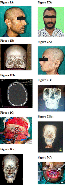
Imaging: CT imaging revealed an isolated, displaced anterior table fracture with no posterior wall involvement or frontonasal duct injury (Figure 1B).
Management: The Bicoronal Incision was used as a surgical access point. The fractured fragments were reduced and fixed with 1.3 mm low-profile titanium microplates. Bone gaps were reconstructed with contoured titanium mesh. No sinus obliteration was performed (Figure 1C).
Outcome: Healing was uneventful. Aesthetic contour was restored, and the scar was barely noticeable at 6-month follow-up. The patient reported high satisfaction (Figure 1D).
Case 2: A 32-year-old Female – Fall from Height
Injury Mechanism: A 32-year-old female, slipped and fell from approximately 2 meters, striking her forehead on concrete.
Clinical Findings: She presented with periorbital swelling and a superficial laceration on the left forehead. Palpable depression and step-off in the frontal bone raised suspicion of a fracture.
Imaging: CT confirmed a unilateral anterior table fracture with minor comminution and sinus preservation. The frontonasal duct and posterior table were intact.
Management: A suprabrow incision was used to access the fracture. Reduction was achieved with fine elevator instruments, and internal fixation was accomplished using titanium microplates. The incision was carefully closed with layered suturing to minimize scarring.
Outcome: No complications occurred. CT at 3 months showed intact sinus architecture. The scar was nearly invisible and contour symmetrical. The patient rated her satisfaction as “excellent.”
Case 3: A 45-year-old Male – Industrial Injury
Injury Mechanism: Patient sustained blunt force trauma from falling metal equipment while working on a construction site.
Clinical Findings: A laceration on the upper mid-forehead was associated with swelling and contour irregularity. No neurological deficits were observed (Figure 2A).
Imaging: CT imaging showed a depressed anterior table fracture with associated bone fragmentation but no extension to the posterior table (Figure 2B, 2Bb).
Management: The Existing Laceration was extended for exposure. Fragments were stabilized with a combination of titanium mesh and 1.5 mm screws. No grafting was necessary. The frontal sinus was left intact (Figure 2C).
Outcome: At 6-month follow-up, the contour was smooth, and the scar healed well. No signs of sinusitis or infection were noted. Patient satisfaction was rated as “very good” (Figure 2D).
Case 4: A, 29-year-old Female – Assault Injury
Injury Mechanism: She suffered direct frontal trauma from a blunt object during an assault.
Clinical Findings: Swelling and ecchymosis obscured a subtle depression. No external lacerations were present, and the skin was intact.
Imaging: CT revealed a depressed but non-comminuted anterior table fracture with intact posterior wall and patent frontonasal duct.
Management: A hidden incision in the hairline allowed access. The bone segment was elevated and secured with a single low-profile microplate. Soft tissue handling was minimized.
Outcome: Healing was uneventful. The incision was completely hidden, and the forehead shape restored. The patient expressed great satisfaction, citing both cosmetic and emotional recovery.
Case 5: A, 38-year-old Male – Sports Injury
Injury Mechanism: Patient was struck by an elbow during a competitive basketball match.
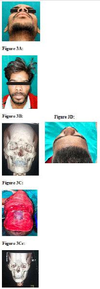
Clinical Findings: There was visible contour flattening over the right supraorbital area. No skin breach was present, and neurological examination was normal (Figure 3A).
Imaging: CT showed a depressed anterior table fracture limited to the right side without sinus obliteration or duct injury (Figure 3B).
Management: A Bicoronal incision was used. Reduction was straightforward, and fixation required only a single titanium mesh. The periosteum and skin were carefully closed. (Figure 3C, 3Cc)
Outcome: Follow-up at 6 months showed excellent healing and restoration of symmetry. The incision healed with no visible scar, and the patient returned to sport within 4 weeks (Figure 3D).
Results
Demographics and Injury Patterns
Five patients (3 males, 2 females) aged between 26 and 45 years (mean age: 34 years) with isolated anterior table frontal sinus fractures were included. The mechanisms of injury varied: road traffic accident (n=1), fall from height (n=1), industrial trauma (n=1), assault (n=1), and sports-related injury (n=1).
Surgical Access and Fixation
Access approaches included
• Existing laceration extension (n=2)
• Bicoronal Incision (n=3)
Fixation techniques:
• Titanium microplates and screws (n=3)
• Titanium mesh augmentation (n=2)
• No cases required sinus obliteration or cranialization.
All procedures were completed successfully without intraoperative complications.
Functional and Radiological Outcomes
Postoperative CT scans at 3 months showed:
- Restoration of frontal bone contour in all patients
- Preservation of frontal sinus volume and frontonasal duct patency
- No evidence of hardware migration or sinus mucosal disruption
No patient developed mucocele, sinusitis, or chronic frontal pain during the 6-month follow-up.
Aesthetic Outcomes
Scar quality was evaluated using the Patient and Observer Scar Assessment Scale (POSAS) and patient satisfaction scores. All patients reported high satisfaction with cosmetic results:
- 3 patients rated outcomes as “excellent”
- 2 patients rated outcomes as “very good”
Surgeons also rated scar visibility as minimal or unnoticeable in all cases. No revision surgery was required.
Complications
There were no major complications such as:
- Infection
- CSF leakage
- Hardware exposure
- Neurological symptoms
Minor swelling and bruising resolved within 10–14 days in all cases. One patient reported temporary numbness over the forehead, which resolved spontaneously by the 1-month follow-up.
Summary
This case series demonstrates that with careful case selection and a tailored aesthetic approach, isolated anterior table frontal sinus fractures can be effectively managed without obliteration. Functional outcomes were preserved, and cosmetic results were highly satisfactory. No long-term complications were observed during follow-up.
Discussion
Frontal sinus fractures represent a small but significant subset of craniofacial trauma due to their proximity to vital neurovascular structures and their cosmetic importance. Traditionally, aggressive surgical approaches including sinus obliteration or cranialization were the mainstay of treatment, particularly when sinus outflow tract injury or posterior table involvement was suspected. However, with advancing imaging techniques, improved fixation hardware, and growing emphasis on aesthetic outcomes, there has been a paradigm shift towards conservative, function-preserving management in cases involving isolated anterior table fractures.
Conclusion
This case series highlights the effectiveness of aesthetically driven, function-preserving surgical repair in managing isolated anterior table frontal sinus fractures. All five patients were managed without sinus obliteration or cranialization, using tailored open reduction and internal fixation techniques that prioritized both cosmetic outcomes and structural integrity.
The consistent success across varied mechanisms of injury— ranging from road traffic accidents to sports-related trauma— underscores the versatility of this approach. Patients experienced excellent aesthetic restoration, high satisfaction, and no significant complications during the follow-up period. Surgical access through discreet incisions and careful intraoperative technique were critical factors in achieving these results.
References
- Strong EB, Kellman RM, Rathburn J, Sillers MJ. Frontal sinus fractures: a rational approach to evaluation and treatment. Laryngoscope. 2006; 116: 1355–1360.
- Manson PN, Markowitz BL, Mirvis SE, Towbin R, Dillon W, Yaremchuk MJ. Toward CT-based treatment of frontal sinus fractures: classification and rationale. Plast Reconstr Surg. 1990; 85: 881–895.
- Gonty AA, Marciani RD, Adornato MC. Management of frontal sinus fractures: a review of 33 cases. J Oral Maxillofac Surg. 1999; 57: 372–379.
- Rodriguez ED, Martin M, Bluebond-Langner R, Manson PN. Functional and aesthetic restoration of the frontal sinus. J Craniofac Surg. 2007; 18: 570– 577.
- Bell RB, Dierks EJ, Brar P, Potter BE. A protocol for the management of frontal sinus fractures emphasizing sinus preservation. J Oral Maxillofac Surg. 2007; 65: 825–839.
- Strong EB. Frontal sinus fractures: current concepts. Craniomaxillofac Trauma Reconstr. 2009; 2: 41–48.
- Constantinides M, Galli SK, Miller PJ. Frontal sinus fractures: a 28-year experience. Laryngoscope. 2001; 111: 2111–2117.
- Liu JK, Gottfried ON, Cole CD, Dougherty WR, Couldwell WT. Surgical nuances for the repair of frontal sinus fractures. Clin Neurosurg. 2006; 53: 150–157.
- Gerbino G, Roccia F, Benech A, Ramieri G. Analysis of 158 frontal sinus fractures: current surgical management and complications. J Craniomaxillofac Surg. 2000; 28: 133–139.
- Lucchina AG, Schaller B, Grätz KW, Zimmermann H, Iizuka T. Treatment strategies for frontal sinus fractures: a review of 49 cases. Oral Surg Oral Med Oral Pathol Oral Radiol Endod. 2005; 100: 183–189.
- Choi JY, Yang JD, Chung HY, Cho BC. The use of absorbable plates in treating anterior table frontal sinus fractures. J Craniofac Surg. 2010; 21: 438–442.
- Gunarajah DR, Samman N. Biomaterials for repair of frontal sinus fractures: current status. Br J Oral Maxillofac Surg. 2013; 51: 693–699.
- de Chalain TM. Frontal sinus injuries: a review of 96 cases. J Plast Reconstr Aesthet Surg. 2003; 56: 648–653.
- Albu S, Avram A, Cozlean V. Surgical management of frontal sinus fractures: a review. Acta Otorhinolaryngol Ital. 2014; 34: 219–226.
- Daffner RH, Hackney DB. Imaging of facial trauma. Radiology. 2007; 243: 10–22.