Clinical Image
Phys Med Rehabil Int. 2019; 6(2): 1164.
A Case Study of Radial Nerve Injury after Segmental Humeral Fracture, Followed by Primary Intramedullary Nailing
Karagounis Panagiotis and Vissarakis Giorgos*
Filoktitis Rehabilitation Center, Koropi-Attica, Greece
*Corresponding author: Vissarakis Giorgos, Filoktitis Rehabilitation Center, Koropi-Attica, Greece
Received: June 30, 2019; Accepted: July 11, 2019; Published: July 18, 2019
Clinical Image
Radial nerve injury is one of the most common mononeuropathies of the upper limb, especially as a result of traumatic injuries or fractures, surgical interventions, or compression effect.
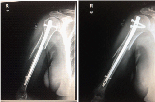
We present the radiological images of a case study of a 78 years old female patient, who sustained a 2 parts segmental humeral fracture, followed by primary intramedullary nailing. Post operatively (9th day post-operation), electro diagnostic studies determined an axonal type of radial nerve injury, included motor nerve conduction study to the extensor indicisproprius, sensory nerve conduction study of the superficial branch of the radial nerve, and needle EMG of radialinnervated muscles and non-radial muscles supplied by the C7 nerve root.

Figure 1:
There are two main anatomic areas where the radial nerve is vulnerable. The first region is the distal third of the humeral bone where the nerve lies directly lateral to the bone and the second area is approximately 6 cm centered around the midshaft of the humerus where deltoid tuberosity is situated.
The main rehabilitation goal of radial neuropathies is to improve hand function for the completion of daily activities.