
Special Article - Pediatric Rehabilitation
Phys Med Rehabil Int. 2018; 5(2): 1144.
Effects of an Articulated Ankle Foot Orthosis on Gait Biomechanics in Adolescents with Traumatic Brain Injury: A Case-Series Report
Rogozinski BM*, Schwab SE and Kesar TM
1Division of Physical Therapy, Department of Rehabilitation Medicine, Emory University School of Medicine, Atlanta, GA, USA
*Corresponding author: Benjamin Rogozinski, Division of Physical Therapy, Department of Rehabilitation Medicine, Emory Rehabilitation Hospital, 1441 Clifton Rd NE, Atlanta, Georgia, USA
Received: March 09, 2018; Accepted: April 05, 2018; Published: April 12, 2018
Abstract
Purpose: To quantify the effects of an articulated ankle foot orthosis on genu recurvatum gait in adolescents with traumatic brain injury (TBI).
Methods: Gait analysis was conducted in 2 individuals with TBI during over ground ambulation with (braced condition) and without (barefoot condition) the AAFO. For each participant, stride-by-stride gait data were compared to assess differences between barefoot and braced walking conditions.
Results: During the braced versus barefoot condition, both participants demonstrated reduced plantar flexion at initial contact, increased knee flexion at initial contact, reduced peak knee extension during stance, and reduced peak and integral of internal knee flexor moment during stance.
Conclusions: The data suggest that the AAFO reduced plantar flexion during stance, therefore attenuating the anterior displacement of the ground reaction force vector (GRFV) relative to the ankle and knee joint axes, and leading to a reduction in knee hyperextension and the internal knee flexor moment during stance. We posit that the reduction in internal knee flexor moment may lead to a more sustainable gait pattern with less potential for mechanical stress on the posterior knee joint capsule.
Keywords: Traumatic Brain Injury; Genu Recurvatum Gait; Gait Analysis; Articulated Ankle Foot Orthosis
Introduction
Traumatic brain injury (TBI) is a leading cause of disability in children and adolescents [1]. Following TBI, survivors present with various neuromuscular impairments, including decreased strength and range of motion (ROM), spasticity, impaired neuromuscular control, impaired proprioception, and/or hemiparesis [2]. These impairments often compromise walking function and can lead to the development of abnormal gait patterns. Genu recurvatum gait is common in individuals with traumatic brain injury (TBI) and is defined as a hyperextension of the tibiofemoral joint during the stance phase of gait [2].
The cause of genu recurvatum gait is multifactorial and may be attributed to the presence of quadriceps weakness, quadriceps spasticity, plantar flexor spasticity or contracture, pre-tibial muscle weakness or paralysis, decreased proprioception, and/ or any combination of the aforementioned impairments [2,3]. In this gait pattern, distal biomechanical factors generally impact the displacement of the ground reaction force vector (GRFV) relative to the ankle and knee joints. Genu recurvatum is often accompanied by excessive ankle plantar flexion early during stance phase. During normal gait, floor contact is made with the ankle in a neutral position. This is followed by a controlled lowering of the foot to the floor through eccentric contraction of the pretibial muscles, allowing for controlled anterior progression of the tibia and the knee. In contrast, excessive ankle plantar flexion at initial contact restricts the normal forward progression of the tibia by redirecting the GRFV anterior to the ankle joint axis and driving the tibia posteriorly. As the body progresses forward, the GRFV is directed further anteriorly with respect to the knee joint axis, increasing the moment arm of the GRFV relative to the knee joint, inducing a large external extensor moment at the knee, and causing a concomitant increase in internal knee flexor moment to control the knee joint [3]. A chronic genu recurvatum gait pattern and the accompanying large and prolonged internal knee flexor moment may cause increased mechanical stress on the posterior knee joint capsule and ligamentous structures of the knee [4,5]. These mechanical stresses may have implications such as structural joint damage, pain, other compensatory gait deviations, and limitations in gait function and speed.
Treating gait dysfunction in patients with TBI focuses on improving the efficiency and sustainability of the gait pattern in the face of diminished selective motor control. In an effort to restore walking function for individuals with hemiplegic gait [6], an articulated ankle foot orthoses (AAFO) is commonly prescribed [7,8]. An AAFO with a plantar flexion stop is a custom fit orthopedic brace externally applied to the foot and ankle that allows for adequate ankle dorsiflexion, while restricting ankle plantar flexion to promote a more normal gait pattern. The ankle plantar flexion block allows the tibia to translate anteriorly during stance, bringing the ground reaction force vector (GRFV) closer to the knee joint center, which subsequently decreases knee hyperextension and the large internal knee flexor moment during stance [9]. Previous literature suggests that an ankle foot orthosis can improve ankle and knee kinematics, kinetics, and energy cost of walking in stroke survivors [10]. Thus, an articulated ankle foot orthosis (AAFO) with plantar flexion stop may be used to control knee recurvatum [11]. In a study investigating multiple configurations of the AAFO on gait parameters in adults with poststroke hemiplegia [12], Fatone and colleagues found that the AAFO decreased plantar flexion at initial contact and mid-swing, changing the peak knee moment in early stance from flexor to extensor [13]. However, research specific to the gait impairments of children and adolescents with TBI is limited. To our knowledge, changes in gait biomechanics caused by an AAFO in children or adolescents with TBI have not been previously examined.
The purpose of this single-subject research case series report was to assess the effect of the AAFO on genu recurvatum gait pattern in adolescents with TBI. We utilized 3-dimensional gait analysis to quantify ankle and knee kinematics and kinetics associated with genu recurvatum gait in adolescents with TBI. Our objective was to evaluate whether the AAFO successfully attenuates genu recurvatum by comparing kinematic and kinetic data between the barefoot (control) and braced conditions for each participant.
We hypothesized that the use of an AAFO in adolescents with TBI demonstrating genu recurvatum gait will (1) decrease ankle plantar flexion during stance phase, producing a more neutral ankle position during stance, and (2) decrease knee hyperextension and internal knee flexor moment during stance by reducing the anterior displacement of the GRFV relative to the ankle and knee joints.
Methods
The study design was a single-subject research comprising 2 case studies, with a repeated-measures comparison of gait biomechanics during over ground walking with versus without the AAFO. The study protocol was reviewed and approved by the Institutional Research Review Committee at Emory University. Fifteen children and adolescents ages 6 to 19 with neurologic impairments were referred to the Emory University Motion Analysis Laboratory between September 2014 and February 2015 for comprehensive quantitative gait analysis. Participants were referred to facilitate clinical and surgical decision-making. All participants participated in a single session comprising clinical examinations and gait analysis. Inclusion criteria for this case series study included a clinical diagnosis of TBI, genu recurvatum gait defined as a knee extension angle = 0° (i.e. hyper-extension of the knee joint) during stance phase while walking barefoot, and use of an AAFO for ambulation. Two participants from the larger group fit the inclusion criteria and were included in the current case series. These participants arrived at the gait laboratory with the orthoses they habitually used, and the participants were not fitted with an AAFO for the purpose of this study. The participants were examined and tested walking barefoot and with the AAFO on the same day in one session (Figure 1). A clinical examination was conducted for each participant, which included a detailed history, range of motion, strength, skeletal alignment, and spasticity (Table 1). All goniometric measurements were made with measures recorded to the nearest 5° increment [14].
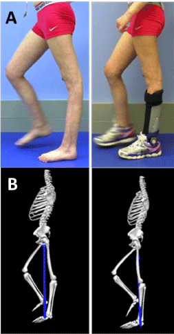
    Figure 1:  Image (A) and biomechanical model reconstruction (B) showing the
position of the lower limb in the barefoot (left) and braced (right) conditions
during stance phase for one of the case study participants. The biomechanical
model (B) also shows the ground reaction force vector and its relationships
with respect to the ankle and knee joint axis.
![]()
    Participant
    TB01
    TB02


    Gender
    M
    F


    Age
    14
    12


    More Impaired Side
    Right
    Left


    Impaired
    Less Impaired
    Impaired
    Less Impaired


    Knee Flexion ROM
    140
    140
    150
    155


    Knee Flexion MMT
    2+
    5
    3+
    5


    Knee Flexion SC
    2
    3
    2
    3


    Knee Extension ROM
    0
    0
    15
    0


    Knee Extension MMT
    4
    5
    4
    5


    Knee Extension SC
    3
    3
    2
    3


    Ankle Dorsiflexion ROM
    5
    15
    10
    30


    Ankle Dorsiflexion MMT
    1
    5
    4
    5


    Ankle Dorsiflexion SC
    0
    3
    2
    3


    Ankle Plantarflexion ROM
    NT
    NT
    40
    35


    Ankle Plantarflexion MMT
    1
    4
    2
    5


    Ankle Plantarflexion SC
    0
    3
    2
    3


    Quadriceps Spasticity
    2
    0
    1
    0


    Hamstring Spasticity
    2
    0
    1
    0


    Clonus
    R gastroc/soleus (unsustained)
    L gastroc/soleus (sustained)


    KEY: Range of Motion (ROM):    Degrees; Manual Muscle Test (MMT): 0-5; Selective Control (SC): 0-3;    Spasticity: 0-2; NT: Not Tested.
Table 1: Relevant clinical examination findings for the two participants.
During gait analysis, 3-dimensional positions of retro-reflective markers attached to the pelvis as well as bilateral hip, knee, and ankle joint segments were collected utilizing a seven-camera motion capture system at 120-Hz (Vicon Inc., Oxford, UK). Two six-degree of freedom force platforms instrumented within a split-belt treadmill were used to record ground reaction forces at 1000Hz (Bertec Inc., USA). Participants were instrumented with 43 passive retro-reflective markers consistent with the gait analysis model previously described [15]. Participants made passes along a seven-meter walkway at a selfselected walking speed until 10 strides were obtained bilaterally for both the barefoot and braced conditions. Participants were instructed to walk at a comfortable walking speed. For both the barefoot and braced conditions, a standing calibration trial was collected prior to gait analysis to establish the relationship between the segmental tracking markers and the corresponding anatomical reference markers. The use of separate standing calibration trials was done to minimize any measurement artifact associated with shoe and/or brace wear by ensuring that consistent anatomical references were used in both barefoot and braced testing conditions. Walking trials with marker gaps greater than six frames or inaccurate kinetic data (i.e. feet not entirely on the respective force plate) were eliminated.
Biomechanical models for each trial were created using Visual 3D (C-Motion Inc., Rockville, MD, USA), a biomechanics analysis and modeling software [9] (Figure 1). Lower limb kinematics were calculated using rigid body analysis and Euler angles. Vertical GRFVs were used to identify gait events (initial contact and toe-off). Strides were time normalized to 100% of the gait cycle and averaged across trials for each participant. The resulting data was processed and utilized to generate metrics evaluating specific kinematic, kinetic, and temporo-spatial data.
Gait kinematic and kinetic outcome variables examined in this study included ankle angle at initial contact, knee angle at initial contact, peak knee extension angle during stance, peak internal knee flexor moment during stance, and internal knee flexor moment integral during stance for the participant’s impaired limb. In case the participants had bilateral impairments, the limb with greater impairment was considered for this study. To maintain consistency of stride-by-stride samples and variability across conditions and participants, data from the first five gait cycles for each participant were used for each dependent variable. Means and standard deviations were calculated for each gait variable and each walking condition (braced and barefoot), and z-scores were utilized to identify and eliminate outliers greater than three standard deviations from the mean. For each participant, paired t-tests were performed on the stride-by-stride data to assess differences between barefoot and braced walking conditions to determine statistical significance (a = 0.05). In addition to the paired t-tests, we computed the 95% CI, which was used to set a threshold for determining whether the change in gait variable caused by the brace was beyond the 95% CI of the stride-to-stride variability for the barefoot condition. The rationale underlying the statistical analysis was that we wanted to evaluate whether the change in a gait variable observed during the braced condition exceeded the magnitude of change expected simply due to chance, measurement error, or physiologic factors affecting stride-tostride variability.
Results
Two participants with TBI and genu recurvatum gait underwent clinical examination (Table 1) and quantitative gait analyses during a single laboratory visit. Gait data were obtained for both barefoot and braced walking conditions for both participants (Figure 2). Table 2 quantifies kinematic, kinetic, and temporospatial data throughout the gait cycle for the barefoot and braced testing conditions for each variable. Both participants demonstrated significant improvements in all the kinematic and kinetic variables between braced and barefoot conditions, as summarized below (Figures 2 and 3).
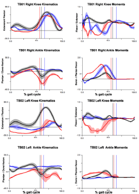
    Figure 2:  Graphs depicting the ankle and knee sagittal plane joint kinematics
(left panels) and joint moments (right panels) throughout the gait cycle for
TB01 (top) and TB02 (bottom). In each graph, the barefoot condition is shown
in red, braced condition in blue, and normative gait data (for comparison) in
black. The shaded area around each line represents the SD for the 5 trials
used to calculate the mean for the braced and barefoot conditions. The x-axis
is normalized to % gait cycle (from initial contact to initial contact). The vertical
lines towards the center of the graph depict toe-off, which designates the
transition from stance to swing phase of gait, and correspond to each of the
three conditions. For ankle and knee angles, positive values indicate dorsiflexion
and knee flexion respectively. For ankle and knee moments, positive
values indicate plantar-flexor and flexor moments, respectively. These
graphs show that in the barefoot condition (red), both participants exhibit
features consistent with genu recurvatum gait, i.e., increased ankle plantar
flexion at initial contact, increased knee extension in stance, and a large
and prolonged internal knee flexor moment in stance when compared to labbased
normative values, (black). For both TB01 and TB02, these variables
showed improvements (change toward the normative value) with the addition
of the AAFO (blue).
![]()
TB01
p
TB02
p
Barefoot
Braced
Barefoot
Braced
ANKLE
Peak Angle at Initial Contact (°)
-13 (1.57)
1 (0.79)*
<.01
-20 (1.21)
7 (1.22)*
<.01
Peak Angle in Stance (°)
4 (1.85)
6 (0.52)
0.22
0.5 (3.18)
7 (1.15)*
0.02
Peak Angle at Toe Off (°)
-14 (6.12)
3 (0.25)*
<.01
-9 (3.43)
6 (1.15)*
<.01
Peak Angle in Swing (°)
-7 (1.96)
4 (0.18)*
<.01
-7 (1.97)
7 (0.77)*
<.01
Peak Moment in Stance (N·m/kg)
1.39 (0.06)
1.3 (0.09)
0.09
0.51 (0.06)
0.03 (0.05)*
<.01
Peak Power in Stance (W/kg·m)
2.01 (0.19)
0.19 (0.07)*
<.01
0.22 (0.09)
0.03 (0.01)*
0.01
KNEE
Peak Angle at Initial Contact (°)
6 (2.19)
12 (1.92)*
0.01
-11 (0.67)
0.8 (4.62)*
0.01
Peak Angle in Stance (°)
-5 (0.77)
-0.5 (0.91)*
<.01
-23 (0.65)
-11 (1.24)*
<.01
Peak Angle in Swing (°)
60 (3.04)
65 (3.10)
0.20
23 (2.97)
15 (4.26)*
0.01
Peak Extensor Moment in Stance (N·m/kg)
0.08 (0.02)
0.51 (0.16)*
<.01
0.10 (0.04)
0.16 (0.10)
0.22
Peak Flexor Moment in Stance (N·m/kg)
-0.90 (0.11)
-0.71 (0.12)
0.11
-0.88 (0.05)
-0.21 (0.04)*
<.01
Flexor Moment Integral in Stance
(N·m·s/Kg)
-0.30 (0.01)
-0.17 (0.03)*
<.01
-0.63 (0.10)
-0.02 (0.01)*
<.01
GAIT PARAMETERS
Gait Speed (m/s)†
1.14
1.18
0.26
0.23
Step Length (Impaired Side) (m)†
0.62 (0.07)
0.51 (0.05)
0.27 (0.04)
0.31 (0.04)
Stance Time (Impaired Side) (s)†
0.62 (0.00)
0.63 (0.02)
1.45 (0.38)
1.60 (0.11)
Note that all values are represented as Mean (Standard Deviation). *Indicates paired t-test comparing braced versus barefoot condition was statistically significant (p< 0.05). †Indicates that a paired t-test was not run for this variable.
Peak Ankle Angle: (-) Plantarflexion (+) Dorsiflexion; Peak Ankle Moment: (-) Dorsiflexor (+) Plantar flexor; Peak Ankle Power: (-) Absorption (+) Generation; Peak Knee Angle: (-) Extension (+) Flexion; Peak Knee Moment: (-) Flexor (+) Extensor.
Table 2: Kinematic and Kinetic Variables for the barefoot and braced conditions for study participants.
Participant TB01
Participant TB01 was a 14-year-old male with a clinical diagnosis of TBI and right hemiparesis. For the barefoot condition, at initial contact, the right ankle was in 13° (1.57°) of plantar flexion (Figure 2 and 3). This significantly improved to an ankle angle at initial contact of 1° (0.79°, p <0.01) of dorsiflexion with the addition of the AAFO. The right knee angle at initial contact changed from 6° (2.19°) of flexion in the barefoot condition to 12° (1.92°, p=0.01) of flexion in the braced condition. Right peak knee extension angle during stance significantly improved from 5° (0.77°) of extension for the barefoot condition to 0.5° (0.91°, p<0.01) of extension for the braced condition (Figure 3). The right peak internal knee flexor moment during stance was 0.90 Nm/kg (0.11) for the barefoot condition and reduced to 0.71Nm/kg (0.12, p=0.11) for the braced condition (Figure 2 and 3). The right internal knee flexor moment integral during stance was 0.30Nm.s/kg (0.01) Nm/kg for the barefoot condition and significantly improved to 0.17Nm.s/kg (0.03, p<0.01) with the addition of the AAFO (Table 2).
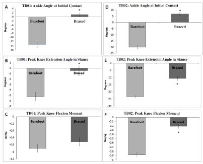
    Figure 3:  Bar plots showing the mean (computed across 5 gait cycles) and 95% confidence interval error bars for three key gait variables measured in our case
series: ankle angle at initial contact (A, D), peak knee extension during stance (B, E), and peak knee flexor moment during stance (C, F) for TB01 (left panels A-C)
and TB02 (right panels D-F). *Represents significant difference detected using a paired t-test between braced and barefoot conditions (p<.05). Note that the paired
t-test for peak knee flexor moment for TB01 was not significant (C), but it met the second criterion for evaluating differences between the two conditions, i.e. the
average value for the braced condition exceeded the 95% CI for the barefoot condition.
Participant TB02
ParticipantTB02 was a 12-year-old female with a clinical diagnosis of TBI and left hemiparesis. For the barefoot condition, the left ankle at initial contact was 20° (1.21°) of plantar flexion, which significantly improved to 7° (1.22°, p<0.01) of dorsiflexion with the application of the AAFO (Figure 3). The left knee at initial contact was in 11° (0.67°) of extension for the barefoot condition and significantly improved to 0.8° (4.62°, p=0.01) of extension for the braced condition. Left peak knee extension during stance significantly improved from 23° (0.65°) of extension for the barefoot condition to 11° (1.24°, p<0.01) of extension for the braced condition (Figure 3). The left peak internal knee flexor moment during stance was 0.88 Nm/Kg (0.05) for the barefoot condition and significantly reduced to 0.21Nm/Kg (0.04, p<0.00) for the braced condition (Figure 3). The left knee flexor moment integral in stance was 0.63 Nm.s/Kg (0.10) for the barefoot condition and significantly improved to 0.02 Nm.s/Kg (0.01, p <0.00) with the addition of the AAFO (Figure 2).
Discussion
The current case series report quantitatively evaluated knee and ankle biomechanics associated with genu recurvatum gait in adolescents with TBI. Consistent with what would be hypothesized for the genu recurvatum gait pattern, in the barefoot condition, both study participants demonstrated increased ankle plantar flexion angles at initial contact, knee hyperextension at initial contact and during stance, and a large and prolonged internal knee flexor moment during stance phase in the barefoot condition. The large and prolonged internal knee flexor moment observed during genu recurvatum gait represents increased mechanical stress on the posterior joint capsule and ligamentous structures of the knee. Over time, the increased internal knee flexor moment may lead to structural changes of the knee joint, symptomatic pain, and decreased function.
The primary goal of this case series was to quantitatively evaluate the effects of the AAFO on gait kinematics and kinetics in adolescents with TBI. During over ground walking in the braced versus the barefoot condition, both participants showed significant improvements in sagittal plane ankle and knee kinematics and kinetics (Figures 2 and 3). During the braced versus barefoot condition, both the participants demonstrated reduced plantar flexion at initial contact, greater knee flexion at initial contact, reduced peak knee extension in stance, reduced peak knee flexor moment in stance, and reduced knee flexor moment integral in stance. The AAFO was effective at providing a more neutral position for the ankle by limiting the amount of plantar flexion during stance. As a result, the tibia had a more vertical alignment and the premature anterior displacement of the GRFV relative to the ankle and knee joints during stance was attenuated. This led to a significant reduction in the amount of knee hyperextension during stance and a reduction in the sagittal plane internal knee flexor moment.
The limitations to this study include the limitations associated with a case-study design (small sample size and the inability to identify clinical examination parameters that may potentially influence the efficacy of the AAFO). Other children and adolescents with TBI and genu recurvatum gait may also benefit from the use of an AAFO to limit the amount of plantar flexion in stance and improve knee hyperextension. Future research with a larger sample size is necessary to gain a better understanding of the biomechanical changes that occur when using an AAFO in adolescents with TBI as well as other neurological impairments. Additionally, future studies should explore whether biomechanical improvements caused by the AAFO are accompanied by long-term improvements in clinical function and participation.
The findings of this case study suggest that in adolescents and children with TBI who demonstrate a genu recurvatum gait pattern, an AAFO may be effective at reducing knee hyperextension during stance phase by improving the foot and ankle position during stance. The resulting reduction in the internal knee flexor moment observed during walking with the AAFO may reduce the mechanical stress on the posterior knee joint capsule. For children and adolescents with TBI, the early abatement of genu recurvatum through the use of an AAFO may lead to a more sustainable gait pattern by reducing the risk of permanent structural damage and future pathologies in the knee joint. This case series report underscores the need for more research investigation into the biomechanical effects as well as clinical factors influencing efficacy of ankle foot orthosis in children and adolescents with TBI.
References
- Katz-Leurer M, Rotem H, Lewitus H, Keren O, Meyer S. Relationship between balance abilities and gait characteristics in children with post-traumatic brain injury. Brain Inj. 2008; 22: 153-159.
- Kerrigan DC, Deming LC, Holden MK. Knee recurvatum in gait: a study of associated knee biomechanics. Arch Phys Med Rehabil. 1996; 77: 645-650.
- JP. Gait Analysis. Thorofare, NJ: SLACK; 1992.
- Loudon JK, Goist HL, Loudon KL. Genu recurvatum syndrome. J Orthop Sports Phys Ther. 1998; 27: 361-367.
- Morgan PM, LaPrade RF, Wentorf FA, Cook JW, Bianco A. The role of the oblique popliteal ligament and other structures in preventing knee hyperextension. Am J Sports Med. 2010; 38: 550-557.
- Boudarham J, Zory R, Genet F, et al. Effects of a knee-ankle-foot orthosis on gait biomechanical characteristics of paretic and non-paretic limbs in hemiplegic patients with genu recurvatum. Clin Biomech (Bristol, Avon). 2013; 28: 73-78.
- Tyson SF, Thornton HA. The effect of a hinged ankle foot orthosis on hemiplegic gait: objective measures and users’ opinions. Clin Rehabil. 2001; 15: 53-58.
- Mulroy SJ, Eberly VJ, Gronely JK, Weiss W, Newsam CJ. Effect of AFO design on walking after stroke: impact of ankle plantar flexion contracture. Prosthet Orthot Int. 2010; 34: 277-292.
- Bleyenheuft C, Bleyenheuft Y, Hanson P, Deltombe T. Treatment of genu recurvatum in hemiparetic adult patients: a systematic literature review. Ann Phys Rehabil Med. 2010; 53: 189-199.
- Tyson SF, Sadeghi-Demneh E, Nester CJ. A systematic review and metaanalysis of the effect of an ankle-foot orthosis on gait biomechanics after stroke. Clin Rehabil. 2013; 27: 879-891.
- Condie DN. The modern era of orthotics. Prosthet Orthot Int. 2008; 32: 313- 323.
- Hogue RE, McCandless S. Genu recurvatum: auditory biofeedback treatment for adult patients with stroke or head injuries. Arch Phys Med Rehabil. 1983; 64: 368-370.
- Fatone S, Gard SA, Malas BS. Effect of ankle-foot orthosis alignment and foot-plate length on the gait of adults with poststroke hemiplegia. Arch Phys Med Rehabil. 2009; 90: 810-818.
- Hislop H MJ. Daniels and Worthingham’s muscle testing: techniques of manual examination. Philadelphia: WB Saunders; 2002.
- Kesar TM, Binder-Macleod SA, Hicks GE, Reisman DS. Minimal detectable change for gait variables collected during treadmill walking in individuals poststroke. Gait Posture. 2011; 33: 314-317.