
Special Article – Cerebral Palsy
Phys Med Rehabil Int. 2016; 3(5): 1097.
Kinematic Motion Analysis in Upper Extremity Cerebral Palsy
Scallon G and Van Heest A*
Department of Orthopaedic Surgery, University of Minnesota, USA
*Corresponding author: Ann Van Heest, MD, Department of Orthopaedic Surgery, University of Minnesota, Administrative Offices 2512 South 7th Street, Suite R200, Minneapolis, MN 55454, USA
Received: August 31, 2016; Accepted: September 23, 2016; Published: September 26, 2016
Abstract
Management of physical impairment in children with upper extremity cerebral palsy is complex and multifaceted. In this patient population, clinical presentation and neurologic involvement is widely variable. Management decision making, particularly with respect to surgical and invasive interventions, is built on comprehensive history, physical examination, and assessment of function. In that cerebral palsy is a disorder of movement and posture, additional methods of assessment of the movement disorder have been developed and validated. Historically, kinematic data in the lower extremities has preceded advancements in the upper extremities due to complexity of motion and multiple body segments. In the upper extremity, motion assessment has been validated for rating videos for joint positioning and functional impairment. Kinematic data collected during functional tasks assesses deficiency. Kinematic data has involved video capture and review, electromyography, and fixed marker tracking. Sophisticated software and technological advancements are promising to aid upper extremity kinematic assessment in children with cerebral palsy. Ultimately, these technologies may play a significant role navigating complex treatment algorithms.
Keywords: Cerebral palsy; Video; Motion analysis; Kinematics; Upper extremity
Abbreviations
CP: Cerebral Palsy; EMG: Electromyography; ROM: Range of Motion; FCU: Flexor Carpi Ulnaris; PT: Pronator Teres; ECU: Extensor Carpi Ulnaris; SHUEE: Shriners Hospital for Children Upper Extremity Evaluation; ECRL: Extensor Carpi Radialis Longus; ECRB: Extensor Carpi Radialis Brevis; BR: Brachioradialis, ECRB: Extensor Carpi Radialis Brevis.
Introduction
Cerebral palsy (CP) is a disorder of development of movement and posture causing activity limitations that are attributed to nonprogressive disturbances the occurred in the developing fetal or infant brain. Wide variability in the manifestation of impairment exists depending on the extent and location of the central nervous system lesion. Abnormal innervation ultimately results in imbalance of muscle forces across multiple joints, affecting different muscles and different joints to varying degrees. The most common manifestations in the upper extremity include shoulder adduction with internal rotation, elbow flexion, forearm pronation, wrist flexion and ulnar deviation, finger flexion, and thumb in palm deformities.
Patient evaluation in the upper extremity includes a careful history and physical examination. History includes a subjective patient and parent assessment of functional limitations. Physical examination includes active and passive range of motion of all joints, as well as assessment of muscle tone and control. Sensibility of the hand with assessment of stereo gnosis function helps understand functional use patterns, as the sensory cortex, as well as the motor cortex, of the brain is commonly affected. Historically, classification of upper extremity involvement in CP has been subjectively based on examiner interpretation, functional tests, and physical examination [1-4].
In that cerebral palsy is a disorder of movement and posture, additional methods of assessment of the movement disorder are necessary. Static and dynamic assessment of limb mechanics informs therapeutic decision making [1]. Analysis of motion in the upper extremity is integral to guiding rehabilitation and management of individuals with CP. Techniques for analysis static and dynamic upper extremity mechanics can include electromyography (EMG), wearable marker kinematic measurement, and motion capture. This review investigates how motion analysis is being used for assessment of upper extremity dysfunction due to CP, as an adjunct for decision making in this complex patient population.
Upper Extremity Cerebral Palsy
Upper extremity involvement in children with CP is pervasive and is commonly assessed by the Manual Ability Classification System (MACS), which describes how children (4-18 years) with cerebral palsy use their hands to handle objects in daily activities. . Of children with CP ages 4-14, approximately 64% are independent with activities of daily living in the upper extremity (MACS I-II) and 14% are completely dependent (MACS V). Thumb-in-palm deformity was noted in 41% of children, with a predilection for those with spastic quadriplegia [3-6]. No or minimal digital flexor spasticity was reported in 69%, moderate spasticity in 23%, and no active wrist or finger extension in 2%. Typical imbalance results in elbow flexion, forearm pronation, wrist flexion, ulnar deviation, finger flexion, and thumb-in-palm deformity. There is some evidence that correction of distal impairments in the upper extremity may improve proximal kinematics [7].
The paradigm for management of upper extremity involvement in children with CP is broad. Non-operative interventions include tone management with medications including botulinum toxin A injections,; therapies including constraint-induced movement therapy, bimanual training, mirror therapy, hand therapy, Kinesio tape, or somatosensory training; as well as splinting, , and electrical stimulation [8]. Surgical intervention has been shown to improve static and dynamic limb positioning as well as improvement on functional testing [4,9-13]. Guidance of surgical intervention in upper extremity procedures relies on careful examination and assessment of muscle imbalance, abnormal limb positioning, and functional impairment [9,11].
Motion Analysis
Delineation of upper extremity kinematics has evolved substantially over the last century. At one end of the spectrum is qualitative description of during routine daily activities. Kinematics of the upper extremity tasks have been assessed since the 1960’s using rudimentary electrogoniometers to measure joint motion and angular velocity [14]. More recent studies have integrated video recordings, EMG, optoelectric and ultrasound tracking, and most recently 3-D pattern recognition. The use of these technologies and the findings in the literature are summarized.
Video Recordings
For many years, video recordings have been used to supplement the evaluation of upper and lower extremity kinematics. Historically, the bulk of this literature has quantified anthropomorphic norms. It has been widely used in sports and assessing repetitive workplace trauma [14]. Video recording of children, particularly in the home environment, has been used to develop several classification systems of upper extremity and hand dysfunction [15,16].
One example of goniometric data extracted from video recordings is demonstrated by Kruelen et al [17,18]. Video recording data is collected from bi-planar cameras on a global coordinate system (Figure 1). Synchronized data is collected of subjects maximally pronating and supinating forearms and lifting a class and a wooden disk at shoulder height. Angular measurements are manually calculated from isolated video frames, which are translated into angular velocity and range of motion (ROM) data. Accuracy of the model is predicted within 5 mm. Ink markings were made over bony prominences about the elbow, wrist, acromion, and sternum. In the first study, the authors examine a cohort of patients with CP and pronation and wrist flexion deformities. They had spasticity of the flexor carpi ulnaris (FCU) and pronator teres (PT) with poor active but good passive ROM. These patients were indicated for PT rerouting and FCU to extensor carpi ulnaris (ECU) transfer. Examination preand postoperatively demonstrates a mean supination increase of 63°, active ROM increase of 23°, and active pronation decrease 40° [17]. The second study using this technique compared 10 patients with hemiplegic CP with impaired supination of the forearm to 10 control subjects. They demonstrate increased lateral and anterior trunk bend as a compensatory mechanism for elbow flexion and pronation deformities.
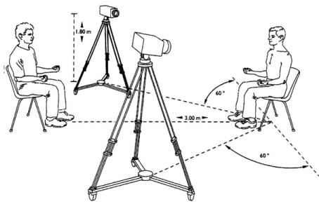
Figure 1: Biplanar video recording coordinate system [17].
In 1996 the Shriners Hospital for Children Upper Extremity Evaluation (SHUEE) was devised to assess upper extremity function in children with hemiplegic CP [2,19]. Standardized tasks of daily living are administered, generally by an occupational therapist. Review of the video recordings allows scoring of thumb, finger, wrist, forearm, and elbow function. A basic survey consisting of demographic information, baseline activities of daily living, independence, goals, and tone is completed first. Passive and active ROM is quantified. Three domains are subdivided by the SHUEE, including Spontaneous Functional Analysis, Dynamic Positional Analysis, and Grasp/Release Analysis. Any standard video recorder can be used in a space large enough to toss a ball and crawl. The camera is repositioned as needed to adequately visualize the desired body segment. Body segments are assessed in sequence through 16 tasks (Table 1). Spontaneous Functional Analysis scores each task based on the modified House Scale from 0 to 5 and aggregates these data. Dynamic Position Analysis is a qualitative description of ROM at each body segment. This functional tool has shown excellent inter observer reliability and correlation with other functional tests. It has demonstrated quantifiable change after surgical intervention [2,9,11,20].
![]()
    Segment Analyzed
    Task
    Dynamic Positional Analysis


    Thumb and fingers
    Pulling    money from wallet
      Folding    paper
      Tearing    paper
      Stringing    bead
    Thumb    – in-palm deformity, web space closed, web space open

      Finger    – flexion, neutral, hyperextension


    Wrist
    Unscrew    bottle cap
      Pull    Play-Doh apart
      Cut    Play-Doh with knife
      Throw    large ball
    Wrist    flexion, neutral, extension, ulnar deviation, radial deviation


    Forearm
    Accept    coins in palm
      Hi-five
      Hand    to mouth
      Touch    contralateral ear
    Forearm    extreme pronation, pronation, neutral, supination


    Elbow
    Don    sock
      Tie    shoe
      Place    sticker on ball
      Crawl
    Elbow    extreme flexion, flexion, extension
Table 1: SHUEE tasks by body segment [2].
Electromyography and Video Capture
Motion capture in conjunction with EMG has been used to delineate phasic control of individual muscles. In the setup described by Van Heest et al [1,10], EMG data is collected from needle electrodes in the PT and FCU. Laboratory setup consists of bi-planar video frames while monitoring EMG data, allowing for assessment of deformity in the coronal and sagittal planes. Surface electrodes are used for the extensor carpi radialis longus (ECRL) and extensor carpi radialis brevis (ECRB). The child performs the Jebsen-Taylor functional test which involves writing, manipulating small objects, stacking, and light and heavy can lifting. EMG firing patterns are collected and simultaneously assessed during functional activity. Normal firing of the FCU produces signal during release and absence of signal during grasp. Continued firing of the FCU implies spasticity and indicates a lack of phasic control. A spastic muscle is a poor donor for tendon transfer, but may be amenable to botox injection or lengthening.
Van Heest et al [21] review 7 patients who underwent FCU to ECRB tendon transfer and PT release with pre- and postoperative video/EMG analysis. This was a group of patients selected for favorable phasic control of the FCU, facilitating transfer to the ECRB. Postoperative findings demonstrated phase change in 1 patient. Postoperatively, 4 patients were able to place a can on a shelf, compared to only 2 preoperatively. The majority pattern was firing of the FCU during release, though now acting as a wrist extensor, and thus not synergistic with finger flexion. A broader study using the same motion lab was performed on 48 patients with hemiplegic CP undergoing surgery for wrist deformity [9]. Surgical decision making based on examination and motion lab findings is outlined in Table 2. Results showed improvement of finger, wrist, elbow, and forearm positioning during functional tasks based on the SHUEE Dynamic Positional Analysis.
![]()
    Motion Laboratory Findings
    Treatment Rendered


    Minimal abnormalities

    No wrist treatment


    Phasic firing of FCU
      Phasic firing of ECRB/ECRL
      Mild wrist flexion/ulnar    deviation positioning
      Full passive ROM of wrist
    FCU Botox injection


    Phasic firing of FCU
      Phasic firing of ECRB/ECRL
      Significant wrist ulnar    deviation (no flexion) positioning
      Full passive ROM of wrist
    Extensor carpi ulnaris to    ECRB transfer


    Phasic firing of FCU
      Minimal/no firing of    ECRB/ECRL
      Significant wrist    flexion/ulnar deviation positioning
      Full passive ROM of wrist
    FCU to ECRB tendon transfer


    Phasic firing of FCU
      Minimal/no firing of    ECRB/ECRL
      Significant wrist    flexion/ulnar deviation positioning
      Limited ROM of wrist
    FCU to ECRB tendon transfer    with proximal row carpectomy


    Spastic firing of FCU
      Phasic of ECRB/ECRL
      Significant wrist    flexion/ulnar deviation positioning
      Limited ROM of wrist
    FCU lengthening or flexor    pronator slide


    Spastic firing of FCU
      Minimal/no firing of    ECRB/ECRL
      Significant wrist    flexion/ulnar deviation positioning
      Limited ROM of wrist
    Lengthening of FCU with    brachioradialis or extensor carpi ulnaris to ECRB


    Minimal motor control of FCU    or ECRB
Table 2: Wrist Treatment Algorithm for Patients Undergoing Motion Laboratory Analysis [9].
EMG findings have been assessed in the upper extremity after traumatic brain injury or stroke [22,23]. Video and goniometric data are collected simultaneously. Out-of-phase and continuous firing of the brachioradialis (BR) is noted, as well as prolonged firing of the biceps with elbow extension (Figure 2). Similar patterns are noted across the wrist; however there is significantly more variability in the presentation. Hoffer et al [24] review 23 patients who underwent EMG analysis for patient with CP presenting with a thumb-in-palm deformity. EMG of the adductor policies informed surgical decision making regarding total or partial muscle release. EMG has been used successfully to facilitate treatment, though it is limited in the wide variability of presentation, cost, and invasiveness.
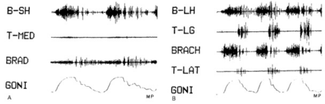
    Figure 2:  EMG data in muscles about the elbow. B-SH/LH: Biceps Short and
Long Heads; T: Triceps; BRAD: Brachioradialis; BRACH: Brachialis; GONI:
Elbow Flexion (deflection up) and extension (deflection down) [22].
Marker Tracking
Wearable markers are subject to interference with adjacent anatomic segments. Wearable devices add weight and alter soft tissue tension, which can alter kinematics [11,25,26]. Prior attempts to capture hand kinematic data in children with CP have been limited by these constraints [1]. Modeling of the hand has typically involved a single marker or a glove with transducers (Figure 3). This methodology has been the gold standard of upper and lower extremity kinematic analysis over the last 2 decades.
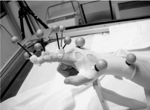
    Figure 3:  Glove optoelectric sensor allowing for capture of index finger
motion segments [26].
Mackey et al [27] examined 10 normal children compared to 10 children with a diagnosis of hemiplegic CP. Optoelectric markers were fixed on bilateral upper limbs and trunk, with a single marker used for the shoulder and a single marker for the hand (Figure 4). The model combined shoulder with scapulothoracic motion and estimated joint by anthropomorphic data. A hand-to-head task, hand-to-mouth task, and reach task were evaluated. Children with hemiplegia demonstrated decreased elbow extension and supination with compensatory trunk flexion in grasping techniques. Additionally, they demonstrated slower peak angular velocities and longer completion times compared to the non-dominant arm in control subjects. Bimanual dexterity in children with hemiplegic CP tended to exacerbate deficiencies in the affected hand when the dominant arm took over. Similar protocols have been described, demonstrating longer duration for task completion with decreased smoothness of trajectory [28-31].
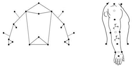
    Figure 4:  Upper extremity modeling of body segments with optoelectric
markers [27].
Fitoussi et al [7] describe a similar protocol in patients with spastic cerebral palsy. Of their patient population, 4 underwent botox injection and 4 underwent surgical lengthening procedures. Children with hemiplegia demonstrated relative shoulder abduction and external rotation, elbow flexion, forearm pronation, wrist ulnar deviation, and lateral trunk bending. After PT, FPL, or adductor polices lengthening, patients had decreased shoulder and trunk compensatory motion. This reinforces the theme of proximal compensation for distal dysfunction, and highlights the importance of collecting sound kinematic data in the more challenging distal body segments.
3-D Motion Capture
Recently time-of-flight cameras have gained significant traction in commercial use. These devices measure the time taken for a projected light signal to reflect off an object. The depth and 3-D structure of the object is mapped. This allows very rapid image capture, though it can be limited by resolution, interference, and background lighting. These commercial models have built-in algorithms for modeling the hand. These biomechanical equations allow for the creation of the 3-D representation of dynamic finger, thumb, wrist, and forearm movement. These devices contain traditional visible light capture cameras which can digitally mined post hoc. The cost for off-the-shelf 3-D cameras is around $150. One such device, the Xbox Kinect™, has gotten significant attention in the last 5 years. Clinical applications have explored involved diagnostic evaluation of autism, CMC arthritis, Parkinson’s disease, sports, stroke, and CP. Significant research is devoted to the use of 3-D motion capture (Figure 5) in rehabilitation, therapy, and biofeedback.
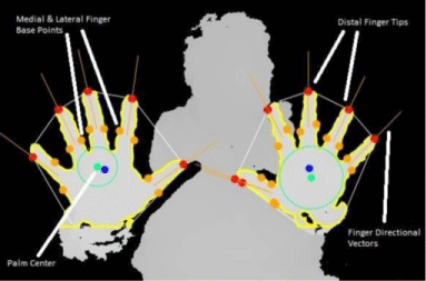
Figure 5: Motion capture technology.
Many studies have investigated the precision and accuracy of the system compared to gold-standard optoelectric marker tracking. Kuster et al [32] examined 20 normal subjects with both optoelectric markers and a Kinect™ system and found accuracy within 3.9 ± 4.0° for shoulder motion and 0.1 ± 3.8° for trunk motion. Accuracy of the system appeared to dip when body segments overlapped in the view of the Kinect™.
Rammer et al [33] published a pilot study of an algorithm developed to quantify upper extremity kinematics during activities of daily living using the Kinect™. They examined 12 normal pediatric subjects administering a SHUEE examination and collecting depth and video data during functional tasks. All subjects had 100% scores on the SHUEE examination. Absolute kinematic data were collected, though variable between the subjects. The examiners were able to extract patterns of motion which seemed to correlate with specific tasks. For example, unscrewing a bottle correlated strongly with wrist ROM, peak wrist velocity, and peak wrist acceleration. It correlated with elbow and shoulder ROM secondarily. These models may ultimately provide normalized kinematic data, and perhaps more importantly, they may illustrate typical patterns of compensation. In the future, this may provide valuable information about compensatory deficiencies in the CP population.
Discussion
Compared to lower extremity kinematics, there are significant challenges to upper extremity analysis. The lower extremities can be simplified to a few anatomic segments and degrees of freedom with closed-chain, weight-bearing force analysis. Upper extremity analysis, on the other hand, involves a much higher number of anatomic segments, each with several degrees of freedom. Additionally, force transduction in the upper extremity is unconstrained and much more difficult to quantify in functional tests. In the upper extremity, there is variability between normal subjects in functional kinematics used to complete a task. There is substantial redundancy in the upper extremities, not found in the lower extremities, which confounds analysis [14]. This redundancy and normal tendency toward compensation suggests that absolute measurements of ROM and angular velocities may be limited in correlation with function, except at extremes. Rather, pattern recognition and categorization of dynamic motion may provide more fruitful in differentiating pathology, and more importantly may inform how individuals may best respond to treatment. Finally, it is speculated that central nervous system plasticity without neurologic impairment allows functional adaptation and compensation [34].
Assessment of dynamic motion is crucial to management of upper extremity involvement in children with CP. This technology is rapidly blossoming in the commercial sector. Advancements of these technologies can evolve into robust clinical decision making for children with upper extremity CP. Standardization of indications for invasive interventions are clearly needed. There is wide variability in clinical manifestations of upper extremity CP as well as non-surgical interventions and operative techniques. Functional assessment tools and current classification schemes can elucidate the quality and magnitude of impairment, but are presently too blunt to guide management [15]. EMG has been used successfully to probe dynamic muscle use, but is limited by expense and invasiveness. Optoelectric markers have been employed successfully in the evaluation of lower extremity CP, but are prone to interference with functional use during the assessment in the upper extremity. This is especially challenging in the hand.
Motion capture technology shows potential as a non-invasive, low-cost, reproducible alternative. Sophisticated video analysis and time-of-flight data may allow classification of dynamic patterns within functional upper extremity limb use. Ultimately, outcomes of surgical and non-surgical interventions may be weighed against the automated motion capture, leading to finely-tuned management indications.
Funding
MERA grant 2014 from Gillette Children’s Specialtycare, St. Paul, MN.
References
- Van Heest AE. Functional assessment aided by motion laboratory studies. Hand Clin. 2003; 19: 565–571.
- Davids JR, Peace LC, Wagner L V, Gidewall MA, Blackhurst DW, Roberson WM. Validation of the Shriners Hospital for Children Upper Extremity Evaluation (SHUEE) for children with hemiplegic cerebral palsy. J Bone Joint Surg Am. 2006; 88: 326–333.
- Eliasson A-C, Krumlinde-Sundholm L, Rösblad B, Beckung E, Arner M, Ohrvall A-M, et al. The Manual Ability Classification System (MACS) for children with cerebral palsy: scale development and evidence of validity and reliability. Dev Med Child Neurol. 2006; 48: 549–554.
- House JH, Gwathmey FW, Fidler MO. A dynamic approach to the thumb-in palm deformity in cerebral palsy. J Bone Jt Surg Am. 1981; 63: 216–225.
- Arner M, Eliasson AC, Nicklasson S, Sommerstein K, H?? gglund G. Hand Function in Cerebral Palsy. Report of 367 Children in a Population-Based Longitudinal Health Care Program. J Hand Surg Am. 2008; 33: 1337–1347.
- Park ES, Sim EG, Rha DW. Effect of upper limb deformities on gross motor and upper limb functions in children with spastic cerebral palsy. Res Dev Disabil. 2011; 32: 2389–2397.
- Fitoussi F, Diop A, Maurel N, Laasel EM, Ilharreborde B, Penneçot GF. Upper limb motion analysis in children with hemiplegic cerebral palsy: Proximal kinematic changes after distal botulinum toxin or surgical treatments. J Child Orthop. 2011; 5: 363–370.
- Shierk A, Lake A, Haas T. Review of Therapeutic Interventions for the Upper Limb Classified by Manual Ability in Children with Cerebral Palsy. Semin Plast Surg. 2016; 30: 14–23.
- Van Heest AE, Ramachandran V, Stout J, Wervey R, Garcia L. Quantitative and qualitative functional evaluation of upper extremity tendon transfers in spastic hemiplegia caused by cerebral palsy. J Pediatr Orthop. 2008; 28: 679–683.
- Van Heest A, Stout J, Wervey R, Garcia L. Follow-up motion laboratory analysis for patients with spastic hemiplegia due to cerebral palsy: analysis of the flexor carpi ulnaris firing pattern before and after tendon transfer surgery. J Hand Surg Am. 2010; 35: 284–290.
- Smitherman JA, Davids JR, Tanner S, Hardin JW, Wagner L V, Peace LC, et al. Functional Outcomes Following Single-Event Multilevel Surgery of the Upper Extremity for Children with Hemiplegic Cerebral Palsy. J Bone Jt Surg. 2011; 93: 655–661.
- Hoffer MM, Perry J, Garcia M, Bullock D. Adduction contracture of the thumb in cerebral palsy. A preoperative electromyographic study. J Bone Jt Surg Am. 1983; 65: 755–759.
- Boyd RN, Morris M., Graham HK. Management of upper limb dysfunction in children with cerebral palsy: a systematic review. Eur J Neurol. 2001; 8: 150–166.
- Buckley MA, Yardley A, Johnson GR, Cams DA. Dynamics of the upper limb during performance of the tasks of everyday living: a review of the current knowledge base. J Eng Med. 1996; 210: 241–247.
- Wagner LV, Davids JR. Assessment tools and classification systems used for the upper extremity in children with cerebral palsy. Clin Orthop Relat Res. 2012; 470: 1257–1271.
- Waters PM, Zurakowski D, Patterson P, Bae DS, Nimec D. Inter observer and Intraobserver Reliability of Therapist-Assisted Videotaped Evaluations of Upper-Limb Hemiplegia. J Hand Surg Am. 2004; 29: 328– 334.
- Kreulen M, Smeulders MJC, Veeger HEJ, Hage JJ, Van Der Horst CMAM. Three-dimensional video analysis of forearm rotation before and after combined pronator teres rerouting and flexor carpi ulnaris tendon transfer surgery in patients with cerebral palsy. J Hand Surg Am. 2004; 29: 55–60.
- Kreulen M, Smeulders MJC, Veeger HEJ, Hage JJ. Movement patterns of the upper extremity and trunk associated with impaired forearm rotation in patients with hemiplegic cerebral palsy compared to healthy controls. Gait Posture. 2007; 25: 485–492.
- Wagner LV, Davids JR, Hardin JW. Selective Control of the Upper Extremity Scale: Validation of a clinical assessment tool for children with hemiplegic cerebral palsy. Dev Med Child Neurol. 2016; 58: 612-617.
- Van Heest AE, Bagley A, Molitor F, James MA. Tendon Transfer Surgery in Upper-Extremity. J Bone Jt Surg - Am Vol. 2015; 529–536.
- Van Heest A, Stout J, Wervey R, Garcia L. Follow-Up Motion Laboratory Analysis for Patients With Spastic Hemiplegia Due to Cerebral Palsy: Analysis of the Flexor Carpi Ulnaris Firing Pattern Before and After Tendon Transfer Surgery. J Hand Surg Am. Elsevier Inc.; 2010; 35: 284–290.
- Kozin S, Keenan MA. Using Dynamic Electromyography to Guide Surgical Treatment of the Spastic Upper extremity in the Brain-Injured Patient. Clin Orthop Relat Res. 1993; 288: 109–117.
- Pinzur MS. Dynamic electromyography in functional surgery for upper limb spasticity. Clin Orthop Relat Res. 1993; 288: 118–121.
- Hoffer BYMM, From D, Jacquelin E, Perry LA, California M, Garcia D, et al. Adduction Thumb Contracture in Cerebral of the Palsy. J Bone Jt Surg. 1983; 65: 755–759.
- Rab G, Petuskey K, Bagley A. A method for determination of upper extremity kinematics. Gait Posture. 2002; 15: 113–119.
- Degeorges R, Parasie J, Mitton D, Imbert N, Goubier JN, Lavaste F. Three-dimensional rotations of human three-joint fingers: an optoelectronic measurement. Preliminary results. Surg Radiol Anat. 2005; 27: 43– 50.
- Mackey AH, Walt SE, Stott NS. Deficits in upper-limb task performance in children with hemiplegic cerebral palsy as defined by 3-dimensional kinematics. Arch Phys Med Rehabil. 2006; 87: 207–215.
- Jaspers E, Feys H, Bruyninckx H, Harlaar J, Molenaers G, Desloovere K. Upper limb kinematics: Development and reliability of a clinical protocol for children. Gait Posture. Elsevier B.V.; 2011; 33: 279– 285.
- Jaspers E, Desloovere K, Bruyninckx H, Klingels K, Molenaers G, Aertbeliën E, et al. Three-dimensional upper limb movement characteristics in children with hemiplegic cerebral palsy and typically developing children. Res Dev Disabil. 2011; 32: 2283–2294.
- Jaspers E, Desloovere K, Bruyninckx H, Molenaers G, Klingels K, Feys H. Review of quantitative measurements of upper limb movements in hemiplegic cerebral palsy. Gait Posture. 2009; 30: 395–404.
- Petuskey K, Bagley A, Abdala E, James MA, Rab G. Upper extremity kinematics during functional activities: Three-dimensional studies in a normal pediatric population. Gait Posture. 2007; 25: 573–579.
- Kuster RP, Heinlein B, Bauer CM, Graf ES. Accuracy of KinectOne to quantify kinematics of the upper body. Gait Posture [Internet]. Elsevier B.V.; 2016; 47: 80–85.
- Rammer JR, Krzak JJ, Riedel SA, Harris GF. Evaluation of upper extremity movement characteristics during standardized pediatric functional assessment with a Kinect??-based markerless motion analysis system. Conf Proc. Annu Int Conf IEEE Eng Med Biol Soc IEEE Eng Med Biol Soc Annu Conf. 2014; 2014: 2525–2528.
- Johanson ME, James M a, Skinner SR. Forearm muscle activation during power grip and release. J Hand Surg Am. 1998; 23: 938–944.