Special Article – Gait Rehabilitation
Phys Med Rehabil Int. 2016; 3(5): 1096.
A Biomechanical Investigation of Selected Lumbo-Pelvic Hip Tests: Implications for the Examination of Running
Bailey R*, Richards J and Selfe J
School of Sport, Tourism & The Outdoors, University of Central Lancashire, UK
*Corresponding author: Bailey RW, School of Sport, Tourism & The Outdoors, University of Central Lancashire, Preston, UK
Received: August 16, 2016; Accepted: September 08, 2016; Published: September 12, 2016
Abstract
Introduction: Lumbo-Pelvic Hip tests are commonly used to examine the components of running. Investigators however have presented very little empirical research in which they have documented the biomechanics of these tests or their relationship to the kinematics of running.
Materials and Methods: 14 male participants who had no pain, injury, or neurologic disorder. Hip and pelvic movements were recorded during the Trendelenburg, Single Leg Squat and Corkscrew tests.
Results and Discussion: The mean and standard deviation of the hip and lumbo-pelvic movements in the sagittal, coronal and transverse planes were reported for the different tests. The pelvic obliquity during the Trendelenburg Test is statistically different to running. Hence the Trendelenburg Test is not an appropriate proxy clinical test for examining the pelvic obliquity component of running. The hip coronal plane range of movement during the Single Leg Squat is similar to that found during of running. The Single Leg Squat is therefore an appropriate clinical test for examining the hip coronal plane range of movement component of running. However the hip flexion range of movement found during the Single Leg Squat and hip rotation during the Corkscrew Test were different to running.
Conclusion: Pelvic obliquity during the Trendelenburg Test, the hip sagittal plane range of movement during the Single Leg Squat, and the hip transverse plane during the Corkscrew Test were different to running. This indicates that the Trendelenburg Test, Single Leg Squat, and Corkscrew Tests are not appropriate to use when examining aspects of the pelvic and hip movements of running. However the hip coronal ranges of movement during the Single Leg Squat was similar to running. Therefore the Single Leg Squat and Corkscrew Tests may be used to examine this component of running. Clinicians may wish to use alternative tests to examine these parameters of gait.
Keywords: Lumbopelvic Hip; Range of motion; Articular; Walking; Biomechanical Phenomena
Introduction
Clinicians commonly use tests including the Trendelenburg [1], Single Leg Squat [2] and Corkscrew Tests during the examination of the Lumbo-Pelvic and Hip complex. These tests are used to examine the movements of the Lumbar, Pelvic and Hip regions in a weight bearing position [1-4]. They may be used in isolation [5,6], or to compliment the examination of functional tasks including running [4]. The clinical assumption is that the Lumbar, Pelvic and Hip movements generated during these tests are similar to those of running [4]. However, there are few biomechanical investigations of the normative kinematics of these tests, and a limited number of previous studies that compare the kinematics of these tests to running.
Running is popular as a recreational [7,8] and competitive sport [9] of its own and forms part of locomotion within other sports [10]. However, running has been associated with developing injuries [11- 13], of the hip [14], and Ilio-Tibial band (ITBS) [15-17]. Previous studies have found differences in Lumbo-Pelvic Hip kinematics between normal, healthy participants and runners. Kelli found that training for running caused participants to exhibit increased stance phase hip adduction range of movement during running (P = 0.05), and a trend towards decreased hip internal rotation range of movement (P = 0.08) when compared to normal healthy participants [18]. Noehren concluded that runners with significantly greater stance phase hip adduction (P>.05) are at increased risk of ITBS, and Zifchock [19] stated that runners with reduced hip internal rotation exhibit increased incidence of injuries. Hreljac found that runners with lower hip flexion range of movement correlated with an increased incidence of hip extensor muscle strains [20]. Similarly, Van Mechelen [21] found runners with reduced hip flexion range of movement (59.4° +/- 8.0, p > 0.001) exhibited higher injury rates. A study by Ferber found that runners with a previous history of ITBS did not regain full hip abduction range of movement following injury (difference 2.47° +/-1.48, P>.05) [16]. In contrast, Van Mechelen and Zifchock found no relationship between ankle sagittal plane mobility [21] and knee valgus angle [19]. Hence the hip appears to have a key role in the development of running injuries, particularly the coronal and transverse plane range of movement. Asymptomatic runners appear to exhibit a hip hypo-mobility cycle. They have less hip mobility when compared to normal, healthy participants. Runners with greater hip hypo-mobility appear to be at higher risk of injuries, and following these injuries hip movement often remains restricted. Whilst the clinical examination of running is acknowledged as one of the most difficult challenges for clinicians in sports medicine [22], the accurate assessment of hip movement, particularly in the coronal and transverse plane, is clearly of clinical importance in preventing and treating running injuries.
Current Lumbo-Pelvic Hip test studies have been confined to walking rather than running gait. There are no previous studies of the relationship of these tests to running. The Trendelenburg Test is interpreted by observing pelvic obliquity during the test [3,23]. Two previous studies have objectively defined when the pelvic drop (obliquity) becomes positive. Asayama stated that a “tilt angle” (pelvic obliquity) of greater than 2° indicated a positive Trendelenburg Test [5]. Westhoff stated that “Pelvic drop to the swinging limb during single stance phase of more than 4° and / or maximum (peak value) pelvic drop in the stance phase of more than 8°” [24] indicated a positive test. There are no published data quantifying sagittal and transverse plane pelvic movement during the Trendelenburg Test. The Single Leg Squat is currently interpreted by observing hip range of movement in the sagittal and coronal planes. Only one author, Livengood, has objectively defined when the Single Leg Squat becomes positive. Hip flexion greater than 65°, hip abduction / adduction greater than 10°, knee valgus / varus greater than 10° [4]. There are no published data for sagittal, coronal and transverse plane pelvic movement during the Single Leg Squat. The Trendelenburg Test requires neuromuscular control of the pelvis in the coronal plane and the Single Leg Squat control of the hip in the sagittal plane. Interestingly there are currently no existing tests for neuromuscular control of the pelvis requiring hip internal-external rotation movement in the transverse plane documented within the musculoskeletal literature. Hence a novel clinical test for the assessment of the Lumbo-Pelvic and Hip region in the transverse plane has started to be used within clinical practice. This test has been termed the “Corkscrew Test”. The method for performing the Corkscrew Test is based upon the Single Leg Squat [4] and its interpretation is based upon the Single Leg Squat criterion in combination with kinematic values found within the walking literature [25-27]. The participant stands on the limb being evaluated, with the contralateral leg lifted off the ground, is as if walking. The participant rotates the weight bearing hip first into maximal hip internal rotation, then external rotation, and returns to the start position in less than 6seconds. The Corkscrew Test is a new test hence there are currently no kinematic data to support its use in clinical practice.
Despite a runnner’s neuromuscular movement control between the lower limb and ground being acknowledged as a factor influencing injury risk [28], there are no existing kinematic studies comparing kinematics of the Lumbo-Pelvic Hip movement tests to running. Establishing normative movement data of the Trendelenburg, Single Leg Squat and Corkscrew Tests for runners may help increase our knowledge of the Lumbo-Pelvic Hip kinematics and develop our understanding of the hip hypo-mobility cycle found within this population. This data may help explain the aetiology of hip [14], and soft tissue injuries [15-17] injuries found within runners, and help clinicians and researchers to develop diagnostic algorithms for their examination and treatment.
The purpose of this study was to investigate the biomechanical characteristics of the Trendelenburg, Single Leg Squat and Corkscrew Tests and their relationship to the kinematics of running. It was hypothesised that, the pelvic obliquity achieved during the Trendelenburg Test would be similar to this parameter of running, but that the hip sagittal and coronal plane range of movement during the Single Leg Squat and the hip rotation range during the Corkscrew Test would be different.
Materials and Methods
Participants
14 healthy male participants were recruited (age 20.5 +/- 2.0 years, 1.76 +/- 0.13m height, mass 73.9 +/- 9.0kg) who had no pain or neuromusculoskeletal disorder. Demographic data were recorded. Data were collected from both limbs of each participant. Volunteers gave written informed consent before data collection. All data collection conformed to the Declaration of Helsinki. The study was approved by the Faculty of Health Research Ethics Committee, University of Central Lancashire.
Instrumentation
Kinematic data were collected using a 10-camera Pro Reflex system (Qualisys Medical AB, Gothenburg, Sweden) at 100Hz. Force data were collected using an AMTI force platform (Advanced Mechanical Technology, Inc, Watertown, MA, model BP400600). Force data was used to define the events of heel strike and toe off.
Modelling of the lower limbs and joints
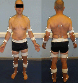
The segments of the lower limbs were modelled based on the calibrated anatomical systems technique (CAST) [29]. The landmarks used included, (Figure 1), medial and lateral femoral epicondyles, greater trochanter, anterior and posterior superior iliac spines of the pelvis. Clusters of 4 markers mounted on rigid plastic shells were attached to each segment, Figure 1.

    Figure 1:  Marker placement based on the Calibrated Anatomical Systems
Technique (CAST).

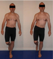
    Figure 2:  Study Trendelenburg Test method; (A) start / finish position (B)
Trendelenburg Test position [3].

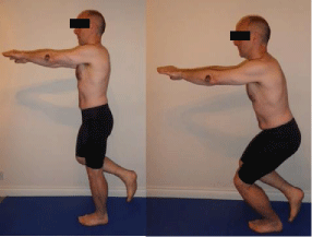
    Figure 3:  Study Single Leg Squat method; (A) start / finish position (B) Squat
position [33].

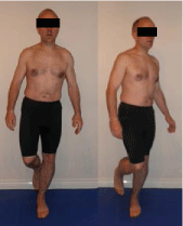
    Figure 4:  Study Corkscrew Test method; (A) start / finish position (B)
Corkscrew position.
After placing all of the markers, a calibration was performed that consisted of data collection for 1 second with the participant standing in the anatomic position. This defined the anatomic coordinate systems that enabled the position and orientation of each segment in space to be identified [29]. Local coordinate systems were defined for all segments of the model, with the y-axis equal to anterior-posterior, x-axis equal to medial-lateral, and z-axis equal to proximal-distal. The centres of the knee and ankle joints were calculated as the mean distance between the medial and lateral joint markers. The centre of the hip joint was calculated based on pelvic depth and width using the regression equations developed by Bell et al [30,31]. Joint kinematics was calculated using a Cardan/Euler method with an XYZ order of rotations.
Procedures
Testing was divided into two groups of tests; the “clinical tests” were the Trendelenburg Test, Single Leg Squat and Corkscrew Test. The “functional test” was running. The order of the clinical tests was randomized using a pseudo-random number generator [32].
Protocol
Prior to commencing the tests, each participant was provided with standardised oral instructions. For the clinical tests participants completed 3 practice trials to become familiar with the procedure, followed by 3 trials of each test.
Clinical tests- Trendelenburg test, single leg squat and Corkscrew Test: Participants were asked to stand on the edge of the laboratory force plates near the centre of the data collection area; this formed the start position for the test. Participants were not instructed which leg to use first during the tests. Participants completed the tests by stepping onto the laboratory force plates, performing the test on both limbs consecutively and stepping back off the force plates to the start position. This reflected how the tests are routinely completed in clinical practice, Figures 2-4.
Data capture commenced when the participant started to step onto the force plate for a duration of; 75 seconds for the Trendelenburg Test, 40 seconds for the Single Leg Squat and 15 seconds for the Corkscrew Test. The participants were allowed 30 seconds rest between clinical tests in order to avoid fatigue. The markers were left in position on the participants between the functional and clinical tests to minimize any errors in marker placement.
Functional running test: Participants were asked to stand at a preset position 5m from the data collection area; this formed the start position for the test. The finish position for the test was 10m from the start position. Participants were not instructed which leg to take the first step with.
Data capture commenced when the participant was approximately 1m outside of the data collection area and stopped when the participant reached the finish position. This ensured the participants were in a steady state of gait. The participants were allowed a 1 minute rest between functional tests in order to avoid fatigue.
Data processing
The movement data were exported toVisual3D (C-Motion, Inc, Germantown, MD) for processing. The movement data were filtered using a second-order, low-pass Butterworth filter with a cut-off frequency of 6 Hz for the clinical tests and 15Hz for running. The trunk, lumbar, thoracic, and hip angles were calculated relative to the local coordinate system, and the pelvic angles were calculated relative to the global coordinate system. The local coordinate system data provides information about movement of one segment relative to the next, whereas the global coordinate system data provides information on the orientation of the segment relative to the ground.
For all tests the data were normalised for time to 101 points. For running this was between heel strike to toe off, for the Trendelenburg Test the range movement starting from maximum pelvic obliquity over 30 seconds duration, for the Single Leg Squat between minimum and maximum hip flexion and for the Corkscrew Test between minimum and maximum hip rotation.
Statistical analysis
Repeated-measures analysis of variance and post hoc pair wise comparisons were used to identify significant differences when comparing the ranges of movement found in the clinical tests with those of running. The Bonferroni adjustment was used to account for multiple comparisons and to reduce the possibility of type I errors. Adjusted P values were reported. The a level was set at .05.
Results and Discussion
The mean and standard deviations for the clinical tests and pair wise comparisons between the clinical tests and running are presented in Table 1. Significant differences were seen between the Trendelenburg Test and running in all ranges of motion except for the transverse plane at the pelvis. For the Single leg squat significant differences were seen in all the range of motion except for thoracic transverse plane, pelvis sagittal and transverse planes and hip in the coronal plane ranges of motion. During the Corkscrew test significant differences were seen in all the ranges of motion except for the pelvis coronal plane range of motion and obliquity.
    Trendelenburg Test
    Single Leg Squat
    Corkscrew
    Running


    Dependent variable
    Plane
    Left Mean (sd)
    Right Mean (sd)
    Left Mean (sd)
    Right Mean (sd)
    Left Mean (sd)
    Right Mean (sd)
    Left Mean (sd)
    Right Mean (sd)


    Lumbar angle range (0)
    Sagittal
    2.1 (0.95)*
    2.2 (1.04) *
    11.7 (5.00)*
    12.6 (5.05) *
    3.6 (2.11)*
    4.0 (2.03) *
    9.00 (3.66)
    8.80 (2.70)


    Lumbar angle range (0)
    Coronal
    1.8 (1.33) *
    2.3 (1.49) *
    4.4 (1.88) *
    4.9 (2.99)
    6.4 (3.50) *
    6.50 (5.55) *
    6.50 (3.24)
    6.30 (2.49)


    Lumbar angle range (0)
    Transverse
    1.6 (0.52) *
    1.5 (0.74) *
    4.8 (2.26) *
    4.90 (1.76) *
    26.1 (17.40) *
    28.8 (16.00) *
    11.80 (2.15)
    12.70 (2.38)


    Thoracic angle range (0)
    Sagittal
    3.3 (2.12) *
    4.4 (2.52) *
    4.8 (2.81) *
    4.8 (3.00) *
    4.1 (1.95) *
    3.9    (1.49)
    7.40 (3.21)
    7.40 (3.21)


    Thoracic angle range (0)
    Coronal
    3.6 (0.78) *
    3.40 (2.03) *
    3.7 (2.01) *
    3.3 (2.07) *
    2.8 (1.34) *
    3.5 (1.70) *
    7.50 (2.93)
    8.00 (2.65)


    Thoracic angle range (0)
    Transverse
    3.0(2.22) *
    3.3 (1.15) *
    6.6 (2.91)
    7.8 (3.82)
    3.6 (1.46) *
    4.3 (1.54) *
    9.70 (4.69)
    9.00 (3.82)


    Trunk angle range (0)
    Sagittal
    2.3 (1.35) *
    2.5 (1.82) *
    8.5 (5.16) *
    8.8 (5.25) *
    5.3 (1.66) *
    6.1 (3.41) *
    10.00 (1.97)
    8.50 (1.84)


    Trunk angle range (0)
    Coronal
    4.0 (1.36) *
    4.5 (1.46) *
    5.9 (2.62) *
    6.0 (2.93) *
    11.6 (7.56) *
    13.0 (9.97) *
    9.90 (3.00)
    10.20 (2.54)


    Trunk angle range (0)
    Transverse
    2.9 (2.60) *
    3.3 (1.40) *
    5.7 (2.58) *
    5.9 (3.12) *
    17.5 (11.78) *
    19.6 (13.26) *
    20.40 (5.01)
    20.30 (3.84)


    Pelvis angle range (0)
    Sagittal
    3.3 (1.75) *
    4.0 (2.32) *
    6.0 (3.11)
    6.9 (4.08)
    4.5 (2.51) *
    4.4 (2.93) *
    7.50 (1.75)
    7.40 (2.18)


    Pelvis angle range (0)
    Coronal
    2.2 (0.92) *
    2.4 (1.14) *
    10.1 (5.56) *
    11.4 (7.20) *
    4.4    (1.68)
    4.8    (2.35)
    5.50 (1.01)
    6.50 (1.50)


    Pelvis angle peak (0)
    Obliquity
    11.3 (4.81) *
    10.8 (4.96) *
    18.9 (9.46) *
    19.5 (11.04) *
    12.6    (5.38)
    13.7    (5.34)
    16.60(5.26)
    17.00(5.88)


    Pelvis angle range (0)
    Transverse
    3.8 (1.43)
    3.7 (2.07)
    3.8 (1.03)
    4.0 (2.19)
    53.6 (17.48) *
    61.5 (18.82) *
    4.30 (3.05)
    5.10 (2.50)


    Hip angle range (0)
    Sagittal
    2.6 (1.60) *
    2.7 (1.98) *
    44.2 (13.70) *
    41.7 (10.89) *
    7.8 (4.30) *
    8.2 (4.09) *
    37.40 (5.12)
    36.50 (4.23)


    Hip angle range (0)
    Coronal
    4.4 (2.61) *
    3.9 (2.19) *
    9.1 (5.76)
    9.0 (4.55)
    5.7 (3.26) *
    5.7 (3.17) *
    10.30 (2.51)
    10.80 (2.78)


    Hip angle range (0)
    Transverse
    3.1 (1.24) *
    3.6 (1.24) *
    5.9 (2.41) *
    5.5 (3.06) *
    8.3 (3.50) *
    6.4    (3.30)
    8.40 (3.22)
    9.90 (4.44)


    Table 1: Results:    clinical tests and running normative data.
      *significant    difference compared to running.
Table 1: Normative data of the clinical tests and pair wise comparisons between the clinical tests and running.
The Trendelenburg Test is currently interpreted by the orientation of the pelvis compared to the horizontal (pelvic obliquity) [3], therefore pelvic obliquity is currently a value normally quoted within research [5,24] and a clinically important parameter for clinicians when examining the components of running [3]. Current research states that the Trendelenburg Test is positive if the pelvic obliquity is between 2° [5] and 4° [24]. The pelvic obliquity found in this study was large and symmetrical for the Trendelenburg Test; left 11.3° (SD= 4.81), right 10.8° (SD= 4.96). The existing evidence base advocates lower values of pelvic obliquity for the interpretation of the test when compared to this study.
This disagreement may be explained by the population studied; Asayama’s participants were post Total Hip Arthroplasty, Westhoff’s study used participants with Legg Calve Perthe’s disease, but this current study was of healthy participants. However if the angle that needs to be achieved is amended to fit within 1 standard deviation of the results of this study then the pelvic obliquity value would become 60 and hence would be in keeping with the previous studies.Based on this current studies results it could therefore be suggested that the Trendelenburg Test should interpreted as positive if the participant is unable to achieve a value of 100 or more for pelvic obliquity.
The Hardcastle and Nade method for performing the Trendelenburg Test does not describe the required position or movements of the other regions during the test. There have been no previous studies that have reported the trunk, lumbar, thoracic, pelvis or hip range of movement in the sagittal, coronal or transverse planes during the Trendelenburg Test. However it is a common clinical assumption that the participant should maintain an upright posture and minimal movement in all planes during the test. This study found the lumbar, thoracic, trunk, pelvis and hip ranges of movement to be small and symmetrical in the three cardinal planes of movement during the Trendelenburg Test. Hence when performing the Trendelenburg Test clinically there should be no observable movement of the participant except at the pelvis in the coronal plane. Consequently during the Trendelenburg Test the participant should appear to be in a position of pelvic obliquity but not moving.
When considering the clinical assessment of running the Trendelenburg Test was found to be an appropriate test for examining the pelvis transverse plane range of movement, and both the lumbar and thoracic coronal plane peak value components, but of note it was not an appropriate test for examining pelvic obliquity Table 1. Trendelenburg originally developed his test to examine the pelvic obliquity component of walking, but clinicians commonly use it to also examine the pelvic obliquity of running. Of clinical importance is that this study has found that the Trendelenburg Test is not an appropriate test for examining the pelvic obliquity component of running as clinicians currently use it. For the Trendelenburg Test to be interpreted clinically as normal the pelvis should achieve a position of at least 100 of pelvic obliquity and there should be no observable movement of the participant in any of the three cardinal planes whilst maintaining this position.
Currently the Single Leg Squat is interpreted as excellent if the individual exhibits over 65° of hip flexion and a coronal plane range of movement of less than 10° [4]. The hip flexion ranges of movement found in this study were large and symmetrical for the Single Leg Squat; left 44.2° (SD=13.7°), right 41.7° (SD=10.89), and moderate and symmetrical in the coronal plane; left 9.1° (SD=5.76), right 9.0° (SD=4.55).
The limited number of previous studies available has advocated higher values for hip sagittal range of movement and similar coronal plane ranges of movement for the interpretation of the test. However the previously published Single Leg Squat papers were not kinematic studies. The values published by Livengood [4] were derived from clinical experience. Interestingly if the hip flexion angle that needs to be achieved is amended to fit within 1 standard deviation then the hip sagittal plane ranges of movement would become 56° but still remain lower than previous studies stated value. Based on the kinematic data generated in this study it could be recommended that the Single Leg Squat should interpreted as normal if the individual is able to achieve 43° of hip sagittal plane range of movement, whilst maintaining under 10°of hip coronal plane movement.
The Livengood method for performing the Single Leg Squat does not describe the required position or movements of the other regions during the test. There have been no previous studies that report trunk, lumbar, thoracic, or pelvic range of movement in the sagittal, coronal or transverse planes during the Single Leg Squat.
However it is a common clinical assumption that participants should maintain an upright posture and exhibit minimal movement in the three cardinal planes. This study found the trunk, lumbar, thoracic, and pelvis ranges of movement to be moderate and symmetrical in the three cardinal planes of movement during the Single Leg Squat. Hence when using the Single Leg Squat during clinical assessment, some movement of the participant in all of the regions is normal with a large movement of the hip in the sagittal plane.
When considering examining running the Single Leg Squat was found to be an appropriate proxy for examining the thoracic transverse plane, pelvis sagittal and transverse, and hip coronal range of movement.This is clinically useful as hip coronal plane movement hypo-mobility has been associated with injury [14], and soft tissue symptoms [15-17] generating substantial problems for runners. Consequently the Single Leg Squat is an appropriate test for both clinical use and development of a diagnostic algorithm for the hip component of running in the coronal plane for runners. However its utility is limited to the coronal plane as it was not found to be a good representation of running for the hip in the sagittal or transverse planes. For the Single Leg Squat to be interpreted as normal the hip should move through 430 in the sagittal plane, not exceed 100 of hip coronal plane movement, and allow a small amount of movement in the trunk and pelvis in all other planes.
There are no previous kinematic studies of the movements occurring during the Corkscrew Test.
The hip transverse plane range of movement values found in this study were large and symmetrical for the Corkscrew Test; left 8.3° (SD=3.50), right 6.4° (SD=3.30). It was presumed a priori that the hip transverse plane movements during the Corkscrew Test and running would be similar, 10°. The hip coronal plane range of movement was predicted to be similar to that observed during the Single Leg Squat, 10°(4). However both the hip transverse plane movements; left 8.3° (SD=3.50), right 6.4° (SD=3.30), and coronal plane movements; left 5.7° (SD=3.26), right 5.7° (SD=3.17), found in this study were smaller than those predicted a priori. Most of the transverse plane movement occurred in the trunk; left 26.1°(SD=17.40), right 28.8° (SD=16.00), and therefore the Corkscrew Test appears to be a greater challenge of trunk rather than hip transverse plane movement. Subsequent to this study the Corkscrew Test could be interpreted as positive if the individual is unable to achieve 6° of hip rotation. However if the angle that needs to be achieved is amended to fit within 1 standard deviation then the hip transverse plane range of movement would become 9°and hence would be in keeping with the values predicted a priori.
The current method for performing the Corkscrew Test does not describe the required position or movements of the other regions during the test. There have been no previous studies that have reported on the trunk, lumbar, thoracic, pelvis or hip kinematics during the Corkscrew Test. However, as the test is becoming more commonly used in clinical practice it is being assumed by clinicians that participants should maintain an upright posture during the test. This study found the sagittal and coronal plane ranges of movement to be symmetrical and either moderate or small for all of the regions during the Corkscrew Test.
When using the Corkscrew Test clinically therefore there should be some observable movement of the participant in each of the regions and cardinal planes with a large amount of movement being observed in the trunk and thoracic spine in the transverse plane.
When considering running the Corkscrew Test was found to be an appropriate test for examining the pelvis coronal plane range of movement, and both the thoracic and pelvis coronal plane peak value components. For the Corkscrew Test to be assessed as normal the hip should move through 60 of rotation, and the trunk through 27° of rotation. There should be some observable movement of the participant in each of the three cardinal planes whilst maintaining this position.
From this study; the Trendelenburg Test, Single Leg Squat and Corkscrew Test were found to examine different, complementary ranges of Lumbo-Pelvic and Hip movement. The clinical application of this is that, when used in isolation these tests do not allow a full examination of the individual. However, when used in combination, these three tests enable examination of the pelvis and hip in all planes except the hip in the sagittal plane. This is particularly useful for runners as hip coronal plane range of movement is of clinical importance when examining these athletes. In contrast the tests were found inappropriate to examine was the thoracic spine, lumbar spine or trunk in all planes but the thoracic spine in the transverse plane. Clinicians may wish to use alternative test to examine these spinal parameters of running.
Conclusion
Clinicians commonly use Lumbo-Pelvic Hip tests to examine components of the running gait cycle [1]. However, little was known about the exact biomechanics of the tests and their relationship to the gait cycle. This study established that, when a clinician uses these clinical tests; for the Trendelenburg Test to be interpreted as normal the pelvis should achieve a position of at least 10° of pelvic obliquity and there should be no observable movement in any of the three cardinal planes whilst maintaining this position. For the Single Leg Squat to be interpreted as normal the hip should move through 43° in the sagittal plane and under 10° in the coronal plane. For the Corkscrew Test to be interpreted as normal the hip should move through 6° of rotation, and the trunk through 27° of rotation. Individuals who exhibit movements in excess of these normative values could be interpreted by clinicians as having hyper mobility in that region; those who demonstrate less movement could be interpreted as being hypomobile. This would aid sub grouping patients in clinical practice leading to targeted interventions which may improve outcome in musculoskeletal patients.
The pelvic obliquity during the Trendelenburg Test is different to running. Hence the Trendelenburg Test is not an appropriate proxy clinical test for examining the pelvic obliquity component of running. The hip coronal plane range of movement during the Single Leg Squat is similar to that found during of running. The Single Leg Squat is therefore an appropriate clinical test for examining the hip coronal plane range of movement component of running. However the hip flexion range of movement found during the Single Leg Squat and hip rotation during the Corkscrew Test were different to running. Therefore the Single Leg Squat and Corkscrew Tests should not be used to examine these components of running. Such information needs to be considered when using these tests clinically. Using the Trendelenburg Test, Single Leg Squat and Corkscrew Tests in combination allows clinicians to more fully examine the Lumbo- Pelvic Hip components of running.
References
- Bailey R, Selfe J, and Richards J. 'The role of the Trendelenburg Test in the examination of gait'. Physical Therapy Reviews. 2009; 14: 190-197.
- Bailey R, Richards J, and Selfe J. 'The Single Leg Squat Test in the Assessment of Musculoskeletal Function: a Review'. Physiotherapy Practice and Research. 2011; 32: 18-23.
- P Hardcastle and S Nade. 'The significance of the Trendelenburg test'. Journal of Bone and Joint Surgery – British. 1985; 67: 741-746.
- L Livengood, MA DiMattia and TL Uhl. '"Dynamic Trendelenburg": Single-Leg-Squat Test for Gluteus Medius Strength'. Athletic Therapy Today. 2004; 9: 24-25.
- Asayama, M Naito, M Fujisawa and T Kambe. 'Relationship between radiographic measurements of reconstructed hip joint position and the Trendelenburg sign'. The Journal of Arthroplasty. 2002; 17: 747-751.
- NA Roussel, J Nijs, S Truijen, L Smeuninx and G Stassijns. 'Low back pain: clinimetric properties of the Trendelenburg test, active straight leg raise test, and breathing pattern during active straight leg raising'. Journal of Manipulative and Physiological Therapeutics. 2007; 30: 270-278.
- E Taunton, MB Ryan, DB Clement, DC McKenzie, DR Lloyd-Smith, and BD Zumbo. 'A prospective study of running injuries: the Vancouver Sun Run In Training clinics'. British Journal of Sports Medicine. 2003; 37: 239-244.
- SE Robbins and AM Hanna. 'Running-related injury prevention through barefoot adaptations'. Medicine and science in sports and exercise. 1987; 19: 148-156.
- P Bale, D Bradbury, and E Colley. 'Anthropometric and training variables related to 10km running performance'. British Journal of Sports Medicine. 1986; 20: 170-173.
- vis Hammonds, KG Laudner, S McCaw, and TA McLoda. 'Acute Lower Extremity Running Kinematics After a Hamstring Stretch'. Journal of Athletic Training (National Athletic Trainers' Association). 2012; 47: 5-14.
- E Taunton, MB Ryan, DB Clement, DC McKenzie, DR Lloyd-Smith, and BD Zumbo. 'A retrospective case-control analysis of 2002 running injuries', British Journal of Sports Medicine. 2002; 36: 95-101.
- Van Middelkoop, J Kolkman, J Van Ochten, SMA Bierma-Zeinstra, and B Koes. 'Prevalence and incidence of lower extremity injuries in male marathon runners'. Scandinavian journal of medicine & science in sports. 2008; 18: 140-144.
- R van Gent, DD Siem, M Van Middelkoop, TA van Os, SS Bierma-Zeinstra, and BB Koes. 'Incidence and determinants of lower extremity running injuries in long distance runners: a systematic review', British Journal of Sports Medicine. 2007; 41: 469-480.
- Fredericson, CLM Cookingham, AMM Chaudhari, BCM Dowdell, NB Oestreicher, and SAP Sahrmann. 'Hip Abductor Weakness in Distance Runners with Iliotibial Band Syndrome. Clinical Journal of Sport Medicine. 2000; 10: 169-175.
- J Hamill, R Miller, B Noehren, and I Davis. 'A prospective study of iliotibial band strain in runners'. Clinical Biomechanics. 2008; 23: 1018-1025.
- R Ferber, B Noehren, J Hamill, and IS Davis. 'Competitive female runners with a history of iliotibial band syndrome demonstrate atypical hip and knee kinematics'. The Journal of orthopaedic and sports physical therapy. 2010; 40: 52.
- B Noehren, I Davis, and J Hamill. 'ASB Clinical Biomechanics Award Winner 2006: Prospective study of the biomechanical factors associated with iliotibial band syndrome'. Clinical Biomechanics. 2007; 22: 951-956.
- KR Snyder, JE Earl, KM OGçÖConnor and KT Ebersole. 'Resistance training is accompanied by increases in hip strength and changes in lower extremity biomechanics during running'. Clinical Biomechanics. 2009; 24: 26-34.
- A Zifchock, I Davis, J Higginson, S McCaw, and T Royer. 'Side-to-side differences in overuse running injury susceptibility: a retrospective study'. Human Movement Science. 2008; 27: 888-902.
- Hreljac, RN Marshall, and PA Hume. 'Evaluation of lower extremity overuse injury potential in runners. / Evaluation du potentiel de blessure par surmenage des membres inferieurs chez les coureurs'. Medicine & Science in Sports & Exercise. 2000; 32: 1635-1641.
- W Van Mechelen, H Hlobil, WP Zijlstra, M De Ridder, and HC Kemper. 'Is range of motion of the hip and ankle joint related to running injuries? A case control study'. International journal of sports medicine. 1992; 13: 605-610.
- E Magrum and RP Wilder. 'Evaluation of the injured runner'. Clinics in Sports Medicine. 2010; 29: 331-345.
- F Trendelenburg. 'Ueber den gang bei angeborener huftgelenksulxation'. Dtsch Med Wochenschrift. 1895; 21: 21-24.
- B Westhoff, A Petermann, MA Hirsch, R Willers, and R Krauspe. 'Computerized gait analysis in Legg Calve Perthes disease-Analysis of the frontal plane'. Gait & Posture In Press, Corrected Proof. 2005.
- P Kadaba, HK Ramakrishnan, and ME Wootten. 'Measurement of lower extremity kinematics during level walking'. Journal of Orthopaedic Research. 1990; 8: 383-392.
- JS Jacobsen, DB Nielsen, H S+©rensen, K Soballe, and I Mechlenburg. 'Joint kinematics and kinetics during walking and running in 32 patients with hip dysplasia 1 year after periacetabular osteotomy'. Acta Orthopaedica. 2014; 85: 592-599.
- Ornetti, D Laroche, C Morisset, JN Beis, C Tavernier, and JF Maillefert. 'Three-dimensional kinematics of the lower limbs in hip osteoarthritis during walking'. Journal of Back & Musculoskeletal Rehabilitation. 2011; 24: 201-208.
- MA Lyle, FJ Valero-Cuevas, RJ Gregor, and CM Powers. 'Control of dynamic foot-ground interactions in male and female soccer athletes: Females exhibit reduced dexterity and higher limb stiffness during landing'. Journal of Biomechanics. 2014; 47: 512-517.
- Cappozzo, F Catani, U la Croce, and A Leardini. 'Position and orientation in space of bones during movement: anatomical frame definition and determination'. Clinical Biomechanics. 1995; 10: 171-178.
- L Bell, RA Brand, and DR Pedersen. 'Prediction of hip joint centre location from external landmarks'. Human Movement Science. 1989; 8: 3-16.
- L Bell, DR Pedersen, and RA Brand. 'A comparison of the accuracy of several hip center location prediction methods'. Journal of Biomechanics. 1990; 23: 617-621.
- Hill and B Wichmann. 'Algorithm AS 183. An efficient and portable pseudo-random number generator'. Applied Statistics. 1982; 31: 188-190.
- MA DiMattia, AL Livengood, TL Uhl, CG Mattacola, and TR Malone. 'What Are the Validity of the Single-Leg-Squat Test and Its Relationship to Hip-Abduction Strength?' Journal of Sport Rehabilitation. 2005; 14: 108-123.