
Review Article
Austin J Pediatr. 2014;1(2): 1006.
Nevus Sebaceous
Alexander K. C. Leung1* and Benjamin Barankin2
1Department of Pediatrics, University of Calgary, Canada
2Toronto Dermatology Centre, Canada
*Corresponding author: Alexander KC Leung, Department of Pediatrics, The University of Calgary, Pediatric Consultant, The Alberta Children’s Hospital, Calgary, Alberta, T2M 0H5, #200, 233– 16th Avenue NW, Canada
Received: April 28, 2014; Accepted: May 26, 2014; Published: May 27, 2014
Abstract
Nevus sebaceous, a hamartoma of the skin and its adnexa, is characterized by epidermal, follicular, sebaceous, and apocrine gland abnormalities. Nevus sebaceous occurs in approximately 0.3% of all newborns. Both sexes are equally affected. The early infantile stage is characterized by papillomatous epithelial hyperplasia. The hair follicles are underdeveloped and the sebaceous glands are not prominent. During puberty, sebaceous glands become numerous and hyperplastic, apocrine glands become hyperplastic and cystic, and the epidermis becomes verrucous. The hair follicles remain small and primordial and may disappear altogether. During adulthood, epidermal hyperplasia, large sebaceous glands, and ectopic apocrine glands are characteristic histological findings. At birth, nevus sebaceous typically presents as a solitary, wellcircumscribed, smooth to velvety, yellow to orange, round or oval, minimally raised plaque. The scalp and face are sites of predilection. Lesions on the scalp are typically hairless. At or just before puberty, the lesion grows more rapidly, becomes more thickened and protuberant, and at times acquires a verrucous or even a nodular appearance. Nevus sebaceous may be complicated by the development of benign and malignant nevoid tumors in the original nevus. Neoplasms occur mostly in the fourth decade of life in approximately 10 to 30% of lesions. The majority of these tumors are benign; less than 1% of nevus sebaceous is complicated by malignant tumors. Because of the potential of malignant transformation, prophylactic full–thickness, complete excision of the lesion with 2 to 3 mm margins or ongoing observation is recommended.
Keywords: Nevus sebaceous; Sebaceous glands; Plaque; Neoplasms; Prophylactic excision
Introduction
Nevus sebaceous, also known as nevus sebaceous of Jadassohn, a hamartoma of the skin and its adnexa, is characterized by epidermal, follicular, sebaceous, and apocrine gland abnormalities [1]. The condition was first described by Josef Jadassohn, a German dermatologist, and now bears his name [2]. Because the lesion involves more than just a sebaceous component, the more encompassing term “organoid nevus” has also been coined [3,4].
Epidemiology
Nevus sebaceous occurs in approximately 0.3% of all newborn infants [1]. Both sexes are equally affected [5]. There is no racial and⁄ or ethnic predominance [2]. Recent studies suggest a possible link with mothers positive for human papillomavirus [4]. The occurrence of the nevus is usually sporadic, although familial cases have rarely been reported. An autosomal dominant mode of inheritance has also been described [6].
Pathogenesis
Recently, it has been shown nevus sebaceous is caused by post zygotic mosaic mutations in the HRAS and KHAS genes [7]. Presumably, nevus sebaceous develops from pluripotent primary epithelial germ cells which have the potential to differentiate into various neoplasms [5]. Deletions in the PTCH tumor suppressor gene on chromosome 9p22.3 may account for the neoplastic potential of the hamartoma [5,8].
There is an increased expression of androgen receptors in nevus sebaceous [9]. The growth of the lesion in adolescence is due to increased production of androgen at the time of puberty and its influence on the pilosebaceous and apocrine elements.
Histopathology
The early infantile stage is characterized by papillomatous epithelial hyperplasia [10]. The epidermis is only slightly acanthotic [11]. The hair follicles are underdeveloped and the sebaceous glands are not prominent [1,10]. Apocrine glands are rarely seen [1]. During puberty, sebaceous glands become numerous and hyperplastic, apocrine glands become hyperplastic and cystic, and the epidermis becomes verrucous [1,10,11]. The hair follicles remain small and primordial and may disappear altogether [1,11]. During adulthood, epidermal hyperplasia, large sebaceous glands, and ectopic apocrine glands are characteristic histological findings [11]. Normal hair follicles are characteristically absent [11]. This stage is also distinguished by the potential development of a variety of adnexal tumors [11].
Clinical Manifestations
Approximately two thirds of cases are present at birth and the rest develop in early childhood. At birth, nevus sebaceous typically presents as a solitary, well–circumscribed, smooth to velvety, yellow to orange, round, oval, or linear, flat or minimally raised, plaque (Figure 1) [2,3]. Lesions on the scalp are typically hairless [12]. Rarely, the lesion may be pedunculated [9]. The lesions are often distributed along Blaschko’s lines and are arrayed in a linear configuration. This may be difficult to appreciate if the lesion is small. The scalp and face are sites of predilection, although the trunk and extremities may also be affected [1–3]. Rare intraoral extension may present as a linear whitish plaque. Characteristically, the lesion grows proportionally with the size of the patient until puberty [13]. At or just before puberty, the lesion grows more rapidly, becomes more thickened and protuberant, and at times acquires a verrucous or even a nodular appearance [1,2,12]. Nevus sebaceous is usually solitary, although multiple and widespread lesions have been reported [14].
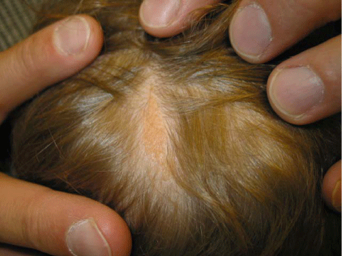
Figure 1: Nevus sebaceous presenting as a yellow-orange, linear, hairless plaque on the scalp.
A new phenotype, known as cerebriform nevus sebaceous, has been described [15]. Cerebriform nevus sebaceous is characterized by large, pedunculated or verrucous, pink, hairless, nodules or tumors in newborn infants [15].
Nevus sebaceous is usually an isolated finding. Syndromes associated with nevus sebaceous include SCALP syndrome and linear sebaceous nevus syndrome (Schimmelpenning syndrome). SCALP syndrome is characterized by sebaceous nevus, central nervous system malformation, aplasia cutis congenita, limbal dermoid, and pigmented nevus. Linear sebaceous nevus syndrome is characterized by nevus sebaceous which is often extensive and distributed along Blaschko’s lines, central nervous system abnormalities, epilepsy, mental retardation, ocular abnormalities, musculoskeletal defects, cardiovascular abnormalities, and urologic abnormalities.
Diagnosis
The diagnosis is usually clinical based on the characteristic features. A tissue biopsy or referral to a dermatologist should be considered if the diagnosis is in doubt. Prenatal diagnosis is feasible by ultrasonography for large and exophytic lesions [16].
Differential diagnosis
The differential diagnosis includes epidermal nevus, aplasia cutis congenita, solitary mastocytoma, and juvenile xanthogranuoloma.
Complications
The lesion can be aesthetically unappealing, especially when it occurs on the face. Large or extensive lesions may be associated with developmental deficits [9]. Centrofacial lesions have an increased association with underlying neurological involvement. The latter may present as mental retardation, seizures, and hemiparesis.
Nevus sebaceous may be complicated by the development of benign and malignant nevoid tumors in the original nevus [1]. The incidence of these tumors increases with age, particularly after puberty [11]. Neoplasms occur mostly in the fourth decade of life in approximately 10 to 30% of lesions [3,5]. Majority of these tumors are benign; less than 1% of nevus sebaceous is complicated by malignant tumors [17]. Malignancy is suggested by the acute appearance of a large, discrete, ulcerating nodule within the lesion [3]. The most common benign tumors are syringocystadenoma papilliferum followed by trichoblastoma [3,13]. Many trichoblastoms were misdiagnosed in the past as basal cell carcinomas [4]. Other benign tumors include trichilemmoma trichoepithelioma, sebaceous adenoma, seborrheic keratosis, sebaceous epithelioma, keratoacanthoma, apocrine cystadenoma, apocrine hidrocystoma, nodular hidradenoma, follicular poroma, spiradenoma, and syringoma [5,11]. The most common malignant tumor is basal cell carcinoma [4,10]. Other malignant tumors include squamous cell carcinoma, apocrine carcinoma, ductal adenocarcinoma, porocarcinoma, anaplastic adnexal carcinoma, trichilemmal carcinoma, syringomatous carcinoma, and sebaceous carcinoma [4,5,11].
Management
Excision of the lesion may be considered at any age for cosmetic reasons. Other than for the sake of cosmesis, some authors suggest watchful observation of the lesion [18]. However because of the potential of malignant transformation, other authors recommend prophylactic full–thickness, complete excision of the lesion with minimum 2 to 3 mm margins [1,2,4]. Because the incidence of malignant transformation in childhood is low, prophylactic excision of the lesion (and the possible need for general anesthetic) prior to puberty may not be justified [13]. Ablative laser and photodynamic therapy may be considered for the occasional cases with inoperable lesions. Any new “bump” within a nevus sebaceous should be examined and biopsy considered.
References
- Terenzi V, Indrizzi E, Buonaccorsi S, Leonardi A, Pellacchia V, Fini G. Nevus sebaceus of Jadassohn. J Craniofac Surg. 2006; 17: 1234-1239.
- Saedi T, Cetas J, Chang R, Krol A, Selden NR. Newborn with sebaceous nevus of jadassohn presenting as exophytic scalp lesion. Pediatr Neurosurg. 2008; 44: 144-147.
- Lountzis N, Junkins-Hopkins J, Uberti-Benz M, Elenitsas R. Microcystic adnexal carcinoma arising within a nevus sebaceus. Cutis. 2007; 80: 352-356.
- Moody MN, Landau JM, Goldberg LH. Nevus sebaceous revisited. Pediatr Dermatol. 2012; 29: 15-23.
- Altaykan A, Ersoy-Evans S, Erkin G, Ozkaya O. Basal cell carcinoma arising in nevus sebaceous during childhood. Pediatr Dermatol. 2008; 25: 616-619.
- West C, Narahari S, Kwatra S, Feldman S. Autosomal dominant transmission of nevus sebaceous of Jadassohn. Dermatol Online J. 2012; 18: 17.
- Happle R. Nevus sebaceus is a mosaic RASopathy. J Invest Dermatol. 2013; 133: 597-600.
- Rosen H, Schmidt B, Lam, HP, Meara JG, Labow BI. Management of nevus sebaceous and the risk of basal cell carcinoma: an 18-year review. Pediatr Dermatol 2009; 26: 676-681.
- Lin HC, Lee JY, Shieh SJ, Hsu CK. Large, papillomatous and pedunculated nevus sebaceus. J Dermatol. 2011; 38: 200-202.
- Arshad AR, Azman WS, Kreetharan A. Solitary sebaceous nevus of Jadassohn complicated by squamous cell carcinoma and basal cell carcinoma. Head Neck. 2008; 30: 544-548.
- Simi CM, Rajalakshmi T, Correa M. Clinicopathologic analysis of 21 cases of nevus sebaceus: a retrospective study. Indian J Dermatol Venereol Leprol. 2008; 74: 625-627.
- Leung AK. Nevus sebaceous. Leung AK, editor. In: Common Problems in Ambulatory Pediatrics: Specific Clinical Problems. New York: Nova Science Publishers, Inc. 2011; 201-204.
- Idriss MH, Elston DM. Secondary neoplasms associated with nevus sebaceus of Jadassohn: a study of 707 cases. J Am Acad Dermatol. 2014; 70: 332-337.
- Chi SG, Kim JY, Kim HY, Lee SJ, Kim do W, Lee WJ. Multiple nevus sebaceous occurring on the scalp and on the contralateral side of the face. Ann Dermatol. 2011; 23: 389-391.
- Correale D, Ringpfeil F, Rogers M. Large, papillomatous, pedunculated nevus sebaceus: a new phenotype. Pediatr Dermatol. 2008; 25: 355-358.
- Dhombres F, Kolanska K, Garel C, Aubry JP, Gonzales M, Jouannic JM. Prenatal diagnosis of exophytic nevus sebaceous of the scalp. Prenat Diagn. 2013; 33: 1305-1307.
- Barankin B, Shum D, Guenther L. Tumors arising in nevus sebaceus: a study of 596 cases. J Am Acad Dermatol. 2001; 45: 792-793.
- Benjamin LT. Birthmarks of medical significance in the neonate. Semin Perinatol. 2013; 37: 16-19.