
Research Article
J Pediatr & Child Health Care. 2023; 8(1): 1058.
Bone Transport in the Management of Post-Osteomyelitis Tibial Defects in Pediatric Population. A Long Term Follow-up Study in Uganda
Loro A*, Franceschi F, Loro F and Brown N
Orthopedic Department, CoRSU Rehabilitation Hospital, P.O.BOC 46, Kisubi, Uganda
*Corresponding author: Loro ADepartment of Orthopedic, CoRSU Rehabilitation Hospital, P.O.BOC 46, Kisubi, Uganda
Received: January 23, 2023; Accepted: February 20, 2023; Published: February 27, 2023
Abstract
Background: This study was designed to evaluate the long-term results and complications of using the bone transport technique in the management of post-osteomyelitis tibial defects in a low income country.
Design: Retrospective study in a cohort of pediatric patients.
Methods: Twenty-two patients were included in the study; all patients were treated between 2005 and 2014. The mean age at time of surgery was 9.5 years. The mean bone defect was 8.2 cm. Transport was done using both ring and monolateral external fixators; the fixator stayed in place for 290 days on average. Clinical and radiological evaluations were performed after a minimum follow-up period of 7 years. Patients were interviewed by social workers with focus on the psychosocial and economic costs of the procedure.
Results: The bone and soft tissues management required a total of ninety-one surgeries, with an average of four surgeries per child. Eradication of infection was obtained in all patients. Bone results were graded as excellent in 10, good in 7, fair in 2 and poor in 3. Functional results were graded as excellent in 8, good in 6, fair in 7 and poor in 1. Major and minor complications were numerous during the bone transport procedure. Three cases of non-union at the docking point were observed. Twelve patients had a limb length discrepancy, four had slight angular deformities. Reduced range of motion was observed both in the ankle (n=4) and knee (n=2). Seven joint fusions were noted, two knees and five ankles.
Conclusion: Bone transport is a valid option in the management of bone defects in children, providing satisfactory long-term outcomes. Bone gaps, soft tissue defects, limb discrepancy and joint disorders can be managed concurrently. The procedure is lengthy and prone to complications.
Keywords: Bone defects; Osteomyelitis; Bone transport; Pediatric osteo-articular infections; External fixator
Introduction
In sub-Saharan Africa, haematogenous osteomyelitis represents a common condition in pediatric populations [1-3]. The tibia is the most commonly affected bone, followed by the femur, radius and humerus. In our facility, children most commonly present with complications of neglected or undertreated chronic osteomyelitis. The uncontrolled clinical course of the disease frequently leads to segmental bone defects that are difficult to manage, especially in settings with limited resources.
Osteoarticular Infections (OAI) display a higher incidence in children from lower socioeconomic groups [3]. In Uganda, with no universal health coverage, all costs related to diagnosis and treatment are taken on by the family; with 41.7% of the population under the international poverty line, OAIs are not always seen as a financial priority [4]. Furthermore, in some regions, OAI are believed to be related to extra-natural forces and so access to health facilities is often delayed by first attending non-medical bone setters or healers. As a result, the disease progresses leading to the complex clinical presentations that attend our institution.
The therapeutic strategies for filling bone gaps are dictated by several clinical and radiological key elements, such as the site of the defect, its size, and the quality of the residual bone stock. Thought must also go to previous surgeries, status of the soft tissues, involvement of the adjacent joints, limb discrepancy and axial deformities [5]. Experienced surgeons working in equipped facilities may choose from various management options, including conventional bone graft [6], Vascularized Fibula Flap (VFF) [7,8], transference of the fibula [9] and internal Bone Transport (BT) [10-13].
Literature exploring the applicability of these different options in low resource settings is limited. Considering the rising pediatric population of sub-Saharan Africa [14] and the significant physical, social and economic implications of neglected osteomyelitis, it is increasingly important to further examine this topic.
This retrospective study was designed to evaluate the outcomes and complications of bone transport in the treatment of pediatric post-osteomyelitis tibial defects managed in our facility in Uganda.
Materials and Methods
The surgical records of two authors (AL and FF) were reviewed in order to identify cases of post-osteomyelitis tibial bone defects that were treated with the bone transport technique between 2005-2014. Twenty-two cases were recorded and included. Clinical notes and radiographs were reviewed; patient characteristics, operative protocols and complications were recorded. Post-operative difficulties were divided into problems, obstacles and complications as per Paley [15].
Patients were asked to voluntarily attend a follow-up consultation in 2021 with our orthopedic team, where the long term bone and functional outcomes were assessed according to the criteria proposed by Paley et al. [13]. Bone outcomes are based on union, infection, deformity, Limb Length Discrepancy (LLD) and cross-sectional area of union of the regenerate bone and docking site. Functional outcomes are based on pain, need for walking aids, joint contracture, loss of range of motion as compared to pre-operative range and ability to return to Activities of Daily Living (ADLs) and/or work [13].
Patient Characteristics
There was a 1:1 male to female ratio, with an average age at presentation of 9.5 years (range 3 to 15). The left tibia was involved in 12 cases.
All bone defects were secondary to debridement for haematogenous osteomyelitis. Six children presented with quiescent infection, referred for limb salvage procedures after initial surgeries were carried out in other institutions. The remaining sixteen presented with active infections; 14 underwent surgical treatments before the BT procedure and 2 received prior conservative treatment.
At presentation, all showed severe clinical signs caused by delay and/or poor management in terms of overzealous debridement (Figure 1-3a). Pathological fractures were seen in 15 children, exposed sequestra with skin loss in 11, joint contracture and stiffness in 10, limb discrepancy in 11, axial deformity in 7 and skin disorders in 8. Nine were unable to walk and a further nine needed walking aids to mobilize safely.
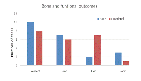
Figure 1: Bone and functional outcomes at the last follow up.
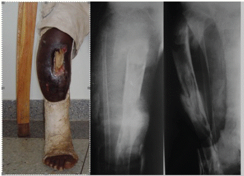
Figure 1a: Clinical and radiographic aspects at presentation in a 9 years old boy. This complicated osteomyelitis may be treated in well-equipped centers which, in low income countries, are few and unable to cope with the rising demand of a growing pediatric population.
Out with the involved segment, two children had osteomyelitis of the contralateral tibia (Figure 2a), one had osteomyelitis of the contralateral femur, four had septic arthritis of the ipsilateral ankle, two of the ipsilateral knee (Figure 3a), one post-infectious deformity of the ipsilateral ankle and elbow joint, and one had osteoarthritis of the contralateral hip and osteomyelitis of contralateral tibia. Joint stiffness of the knee and ankle was observed in 8 children.
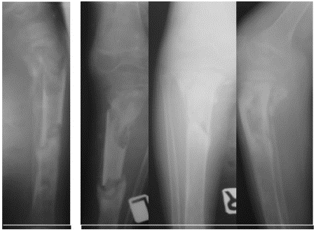
Figure 2a: Radiographs of the tibiae of 8 years old boy at presentation. Both infections started simultaneously 1 year prior to admission. Bone transport was done on the left side, where the sequestrum was exposed, with important loss of skin envelope. Pathological fractures on both sides obliged the mother to carry the child for more than 1 year.
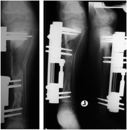
Figure 2b: On the left the first post-operative control. On the right, radiographs at the completion of the transport. To note the collision of the two proximal clamps, not allowing adequate compression of the docking point.
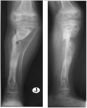
Figure 2c: Radiographs showing the non-union at the docking point.

Figure 2d: The non-union was treated with site debridement, peroneal osteotomy and external fixator. Sound union was achieved.
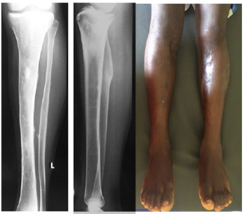
Figure 2e: Clinical and radiographic control of both tibiae 12 years later. To note the outstanding bone remodeling on both sides. Excellent bone and functional results on both sides.
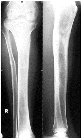
Figure 2f: Clinical and radiographic control of both tibiae 12 years later. To note the outstanding bone remodeling on both sides. Excellent bone and functional results on both sides.
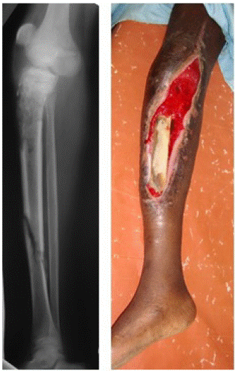
Figure 3a: Clinical and radiological presentation in a 12 years old girl. She had two previous surgeries for debridement and septic arthritis of the knee. There were no features suggestive of involucrum formation on x-ray film.
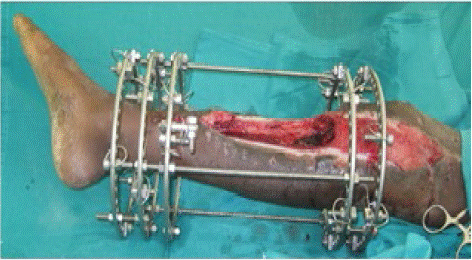
Figure 3b: Immediate post-operative picture taken after partial diaphysectomy and debridement. It shows the ring construct and the extensive bone, skin and soft tissues losses.
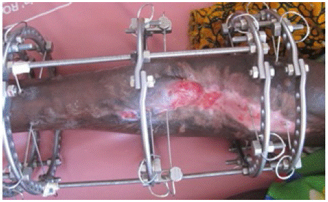
Figure 3c: Clinical aspect of the leg after 90 days of transport. The tension-stress effect had achieved the healing of the ulcer. To note the prominence of the proximal end of the transported segment and the skin invagination which had to be managed surgically.
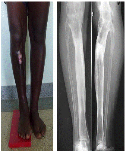
Figure 3d: Clinical and radiographic controls 5 years after completion of bone transport, showing eradication of the infection, the residual limb discrepancy and the knee arthrodesis.
Management Protocol
The reconstructive plan was based on sequestrectomy, when required, radical debridement and application of the external frame. Specimens for culture were not routinely collected due to the lack of equipped microbiology laboratories in our setting.
The bone transport procedure then followed. Ring fixator was used in 9 cases, monolateral in the remaining 13 (Orthofix Rail). Ring fixator was preferred in cases of short residual bone stumps, especially if associated with poor bone quality. In addition, it was the preferred choice when correction of joint deformity or contraction was also required. All constructs were hybrid in nature, mixing half-pins and tensioned wires as required by the mechanical needs. The external fixation time was 290 days on average (range 104 to 730).
The osteotomy for transport was done concurrently in 15 patients, it was delayed in the other 7. The mean delay was of 44 days (range 12 to 105); it was mainly due to the conditions of the soft tissue envelope.
The defect was located in the mid-third in 12 cases, in the upper third in 4, in the mid-distal third in 3 and in the mid-proximal third in 3. The mean defect was 8.2 cm (range 3 to 15 cm).
Plastic surgery was necessary in 6 cases. Before starting the transport procedure, three cases were managed with a fasciocutaneous flap, a gracilis free flap and a gastrocnemius flap, coupled with split skin graft (SSG). In another 3 cases, the wound was left open to granulate and healed by secondary intention during the procedure (Figure 3b-3c).
All cases were treated with a single level transport, distal to proximal in 18 and proximal to distal in 4. Distraction was started on the seventh post-operative day, 1 mm a day in a single step.
The first radiographic evaluation was done between 8 to 10 days post-operatively in order to evaluate the opening of the osteotomyline. Radiographs were then taken, if feasible, every 4 weeks. Compression was usually applied after the docking was reached. In case of non-union, the site was refreshed and compressed, aided by a fibula osteotomy. Physical therapy was usually started in the second or third postoperative day. An elastic band between the shoe and the most proximal ring or clamp, was used to prevent equinism of the foot. Compensation shoes were used for limb length discrepancy above 5 cm.
Results
Outcomes at Final Follow-up
The mean follow-up was 10.8 years (range 7 to 16). The bone and functional outcomes, using the criteria proposed by Paley (13), are shown in (Figure 1).
At the final follow-up, none of the 22 tibiae showed signs of relapsed infection. Sound bone reconstruction was observed in 19 patients, while non-union at the docking point was observed in 3 cases. A major remodeling process was noted in all the tibiae (Figure 2e).
Table 1 highlights the difference in clinical features from initial to latest follow up.
    Clinical    feature

    No.    at presentation

    No.    at latest follow up


    Pathological fracture

    15

    0


    Exposed sequestra with skin loss

    11

    0


    Septic arthritis

    7

    0*


    Joint stiffness/contracture

    8

    6


    Limb length discrepancy

    11

    12


    Axial deformity

    7

    4


    Skin disorders

    8

    0


    Inability to walk

    9

    0


    Requiring walking aids

    9

    2 (only for outdoor activities)
Table 1: Table comparing clinical features present at initial presentation and at the last follow-up. *All seven septic arthritis were complicated by joint fusion at the last follow-up.
    Problems

    Obstacles

    Complication    (minor)

    Complication    (major)


    Pin tract infection

    4

    12


    Delayed consolidation

    1


    Premature consolidation

    2


    Non-union at docking point


    4


    1


    Joint contracture


    2


    Fused joint


    7


    LLD


    12


    Angular deformity


    3

    1


    Loss of axis


    6


    Supracondylar fracture of the femur

    5

    1


    Soft tissue invagination

    4


    Collision of clamps


    2


    Exposure of regenerate with skin loss


    2


    Foot drop


    1


    Superficial breaking down at docking point

    3
Table 2: Problems, obstacles and complications observed during the entire process of BT, from the assembly of the frame to its extraction.
    Condition    addressed (after completion of BT)

    Frequency    of conditions requiring surgery


    Non-union at docking point

    4


    Joint disorder

    2


    Infection in other sites

    5


    Axial deformity of ipsilateral tibia

    4


    Axial deformity of contralateral tibia

    1


    Tibial shortening

    2


    Leg ulcers

    1


    Infection relapse

    1


    Fracture at docking point

    1
Table 3: Conditions that were surgically addressed after the BT completion.
At the latest follow up, twelve patients had LLD with an average of 3.7 cm. Ten had LLD in the range of 1 to 4 cm, one had 5 cm and the remaining one had 7 cm. Three slight axial deformities were seen at the site of docking point, while a 10 degrees varus deformity of the knee was seen in a further case. Six cases in total had a reduced range of movement of at least one joint: four in the ankle and two in the knee. Joint fusion was recorded in 7 cases, two knee and five ankles. All of the patients were able to walk independently, with only two patients requiring walking aids for outdoor activities. Sixteen patients were at school, five were fully employed workers farmers and one was a housewife.
Complications
Three cases experienced no complications during the procedure. A total of 72 difficulties were observed, as outlined in table: 19 problems, 27 obstacles and 27 complications.
Five non-unions occurred and were treated during the procedure itself; four were successfully treated by site debridement and compression. Failure was observed in one case where compression was applied without opening the site.
Loss of axis was mainly due to untimely replacement of loose screws or to incorrect surgical procedure during the replacement itself. All supracondylar femoral fractures were green-stick in nature and were treated conservatively, except in one case in which two supplementary rings were placed in the femur. Accidental fall whilst playing was the cause in all cases.
The joint contractures were both treated with positioning of supplementary rings to the foot and femur.
Surgical site infection, with exposure of the regenerate, required the bone transport to be paused for about 2 weeks.
A total of 93 surgeries were carried out in the selected time period, averaging 4.2 surgeries per child. Sixty-eight surgeries were performed from the index procedure to the removal of external fixator. Twenty-six additional surgeries were required for 12 children long after the removal of the external fixator, in order to address complications related to the BT technique itself and conditions that were concurrently present on admission.
The 4 non-unions were addressed 1, 5, 22 and 24 months after removal of the implant. In one case, the non-union was present both at the docking and trailing points. Refreshing of the site and compression using an external fixator was used in all cases (Figure 2c-d). In one case autogenous bone graft was utilized. In one case, two corrective osteotomies of the ipsilateral tibia were carried out; one at four months and the other at seven years after the removal of the external fixator. In the remaining two cases, the osteotomy was carried out at 3.5 and at 6 years after the completion of transport.Ankle fusion was done 16 months after the completion of treatment, using K wires and cast. The knee fusion was done 6 months after the end of the transport, using an anterior external fixator. In one case, a severe relapse of the bone infection was seen 8 years after completion of transport. Sequestrectomy, bone resection and a new bone transport were put in place, successfully clearing the infection. The stress fracture at docking point was observed 18 months after removal of the frame. It was successfully treated by compression using an external fixator without opening the site.
The ring osteomyelitis was located in the ipsilateral femur as result of knee joint fusion done with external fixator.
Discussion
Debridement is the key element in the management of long-standing osteomyelitis. The aim is to eradicate the infection by removing the sequestered bone and all nonviable tissues so as to obtain a site receptive to reconstructive techniques, should they be needed.
When involucrum formation is defective (Figure 3a), the surgical act leaves behind gaps that call for further procedures which, in severe cases, must address bone reconstruction, soft tissue coverage, joint disorder and limb length discrepancy.
Multiple patient factors must be carefully evaluated before selecting the appropriate method of limb salvage procedure [16]; the status of the soft tissues and the residual bone stock have been the most crucial criteria in our experience.
In low income countries, the options at surgeon’s disposal rely on autogenous bone graft, either free or vascularized, and internal bone transport. However, there is a scarcity of ortho-plastic centers that can routinely offer all methods, and furthermore accessing these facilities is problematic for both logistical and economic constraints. For example, in Uganda’s population of c. 45 million, there are only a few centers where such reconstructions can be performed.
In presence of good skin cover, autogenous cortico-cancellous grafts have been advocated, particularly in small segmental defects usually not exceeding 2 cm. Since large defects require an amount of graft that may not be obtained in small children, fibula graft [6], cortical tibial strut or cortico-cancellous segments of iliac crest have been employed [17]. Healing and graft incorporation are usually slow and so the grafted area must be protected for a long time to allow creeping substitution. Moreover, the morbidity on the donor leg and ilium must be taken into consideration. Displacement, graft fracture and non-union at the graft-host junction have been reported, coupled with recurrence of the infection [6,9,18].
Transference of the ipsilateral fibula has been widely reported in post-traumatic tibial defects, particularly in segmental defects exceeding 8 cm associated with scarred and poorly vascularized skin. Several techniques, either in one or two stages, have been described [6,9,19-20]. Few authors have reported fibula transference in children [6,9,16,20]. They recorded hypertrophy of the grafted fibula and of the tibial remnants. Complications included non-union and fracture of the graft [6]. Vascularized fibula flap can be employed in large skeletal and soft tissue defects following debridement required by exposed sequestra or in cases where surgery leaves behind short epi-metaphyseal bone ends, particularly when associated with poor quality skin. The osseous and osteocutaneous flaps have been used to address bone and soft tissue losses at the same time [7,8]. However, surgical experience and well equipped facilities are required. The microvascular time is crucial, both in harvesting and grafting the flap. Complications are mainly related to vascular disturbances at the anastomosis site but infection, graft fracture, non-union and deformity in the donor leg have been also observed [7,8,21-22].
The literature reporting on limb reconstruction in children using bone transport is scanty [10,12], more so when looking at papers originating from low and middle income countries. In our institution, the method has proved to be rewarding in reconstructing bone and soft tissue, eradicating the infection and addressing the co-existing disorders such as limb length discrepancy and joint contracture, all conditions that all the above-mentioned methods cannot tackle. The local blood supply is increased by the distraction osteogenesis, as reported by Aronson [23] and this translates in better availability of antibiotics and faster healing of soft tissues. Bone transport in the long term can fully suppress the infection in children and heal soft tissues losses.
We used both ring and monolateral fixators. The ring fixator was preferred in presence of short residual bone ends, particularly when the osteopenia was raising doubts about a solid purchase of fixation, and when there was need to correct joint contractures. Sound stability must always be attained when making a construct and must be maintained for the duration of the procedure [24]. Correct positioning of a circular fixator certainly requires a longer surgical time but there are cases where it must be considered as the only alternative to limb amputation (Figure 3a). Surgical skills and creativity are key elements for a good circular construct.
Complications were numerous in our series, mirroring the ones reported by several authors [10,11,13,15,25]. Non-union at the docking point, pin site problems, loss of bone axis and supracondylar fracture of the ipsilateral femur were all experienced both during and after the index procedure.
Non-union can develop when the contact at the docking point is small or when the bone ends are out of axis [15,24-26]. In cases of massive defects, scar tissue or skin invagination may act as barrier between the bone ends, interfering with the union. Grafting of docking point has been recommended in adults but is still debated in children. We didn’t employ this in our series, and it has not been reported in other series in pediatric populations [10,12]. However, the rate of non-union at the docking point was rather high in our cohort, indicating that bone grafting could be considered, particularly in long defects or in the presence of invaginated, scarred skin.
Pin site problems represented a common complication of the method. Since stability of the construct is fundamental for success, pin and wire replacements must performed in a timely manner. This is not always possible in low income countries, since logistical and economic factors play an immense role in maintaining compliance with follow-up. Late replacement of loose pins or wrong positioning can lead to loss of axis or collision of clamps/rings (Figure 2). These can act as cofactors in cases of non-union or slow consolidation of the regenerate. In the early cases, fixator adjustments had to be done without the guidance of intraoperative fluoroscopy. Availability of the C-arm led to reduction of intraoperative errors in the assembly revision surgery in the following years. Additional surgeries were done in over half of the children after completion of the bone transport procedure. This possibility must be properly ex plained to the child’s relatives, particularly in severe cases at presentation, when joint involvement, limb discrepancy and physeal damages may be seen as predictive findings for further surgeries. Children well tolerated the fixator, and all were ambulant just a few days after the procedure. Physical and occupational therapists were key figures in the rehabilitation process, assisting in weight bearing and preventing contracture of the joints. The high number of supracondylar fractures of the ipsilateral femur was most likely due to good adaptation to the frame facilitating children to play and interact with their companions. We opted for conservative treatment, considering the greenstick nature of the fracture and the expected remodeling. Besides the clinical elements, the length of the procedure had dramatic financial repercussions on the children’s families. The surgeries, follow-up appointments, transport and fixator care all required huge financial efforts. The families resorted to bank loans, property sales, grants from good Samaritans or religious organizations. School fees for patient’s siblings were sometimes not affordable in conjunction with the cost of surgeries, halting their education process. Of note, supporting charities helped in this regard and once a child was started on the bone transport process, it was never abandoned purely due to economic reasons. The consultations at last follow-up highlighted the fact that eradication of the infection and the restoration of independent mobility were patient’s priorities. Less importance was assigned to residual limb discrepancy or loss of joint function once infection and bone loss were managed. The erratic compliance with the scheduled follow-up may be seen as a confirming element. Being able to return to school and to carry out the activities of daily life (fetching firewood or water, caring for the siblings, cooking) were achievements emphasized by all patients.
Conclusion
This study showed satisfactory outcomes using the bone transport procedure for the management of children with complex segmental post-osteomyelitis tibial defects in a low income country. In the long term, bone infection was eradicated and function restored, allowing familiar and social reintegration of the patients. The procedure is long and prone to complications, quite often requiring multiple surgeries. It must be carried out in well-equipped facilities and managed, when possible, by ortho-plastic teams. The financial costs of the reconstruction may represent a serious obstacle in some cases.
References
- Beckles VL, Jones HW, Harrison WJ. Chronic haematogenous osteomyelitis in children: a retrospective review of 167 patients in Malawi. Journal of Bone and Joint Surgery. British. 2010; 92: 1138–1143.
- Stanley CM, Rutherford GW, Morshed S, Coughlin RR, Beyeza T. Estimating the healthcare burden of osteomyelitis in Uganda. Trans R Soc Trop Med Hyg. 2010: 104: 139–142.
- Baldan M, Gosselin RA, Osman Z, Barrand KG. Chronic osteomyelitis management in austere environments: the International Committee of the Red Cross experience. Trop Med Int Health. 2014: 19: 832–837.
- Poverty & Equity brief: Uganda. World Bank Group. April 2020.
- Verhelle N, Van Zele D, Liboutton L, Heymans O. How to deal with bone exposure and osteomyelitis: an overview. Acta Orthop Belg. 2003; 69: 481-94.
- Daoud A, Saighi-Bouaouina A. Treatment of sequestra, pseudoarthroses and defects in the long bones of children who have chronic haematogenous osteomyelitis. J Bone Joint Surg. 1989; 71-A: 1448-68.
- Loro A, Hodges A, Galiwango G, Loro F. Vascularized fibula flap in the management of segmental bone loss following osteomyelitis in children at a Ugandan hospital. J Bone Jt Infect. 2021; 6: 179-187.
- Zalavras CG, Femino D, Triche R, Zionts L, Stevanovic M. Reconstruction of large skeletal defects due to osteomyelitis with the vascularized fibular graft in children. J Bone Joint Surg. 2007; 89: 2233-2240.
- Agiza AH. Treatment of tibial osteomyelitic defects and infected peudoarthroses by the Huntington fibular transference operation. J Bone Joint Surg. 1981; 63-A: 814-9.
- Kucukkaya M, Kabukcuoglu Y, Tezer M, Kuzgun U. Management of childhood chronic tibial osteomyelitis with the Ilizarov method. J Pediatr Orthop. 2002; 22: 632-7.
- Patil S, Montgomery R. Management of complex tibial and femoral nonunions using the Ilizarov technique and its cost implications. J Bone Joint Surg Br. 2006; 88-B: 928-32.
- Liu T, Xiangsheng Z, Li Z, Peng D. Management of combined bone defects and limb-length discrepancy after tibial chronic osteomyelitis. Orthopedics. 2011; 34 e: 363-7.
- Paley D, Maar CD. Ilizarov bone transport treatment for tibial defect. J Orthop Trauma. 2000; 14: 76-85.
- Children in Africa: Key statistics on child survival and population. United Nations, Department of Economic and Social Affairs. New York 2017.
- Paley D. Problems, obstacles and complications of limb lengthening by the Ilizarov technique. Clin Orthop. 1990; 250: 81-104.
- Rasool MN. The treatment of tibial defects following chronic pyogenic haematogenous osteomyelitis in children. South African Orthop J Summer. 2008; 34-42.
- Versfeld GA. Management of massive bone defects due to acute pyogenic osteomyelitis. In proceedings of the South African Orthop Ass. J bone Joint Surg. 1987; 69-B: 683.
- De Oliveira JC. Bone grafts and chronic osteomyelitis. J Bone Joint Surg. 1971; 53-B: 672-83.
- Fowles JV, Lehoux J, Zlitni M, Kassab MT, Nolan B, Tibial defects due to acute haematogenous osteomyelitis. Treatment and results in twenty-one children. J Bone Joint Surg. 1979; 61-B: 77-81.
- Zahiri CA, Zahiri H, Tehrany F. Limb salvage in advanced chronic osteomyelitis in children. Int Orthop 1997; 21: 249-52.
- Minami A, Kasashima T, Iwasaki N, Kato H, Kaneda K. Vascularized fibula grafts: an experience of 102 patients. J Bone Joint Surg. 2000; 82-B; 1022-5.
- Vail TP, Urbaniak JR. Donor site morbidity with use of vascularized autogenous fibula grafts. J Joint Bone Surg. 1996; 78-A: 204-11.
- Aronson J. Temporal and spatial increases in blood flow during distraction osteogenesis. Clin Orthop. 1994; 301: 124-31.
- Aronson J. Limb-lengthening, skeletal reconstruction and bone transport with Ilizarov method. J Bone Joint Surg (Am). 1997; 79: 1243-58.
- Murray JH, Fitch RD. Distraction histiogenesis: Principles and indications. J Am Acad Orthop Surg. 1996; 4: 317-27.
- Ilizarov GA. Pseudoarthroses and defects of long tubular bones. In: Ilizarov GA, ed. Transosseous osteosynthesis. Berlin: Springer-Verlag. 1992: 453-94.