
Abstract
A mucocele is a salivary gland cyst, which contains mucous material. It usually arises from the minor salivary glands and is broadly of two types: mucus retention cysts and mucus extravasation phenomenon. A wide variety of histological features have been documented in the literature such as myxoglobulosis, mucoceles presenting papillary synovial metaplasia-like changes, superficial mucoceles, and clear cell changes. The present case shows histological features of myxoglobulosis and papillary synovial metaplasia like changes (PSM like changes).
This paper reports a case of a 17 years old male patient with a chief complaint of a painless swelling on the lower lip since one and half year. Upon a provisional diagnosis of mucocele, surgical excision was carried out. Histopathological examination revealed a mucus extravasation cyst having lumen filled with unique mucinous globular structures, myxoglobulosis and papillary synovial metaplasia like changes.
Keywords: Myxoglobulosis; Papillary synovial metaplasia-like changes; Superficial mucoceles; Clear cell changes
Introduction
An oral mucocele is a harmless, benign, fluid-containing, salivary gland cyst. Mucocele belongs to the category of reactive lesions affecting the salivary gland related to obstruction or trauma of the salivary glands [1]. It can occur in the oral cavity, appendix, bladder, paranasal sinuses, and lacrimal sac [2]. They represent the 17th most common lesion of oral cavity. Oral mucoceles are believed to affect patients of all ages, with the highest incidence in the second decade of life. Teenagers and children are most commonly affected by mucoceles [3]. There are 2 types: the phenomenon of mucus retention and mucus extravasation; the latter is caused by a ruptured duct. Oral mucoceles are common lesions that appear as a painless swelling which measures 0.1 to 2cm in diameter and have a blue to normal mucosa color [1]. These lesions result from rupture of a salivary gland duct and extravasation of mucus into the connective tissue. Ductal rupture often is caused by trauma; indeed the lower lip is a site prone to trauma, and hence the most common location for oral mucocele development. The classic microscopic finding is mucin spillage surrounded by an inflamed granulation tissue response [4].
Despite relatively common histopathological findings, some mucoceles can exhibit wide histological diversity. Within this context, studies have shown some uncommon histopathological features of mucoceles, such as myxoglobulosis, mucoceles presenting papillary synovial metaplasia-like changes, superficial mucoceles, and clear cell changes [2].
Here we report an unusual case of oral mucocele exhibiting histopathologic features of myxoglobulosis and papillary synovial metaplasia like changes. We propose that this case represent rare histopathologic variant of a common oral lesion.
Case Report
A 17 years old male patient presented with painless swelling on lower lip since one and half years with history of a lip bite. Intraoral examination revealed a painless, fluid filled, soft, dome-shaped, bluish, solitary swelling measuring about 1.5cm×1.5cm on left side of lower lip with an intact overlying mucosa.
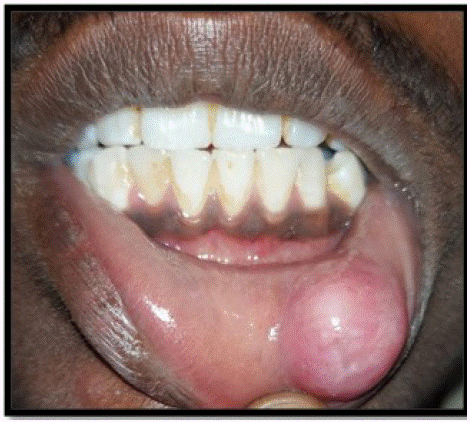
Figure 1: Intraoral examination revealed a painless, fluid‑filled, soft, dome-shaped, bluish, solitary swealling measuring about 1.5cm×1.5cm on left side of lower lip.
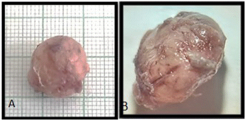
Figure 2: A. Gross examination B. Under scanner microscope.
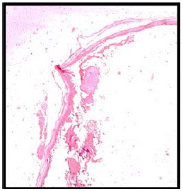
Figure 3: Collagenous and hyalinized globular structures free floating in the lumen compatible with myxoglobulosis.
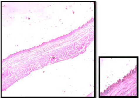
Figure 4: Papillary synovial metaplasia-like changes.
The swelling was well defined and soft to firm in consistency. Provisional diagnosis was given as Mucocele and surgical excision was advised.
Gross examination showed fluid filled single soft tissue which was soft to firm in consistency measuring 1.5cm×1.5cm with smooth surface. The specimen under scanner microscope showed surface irregularities and few erythematous areas.
Histopathological examination showed, H & E stained section of single soft tissue bit exhibits cystic cavity containing mucinous material and inflammatory cells at places. Globular structure containing hyalinized mucinous material arranged in concentric manner suggestive of myxoglobulous structure seen in luminal part. The cystic lining showed Synovial metaplasia like changes towards lumen at places. Adjacent connective tissue is fibro cellular consisting loosely arranged collagen fibre bundle with entrapped mucin. Moderate degree of chronic inflammatory cell infiltrate and mucinophages and vascularity was evident. Towards periphery collagen fibre bundles were parallely arranged with fibroblasts and fibrocytes. Mucous salivary gland acini and dilated ducts were evident in the vicinity.
Overall features were suggestive of “Mucocele with myxoglobulosis and papillary synovial metaplasia-like changes”.
Discussion
Mucoceles (also called sialocele and ptyalocele) are one of the most common benign soft tissue lesions of the oral cavity. A mucocele is defined as the pooling of mucin in a cystic cavity. Two types of mucoceles are recognized: (1) The retention type, and (2) The extravasation type.
The extravasation type is a pseudocyst without defined walls and are caused due to mechanical trauma to the excretory duct of the gland leading to transection or rupture, with consequent extravasation of mucin into the connective tissue stroma and are seen frequently on lower labial mucosa, buccal mucosa and retromolar area; they are not lined by epithelial lining. The mucus extravasation triggers a secondary inflammatory reaction. Many patients report the periodic discharge of viscous fluid from the lesion.
In mucous retention phenomena, mucus may be retained in the duct and/or acini as a result of duct obstruction by sialolith or strictures. The ductal narrowing can occur due to frequent mouth washing with hydrogen peroxide, deodorant mouthwashes, tartar-control toothpastes or anti-plaque solutions, which are possible causes of irritation [3].
The lower lip is the most commonly affected site, followed by the floor of the mouth and ventral tongue which is congruent with our finding of presence of mucocele in lower lip. Patients present with a dome-shaped swelling, often with a blue hue due to the extravasated mucin. If small, the lesion may appear as a blister. Within the floor of mouth, the term ranula may be applied to mucin which dissects into the mylohyoid muscle, resulting in chin or upper neck swelling (rana in Latin means frog, and ranula is used since it looks like a frog’s underbelly clinically) [5].
The lesions can be located directly under the mucous membrane (superficial mucocele) or in the upper submucosa (classical mucocele). Oral mucoceles may be located either as a fluid‑filled vesicle or blister in the superficial mucosa or as a fluctuant nodule deep within the connective tissue. Spontaneous drainage of the inspisatted mucin, especially in superficial lesions followed by subsequent recurrence, may occur. The surface of long‑standing lesions may show fibrosis as seen in this case. The superficial lesions appear as thin-walled, bluish swellings that rupture easily while the deeper lesions are well circumscribed swellings usually covered by normal appearing oral mucosa [3].
Broadly, salivary gland pathologies are classified into neoplastic and non-neoplastic diseases. The latter poses diagnostic challenges to pathologists owing to their clinical behavior and varied histological features. Thus, understanding the associated pathophysiological features is vital to distinguish between lesions and better insight into the behavior in order to provide nobler therapeutics. The pathogenesis of oral mucoceles has been attributed to the damage of excretory ducts of minor salivary glands following traumatic injuries which leads to extravasation of mucin into the stroma triggering an inflammatory reaction. Alternatively, it was suggested that mucous extravasation may arise from the traumatic destruction of a large amount of glandular acini followed by continuous secretion from the remaining acini and pooling of mucin. However, the exact explanation of pathogenesis of mucoceles remains inchoate [6].
Histologically, the presence of a well‑demarcated interstitial mucin surrounded by a granulation tissue containing neutrophils and multinucleated giant cells in the submucosa serves as a peculiar feature to diagnose mucus extravasation phenomenon [6]. Some unusual histopathological features such as myxoglobulosis, papillary synovial metaplasia-like changes, superficial mucoceles, and clear cell changes have been described in the literature and accounts for 19.1% of mucocele histopathological variants [2].
Myxoglobulosis appears as a globular structure that lacks epithelial tissue and whose center contains eosinophilic, lamellar, and amorphous or fibrillary material. Myxoglobulosis is a specific type of mucocele consisting of mucoid material. It is characterized by opaque, transparent globules that resemble “fish eggs” or “frogspawns”. These spherules can be found either in the lumen or in the connective tissue surrounding the mucin extravasation. In our case, these spherules are seen in lumen at places [2]. PAS stain was positive and confirmed the presence of mucinous material in these globules.
Shah et al (2003) reported that the dissociated collagen fibers become surrounded by macrophages and acquire a globular appearance, which may be the result of mechanical fragmentation due to the presence of mucin or enzymatic break down mediated by macrophages. Before fragmentation, the globular structures also contain inflammatory cells and vessels, corresponding to the early stage of myxoglobulosis [8]. Paremala et al emphasized that, although the diagnosis of mucus extravasation phenomena is not challenging for the pathologist, the intraluminal globular organization surrounded by granulation tissue is an unusual histopathological feature of these lesions [9].
Mucoceles with papillary synovial metaplasia-like change are so called as metaplastic changes resembling synovium in non-joint spaces characterized by the partial replacement of the granulation tissue surrounding the mucin extravasation with a folded membrane exhibiting villi. These papillary synovial metaplasia-like changes are seen in histopathological picture of our case at places. The more superficial portion of this membrane consists of a thin amorphous and eosinophilic matrix with an underlying condensation of histiocytes, fibrohistiocytes, and/or multinucleated giant cells. Within this spectrum, marked reduction in the cavity due to the presence of broad papillae that project into the lumen is seen. This presentation is known as collapsed lumen [2].
According to Chi et al (2011), it is important that the pathologist recognizes this rare histopathological variant of oral mucocele to avoid a misdiagnosis. Particularly, Warthin tumor should be included in the differential diagnosis because of its cystic papillary growth and eosinophilic surface. However, the authors emphasize that features such as the preferential location of mucoceles in the lower lip, in contrast to salivary tumors that rarely occur at this site, and the presence of oncocytic cells with extensive eosinophilic cytoplasm in Warthin tumor contribute to the differentiation between these lesions [11].
Superficial mucocele was first described by Eveson et al (1988) [10]. Clinically, this lesion appears as a small translucent vesicle measuring less than 5mm. Microscopic features include a mucin-containing subepithelial blister, atrophic surface epithelium, partial epithelial regeneration at the base of the mucin-filled cavity, and the absence of signs of subepithelial separation at the periphery of the lesion.
Superficial mucoceles can often be misdiagnosed as benign pemphigoid of the mucous membranes or bullous lichen planus. However, the clinical appearance of superficial mucoceles and immune-mediated lesions are somewhat different. In bullous pemphigoid and oral bullous lichen planus, the blisters are generally larger, multiple, flaccid, and opaque while in most superficial mucoceles, the blisters are translucent, single and have a tense surface. The disease is diagnosed on the basis of clinical, histologic, and immunologic findings by direct and indirect immunofluorescence [11].
Piña et al (2013) reported an unusual variant of oral mucocele composed almost exclusively of macrophages that exhibited extensive clear cytoplasm and signet ring alterations. This uncommon presentation of oral mucocele was described by the authors as clear cell change [12].
These clear cells exhibited cytoplasmic vacuolization with shrunken pyknotic peripheral nuclei and a signet ring appearance, intermingled with a delicate network of small vessels. Such cases are difficult to diagnose because of the similarity with clear cell tumors and signet ring cell lesions, such as salivary gland tumors or metastatic neoplasms. Although most of them exhibit distinctive microscopic characteristics, histochemistry and immunohistochemistry can be required with relevant clinical information to achieve the correct diagnosis [11].
Conclusion
As mentioned earlier, considerable variation exists in the prevalence of uncommon histopathological findings in oral mucoceles. Except for myxoglobulosis, there are no explanations for the histological appearance of most of these alterations. These histological features are accidental findings, and the clinical presentation of these lesions does not differ from oral mucoceles exhibiting the usual histology [3], except for superficial mucoceles. However, in some cases, these changes may be prominent enough to cause important diagnostic difficulties. Knowledge of the possible histopathological variations of oral mucoceles is therefore essential.
References
- Tegginamani AS, Sonalika WG, Vanishree HS. Oral mucocele: A clinicopathological analysis of 50 cases. Archives of Medicine and Health Sciences. 2016; 4: 40.
- de Brito Monteiro BV, Bezerra TM, da Silveira ÉJ, Nonaka CF, da Costa Miguel MC. Histopathological review of 667 cases of oral mucoceles with emphasis on uncommon histopathological variations. Annals of Diagnostic Pathology. 2016; 21: 44-6.
- Jani DR, Chawda J, Sundaragiri SK, Parmar G. Mucocele--A study of 36 cases. Indian J Dent Res. 2010; 21: 337-40.
- Chi AC, Lambert III PR, Richardson MS, Neville BW. Oral mucoceles: a clinicopathologic review of 1,824 cases, including unusual variants. Journal of Oral and Maxillofacial Surgery. 2011; 69: 1086-93.
- Thompson LD. Mucocele: retention and extravasation types. Ear, Nose & Throat Journal. 2013; 92: 106-8.
- Pandiar D, Anand R, Kamboj M, Narwal A, Devi A. Papillary synovial metaplasia-like change in oral mucoceles: A retrospective institutional study of 105 cases. Journal of Oral and Maxillofacial Pathology: JOMFP. 2022; 26: 283.
- Dos Santos-Leite ÉG, Silva ÂN, Leonel AC, Bezerra HK, Carvalho EJ, et al. ORAL MUCOCELES: A CLINICOPATHOLOGICAL STUDY OF 401 CASES WITH EMPHASIS ON UNUSUAL HISTOLOGICAL VARIANTS. Oral Surgery, Oral Medicine, Oral Pathology and Oral Radiology. 2022; 134: e190.
- Shah KA. Myxoglobulosis in oral extravasation mucocele: an entity? Histopathology. 2003; 43: 291–6.
- Paremala K, Radhika MB, Thambiah LJ. Myxoglobulosis of lower lip: report of two cases. J Oral Maxillofac Pathol. 2011; 15: 232–5.
- Eveson JW. Superficial mucoceles: pitfall in clinical and microscopic diagnosis. Oral Sur Oral Med Oral Pathol. 1988; 66: 318–22.
- Chi AC, Haigney II RJ, Spagnoli DB, Neville BW, Richardson MS. Papillary synovial metaplasia-like change in oral mucoceles: a rare and previously undescribed histopathologic variant of a common oral lesion. Oral Surg Oral Med Oral Pathol Oral Radiol Endod. 2010; 109: 268–73.
- Piña AR, Almeida LY, Andrade BA, Leon JE. Clear cell change in a lower lip mucocele. J Oral Maxillofac Pathol. 2013; 7: 318