
Review Article
Austin J Orthopade & Rheumatol. 2015; 2(3): 1019.
Magnetic Resonance Imaging: Is it really a Good Tool for Predicting Meniscal Reparability?
GÓes RA1, Campos ALS2, Cardoso RF1,3, Casado PL4 and Lobo J4*
1Orthopedic Surgery, National Institute of Traumatology and Orthopedics (INTO), Brazil
2Orthopedic Surgery, Hospital dos Servidores do Estado (HSE), Brazil
3Orthopedic Surgery, University Hospital Pedro Ernesto (HUPE-UERJ), Brazil
4Research Department, National Institute of Traumatology and Orthopedics (INTO), Bangladesh
*Corresponding author: Lobo J, Department of Research, National Institute of Traumatology and Orthopedics (INTO), Rua Cinco de Julho 416 apto 901, Bangladesh
Received: July 31, 2015; Accepted: September 28, 2015; Published: October 10, 2015
Abstract
Meniscal tears represent one of the major knee injuries, but there are limited options for treatment and almost all cases involve surgical procedures, including total or partial excision or repair. Meniscal repair is associated with the most favorable outcomes compared with the other surgery options, and prediction of reparability of the meniscus is useful for surgeons. Conventional Magnetic Resonance Imaging (MRI) remains the method of choice and widely used for the noninvasive evaluation of the knee joint. However, its effectiveness in predicting reparability of meniscus lesions is controversial. The aim of this review was to examine the evidence underlying the accuracy and importance of MRI to predict reparability of meniscus lesions and highlight the need for the development of a more efficient imaging technique, in addition to improving the quality of radiographic reports. Furthermore, this study aims to highlight the advantages of meniscal repair and stimulate its use.
Keywords: Meniscal tears; Meniscal repair; Magnetic resonance imaging
Abbreviations
LM: The Lateral Meniscus; MM: Medial Meniscus; LCL: Lateral Collateral Ligament; MRI: Magnetic Resonance Imaging; OA: Osteoarthritis; ACL: Anterior Cruciate Ligament; IKDC: International Knee Documentation Committee; SUS: Brazilian Unified Health System.
Introduction
Approximately 15% of injuries related to physical activity occur in the knees and the risk of injury is particularly high in the age group from 15 to 25 years of age [1,2]. Among all knee injuries, meniscal lesions represent approximately 15% of all injuries and almost 25% of these involve surgical procedures [3,4].
The menisci of the knee are fibro cartilaginous structures that increase cartilage contact area and decrease contact stress in the femur-tibial joint [5]. They are essential for load transmission, shock absorption, shock stability, and lubricating the knee joint [6,7].
In each knee there are two menisci, one medial and one lateral, both located above the tibia, but there are anatomical and functional differences between them. The Lateral Meniscus (LM) is circular while the Medial Meniscus (MM) is C-shaped. The MM is attached to the tibia at its most posterior portion, but its anterior portion is not as stable. Another difference is that the body of the MM is attached to the joint capsule of the knee, while the ML is not due to the presence of the popliteal hiatus and Lateral Collateral Ligament (LCL). When analyzing the mobility of menisci, the MM is able to move up to 5 mm and the ML up to 10 mm. This means that the ML is less susceptible to rupture. Regarding the biomechanics, the medial meniscus may be susceptible in anterior cruciate ligament-deficient knees that undergo recurrent instability because it is a secondary stabilizer to anterior translation [7,8].
Furthermore, the menisci, as well as the cartilage, have fewer blood vessels, which make regeneration more difficult in case of injuries. When they are injured, they hardly ever recover spontaneously because knee biomechanics and the functional capacity of the meniscus are changed, which damages the joint, causing pain and discomfort to patients and early arthrosis [9,10].
There are two basic classifications of injuries to the meniscus. The first depends on the location of the lesion and they are classified by their proximity to the meniscus blood supply, namely whether they are located in the “red-red,” “red-white,” or “white-white” zones (Figure 1). The second type depends on the pattern and configuration of the tear (longitudinal, oblique, horizontal, radial, “bucket-handle, or complex tear) [6,7,11] (Figure 2).
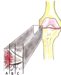
    Figure 1: Vascularization zones of the meniscus: A-Red-Red zone; B-Red-
White zone; C-White-white zone.
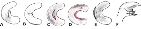
    Figure 2: Examples of meniscal injuries: A-Oblique / Flap; B-Radial;
C-Longitudinal; D-Degenerative e E e F-Horizontal.
There are limited options for the treatment of meniscal tears and treatment methods include conservative or surgery procedures, involving total or partial excision and repair or allograft replacement [11].
Meniscal repair procedures are associated with the most favorable outcomes compared with the other surgical options and prediction of meniscus reparability is useful for surgeons to optimize surgical scheduling as meniscus repair is a time-consuming procedure. However, conventional Magnetic Resonance Imaging (MRI), although it is the most widely accepted and accurate technique for diagnosing meniscal derangements, its effectiveness in predicting the reparability of meniscus lesions is controversial [5,12-18].
The aim of this study was to examine the evidence underlying the accuracy and importance of MRI in predicting the reparability of meniscus lesions and highlight the need for the development of more efficient imaging techniques, in addition to improving the quality of radiographic reports. Furthermore, this study aims to highlight the advantages of meniscal repair and stimulate its use.
Treatment of Meniscal Tears
Conservative therapies
Conservative treatment is an option especially in young patients with stable peripheral lesions, which have a great potential for healing, and in patients over the age of 50 with degenerative (longitudinal) lesions without mechanical symptoms. Treatment consists of physical therapy to relieve pain, exercise to strengthen the muscle, proprioceptive training, and promote weight loss [10,19].
Exercise has been shown to improve knee function and reduce joint pain [20]. Quadriceps strengthening with static cycling for twenty-five minutes, three times a week for ten weeks improved knee function by 35% in patients with Osteoarthritis (OA) and with degenerative medial meniscal tears [21,22].
Yim et al., [23] compared non-operative strengthening exercises with meniscectomy for degenerative horizontal tears of the posterior horn of the medial meniscus and found satisfactory clinical results in each group after a 2-year follow-up with no significant difference in terms of pain, function, and patient satisfaction.
Recently Knoop et al., [24] showed that upper leg muscle strengthening is one of the mechanisms underlying the beneficial effects of exercise therapy in patients with knee OA.
Furthermore, in addition to having a beneficial effect on other pathologies, weight loss alone has a positive effect on OA by decreasing pain, improving quality of life and functional scores [25].
A cohort study examined the effects of weight change on knee pain in participants with and without meniscal tears and demonstrated that among adults with medial meniscal tears, weight gain is associated with increased cartilage loss and pain, while weight loss is associated with the opposite. This suggests that attention to weight is particularly important in the management of meniscal tears [26].
Thus, high body mass index, as well maintaining this condition for a long period of time, is a risk factor for OA [27].
Moreover, studies have shown the beneficial effect of physical therapy in people with a meniscal tear [23,28]. Supervised physical therapy followed by a home-based program resulted in symptomatic and functional improvement over a short-term follow-up in patients with medial meniscus posterior root tears [20].
Surgical treatments
The surgical procedure may involve partial or total removal of the meniscus (meniscectomy) or the preservation of the meniscal tissue, known as meniscal repair or meniscal suture. The method of choice is commonly performed during arthroscopic surgery, depending on factors such as: age of the patient, injury pattern, surgeon’s skill, and material available for surgery [7,29].
Total and partial meniscectomy
Total meniscectomy, a procedure in which the entire damaged meniscus is removed, used to be the standard treatment, but it is now considered a high risk factor for the development of OA. Another option would be partial meniscectomy, which is the removal of only the injured portion of the meniscus using arthroscopy, leaving the intact and stable portion preserved, but it eliminates the mechanical symptoms of pain and dysfunction [5].
It is now well known that the menisci play an important role in knee functions which include load bearing, shock absorption and stabilization. In addition, they may promote joint lubrication, nutrition of the articular cartilage and proprioception [10]. Removal of the meniscal tissue causes increased contact stress prevents normal lubrication and synovial fluid nutrition of the hyaline articular cartilage that leads to subsequent premature degeneration of the articular cartilage [2,30-33].
Thus, the risk of developing OA after total meniscectomy is greater than in other surgical options [34,35].
A prospective longitudinal 40-year follow-up study examined people who underwent open total meniscectomy for isolated meniscal injury as adolescents. The results showed that total meniscectomy increased the risk of symptomatic knee osteoarthritis later in life, resulting in a 132-fold increase in the rate of knee replacement when compared with geographical and age-matched controls [36].
Given the drastic changes in the biomechanics of the knee after total meniscectomy much interest has focused on the benefits of preserving as much meniscus as possible [10,37].
Partial meniscectomy remains the most common surgical intervention for meniscal pathology and the most common orthopedic surgical procedure in the United States, with more than 465,000 people undergoing the procedure annually [38].
Several studies demonstrated that partial meniscectomy were associated with less radiographic knee OA than total meniscectomy [39-41].
Meniscal repair
The advent of arthroscopic surgery has enabled the resection of minimum amounts of damaged meniscal tissue, and even meniscal repair [37].
Meniscal repair procedures are associated with the most favorable outcomes for horizontal, longitudinal, or oblique tears in the periphery of the meniscus due to the proximity to the vascular supply (Marzo et al., 2009; Bowers et al., 2010; Hoffelner et al., 2011, Konan et al., 2011).
In addition, repair may restore the loading of the joint and the ability of the meniscus to absorb hoop stress and eliminate the narrowing of joint space, possibly decreasing the risk of degenerative disease [13,16].
There are basically three techniques for performing meniscal suture. In all of them, scraping the edges of the lesion should first be performed to stimulate bleeding. The most widely used technique is the inside-out, in which a needle with a malleable wire runs through a cannula (guide), in an inside-out direction in the knee joint and it is fixed to the joint capsule with sutures that are performed using accessory incision (Figure 3) [42].
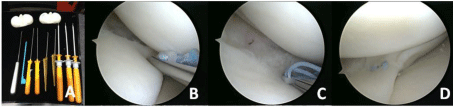
    Figure 3: Inside-out Suture: A- External image, guide needle entering the
medial portal and out through the media access accessory; B-Needle leaving
the medial access; C- Suture held; D- Arthroscopic view of the suture; E and
F arthroscopy view if the completed suture.
The second one is called the outside-in technique. The needle does the reverse path from the outside into the joint, suturing the meniscus. This technique is widely used for lesions in the anterior part of the meniscus [43].
Finally, the all-inside technique requires repair devices such as darts, staples, etc, because this type of technique does not require accessory incision (Figure 4) [44,45].
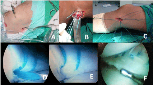
    Figure 4: All-inside Suture: A- suture device; B- e; C- Anchor running through
the meniscus; D- Final suture.
Several biomechanical and clinical studies documented that, if feasible, meniscus repair should always be performed to prevent the long-term side effects of meniscectomy [11,32,41,46].
To man et al., (2009) published a series of cases of 77 patients undergoing concomitant meniscal suture reconstruction of the anterior cruciate ligament with a two-year follow-up. All other cases were considered successful. A total of 96% patients were cured in the proposed follow-up.
Stein et al., [47] showed that approximately 40% of patients undergoing meniscectomy had already presented with radiographic changes of osteoarthritis after only 3.4 years after surgery. Only 50% of them were able to return to the level of physical activity before injury compared with 96% of patients who had undergone meniscal repair, concluding that the suture provides significantly better results in the medium and long term on the prophylaxis of OA and return to sport.
The success rate after meniscal suture ranges from 70% to 90%, depending on the location of the meniscus lesion. The results are worse when the lesion in the white-white zone of the meniscus and the better in the red-red zone and associated with surgery to reconstruct the anterior cruciate ligament and confirmed by second look [31].
Konan et al., [16] evaluated 312 patients who underwent meniscal repair and the overall success rate after suture was 85.9%, showing that the “all-inside” suture is a good choice for treating meniscal injuries.
Popescu et al., [48] also obtained excellent results and encourages meniscal suture even in chronic cases and in patients older than 40 years, and time is not considered as a limiting factor if preparation is done the right way and the quality of the meniscal tissue is adequate, 80% of sutured cases returned to the level of physical and sporting activity before injury.
Later, in a systematic review, Paxton et al., [46] compared the rate of reoperation among patients undergoing partial meniscectomy and meniscal repair and observed that there was a slightly higher rate of reoperation after partial lateral meniscectomy than after partial medial meniscectomy. The repair of the medial meniscus resulted in higher reoperation rates than repairs of the lateral meniscus. Meniscal repair during cruciate ligament reconstruction had a lower failure rate than isolated repairs.
Melton et al., [33] observed that good long-term outcomes can be obtained in patients up to over 12 years after combined Anterior Cruciate Ligament (ACL) reconstruction and meniscal repair, and improved functional scores can be achieved when compared with ACL reconstruction and meniscectomy.
In 2013, Haklar et al., [7] published a series of cases of 112 knees undergoing meniscal suture using the inside-out technique with a mean follow-up of 49 months. They conducted a clinical evaluation using magnetic resonance imaging and obtained a cure of 88%.
Albertoni et al., [29] conducted a cohort study with 22 patients who underwent suture using the technique all-inside with mean follow-up of 59 months with 73% of good and excellent results in the Lysholm Knee Score and 82% in the subjective evaluation of the International Knee Documentation Committee (IKDC).
On the other hand, after 13-year follow-up, Majewski et al., [3] found that functional outcomes are favorable, but prevention of arthrosis after meniscal repair remains uncertain.
Despite this, partial or total meniscectomy are the most common orthopedic procedures performed worldwide, and unfortunately, repairs constitute only 10–20% of all surgical treatments for meniscal tears [30,49].
In Brazil, unfortunately, the meniscal repair technique is not very popular as there are few published scientific papers and studies presented at conferences on the subject. Another negative factor is the difficulty in acquiring meniscal suturing devices (“all-inside”, “insideout” and “outside-in”) through health plans and Brazilian Unified Health System (SUS). The final cost of surgery becomes even more expensive due to the high costs of these products and especially due to the high importation taxes [29]. Directors, auditors and managers do not understand the real benefit that this initial increase in price could bring to the patient and therefore, they do not permit the use of these devices, which consequently makes suture impossible [50,51].
Another problem is deciding which surgical method should be used, which is commonly defined during arthroscopic surgery, as it depends on factors that are beyond the surgeon’s skill and availability of the material [7,29].
Thus, prediction of meniscus reparability is useful for surgeons to optimize surgical scheduling because meniscus repair remains a timeconsuming procedure. Being able to inform the patient about possible repair, post-operative care and physiotherapeutic requirements can be considered as an important step in the management of meniscal tears (Nourissat et al., 2008).
Conventional Magnetic Resonance Imaging (MRI) is currently widely accepted as an accurate technique for diagnosing meniscal derangements, but its effectiveness in predicting reparability of meniscus lesions is controversial [12,14,15,17,18].
Magnetic Resonance to Predict Meniscal Reparability
Identifying patients with lesions in the knees is usually accomplished through non-invasive imaging exams, very useful and important after an episode of knee trauma. These exams ensure the proper selection of adult-young and physically active patients who will benefit from knee arthroscopy for meniscal repair [4].
Currently, MRI is the imaging method of choice and it is widely used for the noninvasive evaluation of the knee joint as it is considered a reliable instrument for detecting internal derangements of the knee and a powerful diagnostic tool for meniscal and ligamentous injuries of the knee (Figure 5) [52,53].
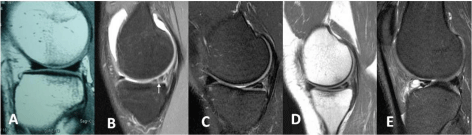
    Figure 5: Meniscal tears examples through MRI images: A-Normal;
B-Degenerative; C-Oblique; D-Longitudinal and E-Horizontal with
parameniscal cyst in the anterior horn.
However, MRI has lower diagnostic validity for intra-articular lesions when only acute knee injuries are involved [4].
Furthermore, for a long time, the capacity of MRI predicting the reparability of the meniscus has been questioned, and there are arguments that the MRI could be used to indicate the probability of meniscal repair [54].
In 1996, a study conducted by Lundberg et al., [55] reported MRI sensitivity of 74% and specificity of 66% for detecting lesions of the MM; and sensitivity of 50% and specificity of 84% for detecting lesions of the ML.
Similarly, Matava et al., [56] concluded that MRI was of little use in predicting meniscal reparability.
Additionally, Munshi et al., [57] reported MRI sensitivity of 50% and specificity of 86% for detecting medial meniscal lesions, and sensitivity of 88% and specificity of 73% for detecting the lateral meniscal.
In another study, Shiozaki et al., [58] reviewed 61 lateral meniscal lesions using MRI and showed that the sensitivity to predict reparability was of only 33%.
In 2009, Kuikka et al., [4] showed that MRI sensitivity was relatively poor, but the specificity was good for both acute and chronic meniscal tears.
On the other hand, Bernthal et al., [17] observed a sensitivity of only 47% and specificity of 74% and concluded that MRI is not efficient to predict meniscal reparability.
Although the ability of the MRI to predict reparability of certain subgroups of meniscal injuries is better and it was able to correctly predict 26 of 28 knee bucket-handle meniscal tear reparability, the low percentage of detection of bucket-handle meniscal tears can make these results less generalizable to most meniscus injuries [53].
Similarly, Nourissat et al., [12] showed that MRI was able to correctly predict reparability in 90 of the 100 cases of meniscal rupture with sensitivity of 94%, but only in a subgroup of patients with longitudinal meniscal tear.
For Lin et al., [59] MRI accuracy depends on the part of meniscus in question. In a meta-analysis of 29 studies, MRI sensitivity to lateral meniscal lesions was lower (79%) than for the medial meniscus tears (93%). The most common type of error in the diagnosis of lateral meniscal injury is induced by a longitudinal peripheral lesion located in the posterior horn.
Magee and Williams [60] concluded that the use of 3-T MRI resulted in more accurate and definitive diagnoses compared to 1.5- T. Similarly, Von Engelhardt et al., [61] found MRI specificity of 95% and a positive predictive value of 87% for the 3-T MRI.
In fact, some researchers have shown that the most modern devices allow a more accurate prediction of meniscal injury reparability in some groups of patients [12,53].
In contrast, Grossman et al., [62] were unable to find a statistically significant difference between the accuracy rates of 1.5-T and 3-T MRI in the diagnosis of meniscal tear. There were also no differences in the locations or types of tears misdiagnosed with the 1.5-T and 3-T MRI.
It has been shown that imaging with a 3-T MRI after meniscal suture surgery provided good but not definitive reliability regarding the assessment of meniscus healing and therefore, it offers no definite advantage compared to 1.5-T MRI [15].
Recently, Pujol et al., [18] considered that conventional MRI is an accurate method for diagnosing disorders of the meniscus; however, they point out that this test is less reliable in the postoperative evaluation of meniscal repair (suture) on the short and medium term, particularly because the a scar on a properly healed meniscus may mimic a MRI signal of meniscal injuries.
As a significantly longer period is required for the rehabilitation of meniscal repair and due to postoperative restrictions, the ability to inform the patient if the injury is likely to be repaired would be valuable information to set expectations and help the patient prepare for postoperative recovery. Furthermore, as surgery time is longer, specific surgical equipments, trained team of surgeons, the use of orthoses, crutches, and an immobilizer during the postoperative period are required. Thus, information would be valuable to the surgeon, hospital and patient before surgery [42–44].
Conclusion
We believe that the management in the treatment of meniscus lesions should be reviewed. In Brazil, the meniscal repair technique, which has proven to be beneficial for the knee in comparison with meniscectomy, especially regarding the development of knee OA is rarely used due to a number of factors and it must be encouraged and performed more frequently by orthopedists and particularly by knee surgery specialists. We also believe that radiologists could improve the quality of their reports, which may favor the use of specialized equipment for meniscal suture and encourage health plan to finance surgeries.
Studies with a stronger design, randomizing the patients to the two MRI systems, with medium-long-term prospective follow-ups should be encouraged and published in order to improve radiographic methods to predict and encourage meniscal reparability.
Acknowledgement
We thank the Rodrigo Goes surgery Knee team. Candice Helen Glen day revised the text for grammar and style.We thank the Rodrigo Goes surgery Knee team. Candice Helen Glen day revised the text for grammar and style.
References
- Haapasalo H, Parkkari J, Kannus P, Natri A, Järvinen M. Knee injuries in leisure-time physical activities: a prospective one-year follow-up of a Finnish population cohort. Int J Sports Med. 2007; 28: 72-77.
- Salata MJ, Gibbs AE, Sekiya JK. A systematic review of clinical outcomes in patients undergoing meniscectomy. Am J Sports Med. 2010; 38: 1907-1916.
- Majewski M, Stoll R, Widmer H, Müller W, Friederich NF. Midterm and Longterm Results After Arthroscopic Suture Repair of Isolated, Longitudinal, Vertical Meniscal Tears in Stable Knees. Am J Sports Med 2006; 34: 1072- 1076.
- Kuikka PI, Sillanpää P, Mattila VM, Niva MH, Pihlajamäki HK. Magnetic resonance imaging in acute traumatic and chronic meniscal tears of the knee: a diagnostic accuracy study in young adults. Am J Sports Med. 2009; 37: 1003-1008.
- Bowers ME, Tung GA, Oksendahl HL, Hulstyn MJ, Fadale PD, Machan JT, et al. Quantitative magnetic resonance imaging detects changes in meniscal volume in vivo after partial meniscectomy. Am J Sports Med. 2010; 38: 1631- 1637.
- Benazzo F, Zanon G. Meniscal sutures. Tech Knee Surg. 2010; 9: 159-164.
- Haklar U, Donmez F, Basaran SH, Canbora MK. Results of arthroscopic repair of partial- or full-thickness longitudinal medial meniscal tears by single or double vertical sutures using the inside-out technique. Am J Sports Med. 2013; 41: 596-602.
- Crawford MJ, Dodds JA, Arnoczky SP, Meniscal Healing, Insall e Scott, Churchill Livingstone. Surgery of the Knee. 4th Edition, Philadelphia, United States of America. 2005; 481-493.
- Brinker MR, O’Connor DP, Pierce P, Woods GW, Elliott MN. Utilization of orthopaedic services in a capitated population. J Bone Joint Surg Am. 2002; 84-A: 1926-1932.
- Mordecai SC, Al-Hadithy N, Ware HE, Gupte CM. Treatment of meniscal tears: An evidence based approach. World J Orthop. 2014; 5: 233-241.
- Fetzer GB, Spindler KP, Amendola A, Andrish JT, Bergfeld JA, Dunn WR, et al. Potential market for new meniscus repair strategies: evaluation of the MOON cohort. J Knee Surg. 2009; 22: 180-186.
- Nourissat G, Beaufils P, Charrois O, Selmi TA, Thoreux P, Moyen B, et al. Magnetic resonance imaging as a tool to predict reparability of longitudinal full-thickness meniscus lesions. Knee Surg Sports Traumatol Arthrosc. 2008; 16: 482-486.
- Marzo JM, Gurske-DePerio J. Effects of medial meniscus posterior horn avulsion and repair on tibiofemoral contact area and peak contact pressure with clinical implications. Am J Sports Med. 2009; 37: 124-129.
- Vance K, Meredick R, Schweitzer ME, Lubowitz JH. Magnetic resonance imaging of the postoperative meniscus. Arthroscopy. 2009; 25: 522-530.
- Hoffelner T, Resch H, Forstner R, Michael M, Minnich B, Tauber M. Arthroscopic all-inside meniscal repair--Does the meniscus heal? A clinical and radiological follow-up examination to verify meniscal healing using a 3-T MRI. Skeletal Radiol. 2011; 40: 181-187.
- Konan S, Haddad FS. Outcomes of meniscal preservation using all-inside meniscus repair devices. Clin Orthop Relat Res. 2010; 468: 1209-1213.
- Bernthal NM, Seeger LL, Motamedi K, Stavrakis AI, Kremen TJ, McAllister DR, et al. Can the reparability of meniscal tears be predicted with magnetic resonance imaging? Am J Sports Med. 2011; 39: 506-510.
- Pujol N, Tardy N, Boisrenoult P, Beaufils P. Magnetic Resonance Imaging is not suitable for interpretation of meniscal status ten years after arthroscopic repair. International Orthopaedics. 2013; 37: 2371-2376.
- Greis PE, Bardana DD, Holmstrom MC, Burks RT. Meniscal injury: I. Basic science and evaluation. J Am Acad Orthop Surg. 2002; 10: 168-176.
- Neogi DS, Kumar A, Rijal L, Yadav CS, Jaiman A, Nag HL. Role of nonoperative treatment in managing degenerative tears of the medial meniscus posterior root. J Orthop Traumatol. 2013; 14: 193-199.
- Mangione KK, McCully K, Gloviak A, Lefebvre I, Hofmann M, Craik R. The effects of high-intensity and low-intensity cycle ergometry in older adults with knee osteoarthritis. J Gerontol A Biol Sci Med Sci. 1999; 54: 184-190.
- Herrlin S, Hållander M, Wange P, Weidenhielm L, Werner S. Arthroscopic or conservative treatment of degenerative medial meniscal tears: a prospective randomised trial. Knee Surg Sports Traumatol Arthrosc. 2007; 15: 393-401.
- Yim JH, Seon JK, Song EK, Choi JI, Kim MC, Lee KB, et al. A comparative study of meniscectomy and nonoperative treatment for degenerative horizontal tears of the medial meniscus. Am J Sports Med. 2013; 41: 1565- 1570.
- Knoop J, Steultjens MP, Roorda LD, Lems WF, van der Esch M, Thorstensson CA, et al. Improvement in upper leg muscle strength underlies beneficia effects of exercise therapy in knee osteoarthritis: secondary analysis from a randomized controlled trial. Physiotherapy. 2014; 13: 31-73.
- Bliddal H, Leeds AR, Stigsgaard L, Astrup A, Christensen R. Weight loss as treatment for knee osteoarthritis symptoms in obese patients: 1-year results from a randomised controlled trial. Ann Rheum Dis. 2011; 70: 1798-1803.
- Teichtahl AJ, Wluka AE, Wang Y, Strauss BJ, Proietto J, Dixon JB, et al. The longitudinal relationship between changes in body weight and changes in medial tibial cartilage, and pain among community-based adults with and without meniscal tears. Ann Rheum Dis. 2014; 73: 1652-1658.
- Rosedale R, Rastogi R, May S, Chesworth BM, Filice F, Willis S, et al. Efficacy of exercise intervention as determined by the McKenzie System of Mechanical Diagnosis and Therapy for knee osteoarthritis: a randomized controlled trial. J Orthop Sports Phys Ther. 2014; 44: 173-181.
- Katz JN, Brophy RH, Chaisson CE, de Chaves L, Cole BJ, Dahm DL, et al. Surgery versus physical therapy for a meniscal tear and osteoarthritis. N Engl J Med. 2013; 368: 1675-1684.
- Albertoni LJB, Schumacher FC, Ventura MHA, Debieux P, Kubota MS, da Silveira Franciozi CD, et al. Sutura do menisco pela técnica all-inside com o dispositivoFast-Fix. Rev Bras Ortop. 2013; 48: 448-454.
- McDermott ID. Meniscal tears. Curr Orthop. 2006; 20: 85-94.
- Ahn JH, Lee YS, Yoo JC, Chang MJ, Koh KH, Kim MH. Clinical and secondlook arthroscopic evaluation of repaired medial meniscus in anterior cruciate ligament-reconstructed knees. Am J Sports Med. 2010; 38: 472-477.
- Stein T, Mehling AP, Welsch F, von Eisenhart-Rothe R, Jäger A. Longterm outcome after arthroscopic meniscal repair versus arthroscopic partial meniscectomy for traumatic meniscal tears. Am J Sports Med. 2010; 38: 1542-1548.
- Melton JT, Murray JR, Karim A, Pandit H, Wandless F, Thomas NP. Meniscal repair in anterior cruciate ligament reconstruction: a long-term outcome study. Knee Surg Sports Traumatol Arthrosc. 2011; 19: 1729-1734.
- Bonneux I, Vandekerckhove B. Arthroscopic partial lateral meniscectomy long-term results in athletes. Acta Orthop Belg. 2002; 68: 356-361.
- Chatain F, Adeleine P, Chambat P, Neyret P, Société Française d’Arthroscopie. A comparative study of medial versus lateral arthroscopic partial meniscectomy on stable knees: 10-year minimum follow-up. Arthroscopy. 2003; 19: 842-849.
- Pengas IP, Assiotis A, Nash W, Hatcher J, Banks J, McNicholas MJ. Total meniscectomy in adolescents: a 40-year follow-up. J Bone Joint Surg Br. 2012; 94: 1649-1654.
- Mezhov V, Teichtahl AJ, Strasser R, Wluka AE, Cicuttini FM. Meniscal pathology - the evidence for treatment. Arthritis Res Ther. 2014; 16: 206.
- Kim S, Bosque J, Meehan JP, Jamali A, Marder R. Increase in outpatient knee arthroscopy in the United States: a comparison of National Surveys of Ambulatory Surgery, 1996 and 2006. J Bone Joint Surg Am. 2011; 93: 994-1000.
- Jaureguito JW, Elliot JS, Lietner T, Dixon LB, Reider B. The effects of arthroscopic partial lateral meniscectomy in an otherwise normal knee: a retrospective review of functional, clinical, and radiographic results. Arthroscopy. 1995; 11: 29-36.
- Moseley JB, O’Malley K, Petersen NJ, Menke TJ, Brody BA, Kuykendall DH, et al. A controlled trial of arthroscopic surgery for osteoarthritis of the knee. N Engl J Med. 2002; 347: 81-88.
- Englund M, Roos EM, Lohmander LS. Impact of type of meniscal tear on radiographic and symptomatic knee osteoarthritis: a sixteen-year followup of meniscectomy with matched controls. Arthritis Rheum. 2003; 48: 2178-2187.
- Mooney MF, Rosenberg TD, Reparo Meniscal: A Técnica de dentro para fora, Jackson DW, Revinter. Master Techniques in Orthopaedic Surgery. Rio de Janeiro. 2005; 57-71.
- Johnson LL, Reparo Meniscal. A Técnica de fora para dentro, Jackson DW, Revinter. Master Techniques in Orthopaedic Surgery. Rio de Janeiro. 2005; 39-55.
- Morgan CD, Leitman EH, Reparo Meniscal: A Técnica ArtroscÓpica totalmente por dentro, Jackson DW, Revinter. Master Techniques in Orthopaedic Surgery. Rio de Janeiro. 2005; 73-84.
- Kurzweil PR, Rhode BA, Reparo Meniscal com Implantes BioabsorvÍveis, Jackson DW, Revinter. Master Techniques in Orthopaedic Surgery. Rio de Janeir. 2005; 85-91.
- Paxton ES, Stock MV, Brophy RH. Meniscal repair versus partial meniscectomy: a systematic review comparing reoperation rates and clinical outcomes. Arthroscopy. 2011; 27: 1275-1288.
- Toman CV, Dunn WR, Spindler KP, Amendola A, Andrish JT, Bergfeld JA, et al. Success of meniscal repair at anterior cruciate ligament reconstruction. Am J Sports Med. 2009; 37: 1111-1115.
- Popescu D, Sastre S, Caballero M, Lee JW, Claret I, Nuez M, et al. Meniscal repair using the FasT-Fix device in patients with chronic meniscal lesions. Knee Surg Sports Traumatol Arthrosc. 2010; 18: 546-550.
- Brophy RH, Cottrell J, Rodeo SA, Wright TM, Warren RF, Maher SA. Implantation of a synthetic meniscal scaffold improves joint contact mechanics in a partial meniscectomy cadaver model. J Biomed Mater Res A. 2010; 92: 1154-1161.
- Silva JL, Namba MM, Pereira Filho FA, Barbosa MA, Albano M, Martins RO, et al. Sutura meniscal inside-out com agulha de anestesia peridural. Rev Bras Ortop. 2004; 39: 264-269.
- Júnior WL. Evoluç&aTilde;o funcional da reparaç&aTilde;o do meniscopor implante absorvÍvel. Rev Bras Ortop. 2009; 44: 112-119.
- Oei EH, Nikken JJ, Verstijnen AC, Ginai AZ, Hunink MG. MR imaging of the menisci and cruciate ligaments: a systematic review. Radiology. 2003; 226: 837-848.
- Thoreux P, Réty F, Nourissat G, Rivière X, Safa P, Durand S, et al. Buckethandle meniscal lesions: magnetic resonance imaging criteria for reparability. Arthroscopy. 2006; 22: 954-961.
- Diment MT, DeHaven KE, Sebastianelli WJ. Current concepts in meniscal repair. Orthopedics. 1993; 16: 973-977.
- Lundberg M, Odensten M, Thuomas KA, Messner K. The diagnostic validity of magnetic resonance imaging in acute knee injuries with hemarthrosis: a single-blinded evaluation in 69 patients using high- field MRI before arthroscopy. Int J Sports Med. 1996; 17: 218-222.
- Matava MJ, Eck K, Totty W, Wright RW, Shively RA. Magnetic resonance imaging as a tool to predict meniscal reparability. Am J Sports Med. 1999; 27: 436-443.
- Munshi M, Davidson M, MacDonald PB, Froese W, Sutherland K. The efficacy of magnetic resonance imaging in acute knee injuries. Clin J Sport Med. 2000; 10: 34-39.
- Shiozaki Y, Horibe S, Mitsuoka T, Nakamura N, Toritsuka Y, Shino K. Prediction of reparability of isolated semilunar lateral meniscus tears by magnetic resonance imaging. Knee Surg Sports Traumatol Arthrosc. 2002; 10: 213-217.
- Lin E. Magnetic resonance imaging of the knee: clinical significance of common findings. Curr Probl Diagn Radiol. 2010; 39: 152-159.
- Magee T, Williams D. 3.0-T MRI of meniscal tears. AJR Am J Roentgenol. 2006; 187: 371-375.
- von Engelhardt LV, Schmitz A, Pennekamp PH, Schild HH, Wirtz DC, von Falkenhausen F. Diagnostics of degenerative meniscal tears at 3-Tesla MRI compared to arthroscopy as reference standard. Arch Orthop Trauma Surg. 2008; 128: 451-456
- Grossman JW, De Smet AA, Shinki K. Comparison of the accuracy rates of 3-T and 1.5-T MRI of the knee in the diagnosis of meniscal tear. AJR Am J Roentgenol. 2009; 193: 509-514.