
Case Report
Austin J Orthopade & Rheumatol. 2025; 12(1): 1137.
Open Trans-scaphoid Trans-styloid Perilunate Dislocation with Lunate Enucleation: A Case Report
Rachdi A*, Benomar AH, Serraji A, Benyass Y, Boukhris J and Chafry B
Department of Orthopedic Trauma, Mohamed V Military Hospital, University Mohamed V – Souissi, Morocco
*Corresponding author: Rachdi A, Department of Orthopedic Trauma, Mohamed V Military Hospital, University Mohamed V – Souissi, Avenue Al Khanssae, Residence Atissir 9, App 20, Wifak, Temara, Morocco Tel: +212 666 947 629; Email: abdo.rachdi.ar@gmail.com
Received: February 03, 2025; Accepted: February 17, 2025 Published: February 20, 2025
Abstract
Open perilunate dislocations are rare injuries, with trans-scapholunate and trans-radial styloid variants being the most common patterns. We report an exceptional case of a patient with open trans-scapholunate dislocation combined with complete lunate enucleation and scaphoid fragment avulsion. A 27-year-old male presented after a fall from a height. Emergency surgical management included wound debridement, lunate reduction, internal fixation, temporary arthrodesis, and repair of the capsuloligamentous complex. At six months follow-up, despite some limitation in wrist range of motion, the patient achieved satisfactory functional outcomes with a return to daily activities. This case underscores the importance of prompt surgical intervention and adapted rehabilitation in achieving optimal outcomes for complex carpal injuries.
Keywords: Perilunate dislocation; Open carpal injury; Lunate enucleation; Trans-scapholunate dislocation; Scaphoid avulsion
Introduction
Perilunate dislocations represent approximately 7% of all carpal injuries and 0.2% of all dislocations [1]. Trans-scapholunate variants are the most common pattern, while the combination of open dislocation with lunate enucleation and scaphoid fragment avulsion is exceptionally rare. These complex injuries invariably result in significant capsuloligamentous disruption, regardless of whether the dislocation is volar or dorsal [1].
High-energy trauma is the typical mechanism of injury, necessitating urgent surgical intervention to optimize outcomes. Management of these injuries remains challenging due to the need for anatomical reduction and stable fixation. The purpose of this case report is to highlight our management approach and treatment outcomes for this severe carpal injury pattern.
Case Presentation
A 28-year-old right-handed soldier with no significant medical history presented to our emergency department following a fall from the third floor. Physical examination revealed associated polytrauma, including pelvic and open right wrist injuries with exposed bone (Figure 1). Neurovascular examination was intact. A full-body CT trauma scan revealed pelvic fractures involving the right ischiopubic ramus and iliopubic ramus, as well as staggered fractures of the right lumbar transverse processes from L1 to L5. Plain X-ray AP and lateral wrist views (Figure 2) demonstrated complete medial enucleation of the lunate, associated with a fracture of the radial styloid and scaphoid fragment avulsion. A focused CT scan of the wrist with 3D CT-scan (Figure 3) confirmed a trans-radial styloid, trans-scaphoid perilunate dislocation with complete lunate enucleation.
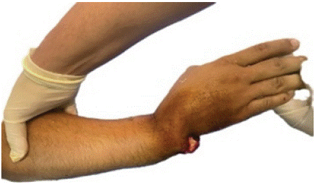
Figure 1: Initial clinical asp
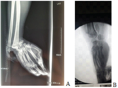
Figure 2: Initial X-ray. AP view (A) and lateral view (B).
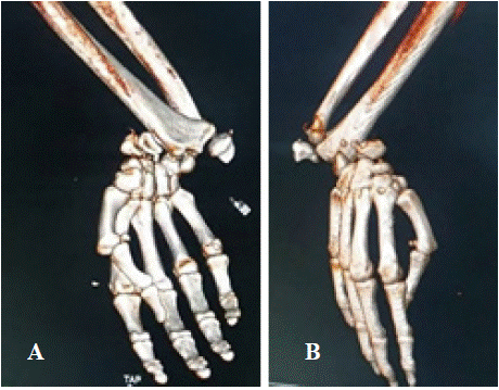
Figure 3: 3D computed tomography scan. AP view (A) and lateral view (B).
The patient was stabilized, and emergency surgery was performed within seven hours of injury. Following extensive lavage and debridement, a dorsal approach to the wrist was performed. The scaphoid was fixed using a headless compression screw, and the lunate was reduced anatomically under fluoroscopic control. Carpal alignment was maintained with four K-wires: two for triquetro-lunate fixation, one for radio-luno-capitate arthrodesis, and one to fix the radial styloid (Figure 4). The wound was closed primarily. Postoperative immobilization was maintained for eight weeks. Passive rehabilitation of the fingers was initiated three weeks post-operatively. Six weeks later, the cast and K-wires were removed, followed by supervised active rehabilitation with three sessions per week to restore joint mobility. The range of motion of our patient was 60° (extension 10°, flexion 50°) at 6 months (Figure 5) and 90° (extension 75°, flexion 15°) at 9 months. At 12 months, the patient presented with ulnar translation of the carpus (Figure 6) but reported no pain.
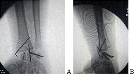
Figure 4: Immediate postoperative X-ray. AP view (A) and lateral view (B).
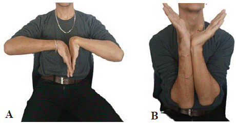
Figure 5: clinical results after six months.
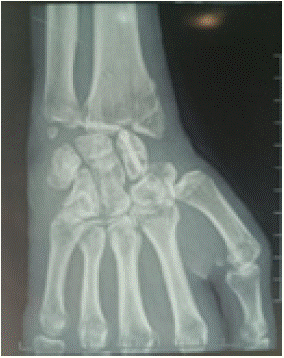
Figure 6: Radiographic result after 12 months.
Discussion
Trans-scaphoid trans-styloid dorsal dislocation with enucleation of the lunate and avulsion of the proximal pole of the scaphoid was first described by Green and O'Brien in 1978 [2]. These severe carpal injuries require thorough diagnostic evaluation, as complementary examinations are critical for accurate classification and treatment planning. Intraoperative X-rays are often recommended to identify associated ligament injuries, which are common in such complex cases [1].
Open dislocations with exposed bone represent particularly severe carpal injuries, posing significant management challenges due to the high risk of contamination. Tillson [3] emphasized that protruding bone should not be reduced into the wound, as this can introduce further contamination. Early and aggressive debridement of necrotic tissue is essential to minimize infection risk and optimize healing [4].
Treatment strategies for perilunate injuries vary, with both closed and open reduction techniques proposed. However, closed reduction carries a higher risk of secondary dislocation and nonunion compared to open techniques. Stabilization of the reduced joint—whether through percutaneous pinning, open pinning, or external fixation—is a critical component of care [1]. Additionally, extrinsic ligament repair is considered the cornerstone of treatment, as it ensures long-term radioulnocarpal stability [5,6].
The surgical approach remains a topic of debate. While anatomical repair is universally emphasized [7], many authors advocate for a combined dorsal and volar approach to achieve comprehensive ligamentous repair [8,9]. In our case, a single dorsal approach was utilized, which limited direct repair of volar ligamentous structures. Nevertheless, the procedure allowed for the release of tendon interpositions and removal o A B f bone or cartilage fragments [1].
Post-operative management typically involves immobilization in a long-arm cast for 3 to 10 weeks to facilitate adequate ligamentous healing [5]. In cases involving distal radioulnar joint injury, elbow immobilization is also necessary. In our case, immobilization was maintained for six weeks, consistent with standard protocols.
Functional outcomes vary significantly between closed and open injuries. Girard et al. and Le Nen et al. reported favorable results in closed fracture-dislocations, with flexion-extension arcs exceeding 85 degrees. In contrast, Nyquist and Stern documented more limited outcomes in open injuries, with an average range of motion of 57 degrees. In our case, despite the severity of the initial injury, the patient achieved a range of motion of 95 degrees at 5 months and was able to return to work within 6 months. In the literature, we found no studies describing long-term radiological and functional results in cases of open dislocation with enucleation of the lunate.
Conclusion
This case demonstrates that favourable outcomes can be achieved in complex open trans-scapholunate dislocation with lunate enucleation through a systematic approach. Key elements for success include prompt surgical intervention to prevent infection, open anatomical reduction with temporary fixation, meticulous repair of the capsuloligamentous complex, appropriate postoperative immobilization, and a structured rehabilitation protocol, as supported by current literature. This management strategy aligns with established recommendations and can lead to satisfactory functional results despite the severity of the initial injury.
References
- Jardin E, Pechin C, Rey PB, Gasse N & Obert L. Open volar radiocarpal dislocation with extensive dorsal ligament and extensor tendon damage: A case report and review of literature. Hand Surgery & Rehabilitation. 2016; 35: 127–134.
- Green DP & O’Brien ET. Open reduction of carpal dislocations: indications and operative techniques. The Journal of Hand Surgery. 1978; 3: 250–265.
- Tillson DM. Open fracture management. Vet Clin North Am Small Anim Pract. 1995; 25: 1093-1110.
- Sagi HC & Patzakis MJ. Evolution in the Acute Management of Open Fracture Treatment? Part 1. Journal of Orthopaedic Trauma. 2021; 35: 449–456.
- Herzberg G, Comtet JJ, Linscheid RL, Amadio PC, Cooney WP & Stalder J. Perilunate dislocations and fracture-dislocations: a multicenter study. The Journal of Hand Surgery. 1993; 18: 768–779.
- Kremer T, Wendt M, Riedel K, Sauerbier M, Germann G & Bickert B. Open reduction for perilunate injuries--clinical outcome and patient satisfaction. The Journal of Hand Surgery. 2010; 35: 1599–1606.
- Bilos ZJ, Pankovich AM, Yelda S. Fracture-dislocation of the radiocarpal joint. The Journal of Bone & Joint Surgery. 1977; 59: 198-203.
- Melone CP Jr, Murphy MS, Raskin KB. Perilunate injuries. Repair by dual dorsal and volar approaches. Hand Clin. 2000; 16: 439-448.
- Souer JS, Rutgers M, Andermahr J, Jupiter JB, Ring D. Perilunate fracturedislocations of the wrist: comparison of temporary screw versus K-wire fixation. J Hand Surg Am. 2007; 32: 318-325.