
Research Article
J Ophthalmol & Vis Sci. 2023; 8(3): 1085.
Pressure Results of Primary Open-Angle Glaucoma Surgery by Trabeculectomy in Kankan, Guinea
Sovogui Md1,2*; Zoumanigui2; Doukoure MB2; Camara F3; Vonor K4
¹Faculty of Health Sciences and Techniques, University of Conakry, Republic of Guinea
²Bartimee Ophthalmological Clinic, Republic of Guinea
³Kankan Regional Hospital, Republic of Guinea
4University of Kara in Togo, Republic of Guinea
*Corresponding author: Maxime Dantouma SOVOGUI Assistant at the Faculty of Health Sciences and Techniques, Gamal Abdel Nasser University. BP: 2525 Conakry, Republic of Guinea. Tel: +224 628 17 93 23 Email: maximesovo79@gmail.com
Received: July 05, 2023 Accepted: August 08, 2023 Published: August 15, 2023
Summary
Purpose: To assess the pressureal outcomes of primary open-angle trabeculectomy surgery for primary open-angle glaucoma.
Materials and Methods: This was a six-month prospective observational study. Patients operated by the trabeculectomy technique and followed during the study period were included. Not included were those who had combined surgery. Recruitment was comprehensive. Our variables were epidemiological, clinical, paraclinical and therapeutic. Epi info version 7.4.0 was used for data analysis.
Conclusion: Glaucoma remains a public health problem in Kankan. Trabeculectomy concluded an overall success in lowering postoperative intraocular pressure with almost no complications. Given these results, it would be better to undertake it in 1st intention.
Keywords: Glaucoma; Trabeculectomy; Guinea
Introduction
Glaucoma is a group of progressive and degenerative optic neuropathies with irreversible loss of visual field (CV) and can lead to blindness [1]. Intraocular hypertension is the main risk factor. Diagnosis and monitoring are based on a joint analysis of structural involvement by clinical examination of the papilla supplemented by Optical Coherence Tomography (OCT) and functional impairment, by the realization of CV. Treatment consists of reducing Intraocular Pressure (IOP), thus slowing the progression of the disease, through medical, physical (laser) treatments and surgical [2]. Surgery is the most effective way to lower IOP. It is usually offered when the maximum tolerated medical treatment is insufficient to achieve a target IOP. Many incisional techniques of variable mode of action facilitate the evacuation of aqueous humor out of the anterior chamber. Some make it possible to remove the trabecular obstacle in whole (trabeculectomy) or in part (deep non-perforating sclerectomy) and others (micro invasive) try to restore the physiological flow of aqueous humor by trabecular and / or uveoscleral route. Trabeculectomy is a reference technique but depends on a filtration bubble and its complications requiring very rigorous and prolonged monitoring for life. The failure of this technique is almost always due to the healing of the conjunctiva and Tenon capsule [3]. In Ireland in 2020, in a study of trabeculectomy with the use of Mitomycin C, IOP was 10.8±4.8 mm Hg representing a 47% reduction in IOP from baseline [4]. In the Democratic Republic of Congo in 2017, after trabeculectomy associated with anti-VGEF in the management of neovascular glaucoma, the mean IOP of 38 mm Hg preoperatively, increased to 15 mm Hg postoperatively [5]. In Togo in 2017, Maneh et al [6] reported in a study of trabeculectomy outcomes in childhood glaucoma, that the mean preoperative IOP was 20.08 mm Hg increasing to 12.86 mm Hg, 6 months later with 62.86% success. In the Republic of Guinea, Sovogui MD et al. [7] in their study of Non-Perforating Deep Sclerectomy (NPPS), report that the mean preoperative IOP was 19±5.73 mm hg versus 11.6±1.86 mm hg on postoperative Day 30. However, it seemed interesting to us to evaluate the functional results of Trabeculectomy at Kankan Regional Hospital.
Materials and Methods
This was a six-month prospective observational study from 01 March 2022 to 31 August 2022. It took place in the ophthalmology department of Kankan Regional Hospital and the Nabaya Yelen application center in Kankan. Due to their administrative positions and equipment, they are the largest ophthalmology departments in the Kankan administrative region. Patients operated by the trabeculectomy technique and followed during the study period were included in this study. Not included were those who received combined surgery (cataract-glaucoma). The recruitment was exhaustive according to our selection criteria. Our variables were epidemiological, clinical, paraclinical and therapeutic. Epi info version 7.4.0 was used for data analysis; Word and Excel software from the Office 2016 pack were used for text and table entry; Zotero version 5.0.96.2 was used for bibliographic references. The confidentiality and anonymity of interviewees were respected in accordance with the principles of ethics and medical deontology.
Results
In all, 2401 patients including 47 patients were received in whom 79 eyes were operated for glaucoma by the technique of Trabeculectomy, a frequency of 1.95%. The figure below shows the considerable normalization of IOP postoperatively, compared to preoperative IOP. Table I shows that poor compliance with treatment was the most frequent operative indication.Table II shows that the 41-60 age group is more represented with a male predominance. Average age 41.88±24.98 extreme years 1 month and 76 years; Sex ratio 1.61.Table III on clinical variables shows that decreased visual acuity was the most common reason, high blood pressure (hypertension) was the most dominant antecedent. Both eyes were more affected at the same time. In Table IV, visual acuity by far without carried correction (AVLSC) =3/10 was the most dominant. According to Table V shows that, the optical fiber thickness at OCT-RNFL was < 88μm plus severe optical disc damage in the majority of cases. On the CV, the Mean Defect was > -12 dB in almost half of the cases. The only complication found postoperatively in this study was hyphema, encountered in only one patient.
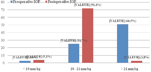
Figure : Comparison of preoperative and postoperative IOP.
![]()
    Directions

    Effective

    Percentage


    Poor therapeutic    compliance

    15

    31,9


    Financial    difficulty

    13

    27,7


    Worsening of the visual field

    10

    21,3


    1st line surgery

    8

    17,0


    Therapeutic    failure

    1

    2,1


    Total

    47

    100
Table 1: Operative indications.
![]()
    Variable

    Effective

    Percentage


    Age in year


    0–20 ans

    8

    17,0


    21–40

    7

    14,9


    41–60 ans

    17

    36,2


    =60

    15

    31,9


    Sex


    Male

    29

    61,7


    Female

    18

    38,3
Table 2: Socio-demographic variables.
![]()
    Variable

    Effective

    Percentage


    Reasons for consultation (47)


    Decreased    visual acuity

    36

    76,6


    Eye pain

    32

    68,1


    Headache

    27

    57,5


    Tearing

    24

    51,1


    Visual blur

    17

    36,2


    Photophobia

    13

    27,7


    Megalocornea

    5

    10,6


    Buphthalmos

    3

    6,4


    Corneal    opacity

    2

    4,3


    Histry ( N = 47)


    Ophthalmological myopia

    2

    4,3


    Medical


    High blood    pressure

    23

    48,9


    Diabetes

    17

    36,2


    Family    history of glaucoma

    4

    8,5


    No history

    16

    34,0


    Laterality (N= 79)


    Right/left    eye

    64

    81,0


    Left eye

    9

    11,4


    Right eye

    6

    7,6
Table III: Clinical variables.
![]()
    Visual acuity

    Right eye

    Left eye


    Effective

    Percentage

    Effective

    Percentage


    <1/10

    10

    21,3

    12

    25,5


    1/10-2/10

    9

    19,1

    10

    21,3


    =3/10

    23

    48,9

    21

    44,7


    Visual    acuity not done

    5

    10,7

    4

    8,5


    Total

    47

    100

    47

    100
Table IV: Visual acuity from afar without preoperative correction (N=47).
Discussions
This study allowed us to demonstrate the effectiveness of Trabeculectomy by a considerable standardization of postoperative IOP in this series of studies, compared to preoperative IOP. It indicates that poor compliance with treatment as well as financial difficulties related to the purchase of anti-glaucomatous drugs were the main surgical indications. It has the advantages of being prospective and having a single surgeon with mastery of the technique used, who has operated on all patients. However, the main limitation was the non-performance of paraclinical examinations by some patients, due to lack of financial means and lack of support from a mutual health insurance company. The success of this technique is confirmed by Kabesha TB et al [5], who report that the average IOP increased from 38 mm Hg preoperatively to 15 mm Hg postoperatively. The frequency of glaucomatous patients who have benefited from the Trabeculectomy technique in the Ophthalmology Department and the Nabaya Yelen Application Center in Kankan, is 1.95% with a male predominance and an average age of 41.88±24.98 years. Sovogui MD et al [7] in their study at the Bartimaeus Ophthalmological Clinic in Conakry, Guinea report similar results, an average age of 45.20±14 years with a sex ratio of 1.21. Table III on clinical variables shows that decreased visual acuity was the most common reason, hypertension was the most dominant antecedent followed by diabetes, and both eyes were more affected at the same time. Kabesha TB et al. [5] report identical results from history, as well as Sovogui MD et al [7] in relation to the reasons for consultations. According to the Swiss Society of Ophthalmology, the first sign of glaucoma is the loss of peripheral vision and CV damage which is noticed only at an advanced stage of the disease when central vision is affected [8]. This explains the results in relation to visual acuity from afar without correction of the patients in this series. As for Table V, the thickness of optical fibers at OCT-RNFL was <88 μm mostly associated with severe damage to the optical disc. On the CV, the Mean Defect was >-12 dB in almost half of the cases. These results are explained by the insidious nature of the disease, manifested by a progressive loss of retinal ganglion cells, morphological changes in the head of the optic nerve and typical visual field involvement [9].
![]()
    Variable

    Effective

    Percentage


    Fiber thickness at OCT-RNFL (N = 94)


    <88μm

    42

    44,7


    88-153μm

    21

    22,3


    Non    réaliser

    31

    33,0


    OCT-DDLS (N = 94)


    01–04 (Normal)

    3

    3,2


    05–06 (Moderate impairment)

    19

    20,2


    07–10 (severe impairment)

    41

    43,6


    Not realized

    31

    33,0


    Mean Defect (N=94)


    MD < -6 dB

    10

    10,6


    MD < -12 dB

    22

    23,4


    > -12 dB

    31

    33,0


    Not realized

    31

    33,0
Table V: Paraclinical variables.
Conclusion
Glaucoma remains a public health problem in Kankan. Trabeculectomy concluded an overall success in lowering intraocular pressure after a one-month setback with almost no complications. Given the effectiveness of this technique, it would be better to undertake it in first intention among Kankan populations.
Author Statements
Conflicts of Interest
The authors do not declare any conflicts of interest in relation to this work.
Author’s Contributions
Authors contributed to one or more levels of manuscript writing since protocol, data collection, and writing. All have read and approved the manuscript.
References
- Institut national d’excellence en santé et en services sociaux (INESSS). The XEN45 gel implant for micro-invasive glaucoma surgery (MIMC). Review by Luc Cloutier, Rania Saidi, François Désy. Québec, Qc: INESSS. 2020; 46.
- Bertaud S, Aragno V, Baudouin C, Labbé A. Primary open-angle glaucoma. Rev Med Interne. 2019; 40: 445-52.
- Hamard P. Surgical treatment of open-angle glaucoma. JFO. 2016; 13: 44437-5.
- Sharpe R, Pham G, Chang P. Comparison of ab interno XEN gelatin stent vs trabeculectomy with Mitomycin C: A retrospective study. J Curr Glaucoma Pract. 2020; 14: 87-92.
- Kabesha TB, Kabesha D, Maloba V, Mwamba C, Chenge B, Mukalay A. Results of trabeculectomy associated with anti-VGEF in the treatment of neovascular glaucoma complicating retinal vein occlusions (about 21 cases, followed at the eye clinic of Bukavu from 1 January to 31 December 2015). J Fr Ophtalmol. 2017; 40: 17-21.
- Maneh N, Vonor K, Diatewa BM, Batomaguela NSK, Amedomé KM, Nagbe YE, et al. Results of trabeculectomy in infantile glaucoma at the CHU campus of Lomé (Togo). Eur Sci J ESJ. 2017; 13: 119.
- Sovogui MD, Baldé R, Zoumanigui C, Grovogui MM, Vonor K, Mermoud. Surgery for chronic glaucoma by the technique of non-perforating deep sclerectomy at the Bartimée ophthalmological clinic: about 50 cases. Acta Sci Sci. 2022; 6: 27-31.
- Swiss Society of Ophthalmology. Glaucoma [cited Jun 11, 2023]. Available from: https://www.sog-sso.ch/fr/pour-patientes/glaucome.html.
- Classification and terminology. In: European glaucoma society terminology and guidelines for glaucoma. 4th ed. Genova: PubliComm. 2014; 79.