
Case Report
J Ophthalmol & Vis Sci. 2023; 8(2): 1081.
Cessation of Recurrent Bilateral Optic Disc Haemorrhages after Successful Trabeculectomy: A Case Report
Ieva Laure; Franz Grehn; Christina Korb; Anna Beck; Alicja Strzalkowska; Bert C Giers; Katrin Lorenz
Department of Ophthalmology, University Medical Center of the Johannes Gutenberg-University Mainz, Germany
*Corresponding author: Katrin LorenzDepartment of Ophthalmology, University Medical Center of the Johannes Gutenberg-University Mainz, Langenbeckstr. 1, 55131 Mainz, Germany. Tel: +49 6131 174069, Fax: +496131 175509 Email: katrin.lorenz@unimedizin-mainz.de
Received: February 17, 2023 Accepted: March 24, 2023 Published: March 31, 2023
Abstract
Purpose: To describe a patient with primary open-angle glaucoma in whom bilateral recurrent optic disc haemorrhages disappeared permanently after trabeculectomy.
Methods: A 67-old male patient was diagnosed with primary open-angle glaucoma and was followed for 10 years including best corrected visual acuity, visual field examinations, slit lamp examinations, Goldmann applanation tonometry, fundoscopy and optic disc photographs.
Results: Optic disc haemorrhages were detected in 8 out of 30 preoperative visits in the right eye and in 3 out of 19 visits in the left eye. Trabeculectomy with intraoperative application of mitomycin C was performed in both eyes due to the advanced visual field loss with an inadequate intraocular pressure control despite maximum-tolerated medical therapy and because of the recurrent optic disc haemorrhages. No further optic disc haemorrhages were observed in either eye after trabeculectomy.
Conclusion: Our case report demonstrates that recurrent bilateral optic disc haemorrhages can cease permanently after successful trabeculectomy.
Keywords: Primary open-angle glaucoma; Optic disc haemorrhage; Intraocular pressure; Progression.
Introduction
Glaucoma is a leading cause of blindness and represents a group of ocular disorders characterized by progressive loss of retinal ganglion cells and their axons. This optic nerve degeneration results in gradual loss of visual field, which may ultimately lead to blindness [1-3]. The pathogenesis of glaucoma is only partially understood. Elevated Intraocular Pressure (IOP) is the most relevant risk factor for disease development or progression and presently the only therapeutically addressable factor [4-8]. Previous studies have shown that optic disc haemorrhages can also be an important risk factor for the progression of glaucoma [9-11]. Nevertheless, the explanation of this observation is still unclear. In addition, the theories of the pathogenesis of optic disc haemorrhages vary considerably. A new approach supports the hypothesis that optic disc haemorrhages are induced by arterial sources and are connected with vascular dysregulation [12]. The latest optical coherence tomography studies found a significant association between the occurrence of optic disc haemorrhages and the focal defects of the lamina cribrosa [13,14]. Lee et al. [15] have suggested a new mechanism, where the reactive gliosis in glaucoma can form fibrous glial scars that can disrupt the vascular wall and lead to the formation of optic disc haemorrhages. To the best of our knowledge, there is only one study mentioned in the literature so far, that evaluates the effect of surgical reduction of intraocular pressure on the recurrence of optic disc haemorrhages [16]. Our case report demonstrates that recurrent bilateral optic disc haemorrhages can cease after successful bilateral trabeculectomy.
Case Presentation
In 2013, a 67 years old white male was diagnosed with Primary Open-Angle Glaucoma (POAG) in our department during a routine examination. The patient was followed-up for 10 years until 2022. A total of 52 ophthalmologic examinations were performed in this patient. There were 19 follow-up examinations prior to trabeculectomy in the left eye and 30 prior to the trabeculectomy in the right eye.
In the beginning his decimal visual acuity was 1.0 in the right eye and 0.8 in the left eye. Visual field examinations were performed with the Humphrey Field Analyzer using the SITA (Swedish Interactive Threshold Algorithm) standard central 24-2 threshold test. The MD values varied during the entire follow-up period (right eye: -11.42dB to -12.97dB in 2013 to -13.52dB in 2022; left eye: -12.73dB to -14.45dB in 2013 to -24.28dB in 2022) and revealed typical advanced glaucomatous visual field defects (Figure 1).
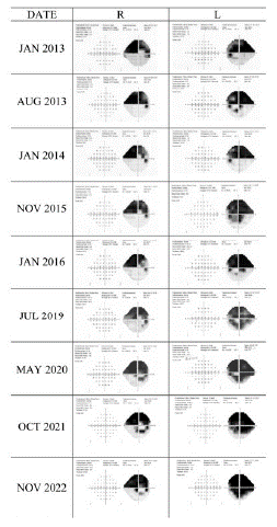
Figure 1: Humphrey visual field examinations.
Visual acuity, MD values of the visual field, as well as Intraocular Pressure (IOP) values and medication are summarized in (Table 1). At the time of diagnosis, vertical cup to disc ratio was 0.7 in the right eye and 0.8 in the left eye. Corneal thickness was 515μm in the right eye and 510μm in the left eye. The maximum IOP was 23mmHg in the right eye and 22mmHg in the left eye, respectively, including diurnal 24-hours IOP profiles. The patient had a family history of glaucoma and was treated for hyperlipoproteinaemia.
![]()
    Date

    VA

    VF(MD)

    IOP(mmHg)

    Medication


    Right

    Left

    Right

    Left

    Right

    Left


    01.2013

    1.0

    0.8

    -12.97

    -12.73

    14

    14

    Both: PGA


    08.2013

    1.2

    1.2

    -11.42

    -14.45

    13

    14

    Both: PGA


    01.2014

    1.0

    1.0

    -10.14

    -11.77

    18

    18

    Both: PGA; Left: BB


    08.2014

    1.0

    1.2

    -8.97

    -12.37

    14

    13

    Both: PGA, BB


    11.2015

    1.2

    1.2

    -10.95

    -16.97

    17

    18

    Both: PGA, BB


    03.2016

    1.0

    1.0

    -10.41

    -18.04

    20

    21

    Both: PGA, BB,CAIs


    10.2016

    1.2

    1.2

    -10.93

    -18.32

    18.1

    14.9

    Both: PGA, BB,CAIs


    12.2016

    Lefteye: Trabeculectomy


    03.2017

    1.2

    1.2

    -10.63

    -18.70

    14

    7

    Right:PGA, BB,CAIs


    11.2017

    Righteye: Trabeculectomy


    01.2018

    0.8

    0.6

    -11.11

    -19.86

    8

    9


    04.2018

    1.0

    1.0

    -12.72

    -21.50

    8

    9


    08.2018

    1.0

    0.8

    -13.61

    -20.46

    8

    8.3


    07.2019

    1.0

    0.8

    -10.97

    -16.66

    7

    13


    05.2020

    0.8

    0.5

    -15.50

    -23.19

    12.6

    18.4

    Left: PGA, BB


    06.2020

    Lefteye: Needling


    07.2020

    1.0

    0.4

    -15.60

    -22.66

    14

    12

    Left: BB


    10.2021

    0.4

    0.32

    -11.71

    -22.20

    22

    22

    Both: BB


    11.2021

    Left eye: Cataract surgery and trabecular aspiration


    12.2022

    Right eye: Cataract surgery and trabecular aspiration


    11.2022

    1.2

    0.5

    -13,52

    -24,28

    13

    13

    Both: BB


    Abbreviation: PGA: Prostaglandin; BB: Beta Blocker; CAIs: Carbonic Anhydrase    Inhibitor; VA: Visual Acuity; VF: Visual Field
Table 1: Visual acuity, MD Values, IOP, Medication.
A sleep apnoea was diagnosed in 2012 and was treated with Continuous Positive Airway Pressure (CPAP) therapy until December 2019. For the first three years, the target IOP of ≤15mmHg was achieved with topical antiglaucomatous eye drops (prostaglandins, later combination therapy with 4 antiglaucoma agents). By 2016, the target IOP was no longer achieved and IOP rose to 20mmHg in the right eye and 27mmHg in the left eye. Due to the advanced visual field loss and recurrent optic disc haemorrhages within adequate IOP control despite maximum-tolerated medical therapy a glaucoma operation was scheduled.
Standardized trabeculectomy with intraoperative application of mitomycin C using a defined volume (100μl), concentration (0.2mg/ml), and duration of application (5 minutes) was performed by 2 experienced surgeons in December 2016 (left eye) and in November 2017 (right eye). The right eye was treated postoperatively with 3 subconjunctival injections of 5-fluorouracil.
Following the suggestion of the European Glaucoma Society guidelines (2020) [17], optic disc haemorrhages were followed-up with ophthalmoscopy and colour photography of the optic disc. An optic disc haemorrhage was defined as an isolated haemorrhage seen on the optic disc tissue or in the peripapillary retina extending to the disc rim [16]. Several optic disc haemorrhages were detected in both eyes during the follow-up period (Figure 2).
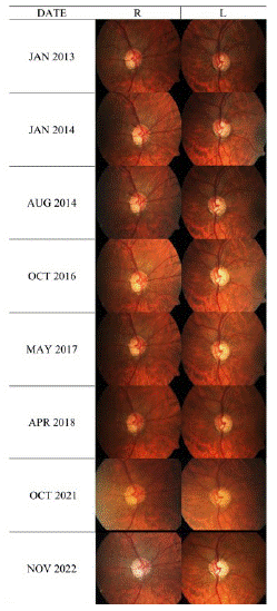
Figure 2: Photo documentation of the optic disc.
Preoperatively, in the left eye optic disc haemorrhages were documented in 3 out of 19 visits. These optic disc haemorrhages were observed twice in the upper temporal position, and multiple small optic disc haemorrhages were present during the third visit. Finally, trabeculectomy was performed in December 2016. On the right eye, the patient was followed-up 30 times, and optic disc haemorrhages were detected at 8 visits. In 6 out of those 8 visits, optic disc haemorrhages were seen in the upper temporal sector. In the remaining 2 visits, once multiple optic disc haemorrhages and once a single optic disc haemorrhage without documented location were observed. In this eye, trabeculectomy was performed in November 2017. No further opticdisc haemorrhages were observed to the present day in either eye after the trabeculectomies (Figure 3).
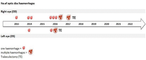
Figure 3: Presence of disc haemorrhages during the follow-up period of 10 years before and after trabeculectomy.
In the left eye, postoperative IOP ranged between 7-13mmHg, without antiglaucomatous eye drops until 2019. In May 2020 the IOP rose to 16-18mmHg. MD values deteriorated from -12.73 to -14.45 in 2013 to -23.19dB in 2020. Prostaglandin analogues and beta-blockers were started and a needling was performed in June 2020.
In the right eye, postoperative IOP ranged between 7 and 14mmHg without any topical therapy until 2020. In October 2021, however, IOP was 22mmHg in both eyes. Beta-blockers were started in the right eye. Because of reduction of best corrected visual acuity and significant cataract formation, visual field changes were attributed to lens changes and cataract surgery in combination with trabecular aspiration was performed in the left eye in November 2021 and in the right eye in December 2021. The last follow-up was in November 2022.
Discussion
In this case report we demonstrate that IOP reduction after trabeculectomy had favourable effects on the incidence of optic disc haemorrhages in glaucoma. Cessation of the optic disc haemorrhages was observed in both eyes of the patient after trabeculectomy and continued until present.
Various studies have reported the prevalence of optic disc haemorrhages to range between 2% and 37% in POAG patients [9,16,18-28], and between 11% and 42% in Normal Tension Glaucoma (NTG) patients [23,25,29-31] and between 0.4% and 10% in patients with ocular hypertension [9,18,21,24-28].
Heijl followed two patients with previously known optic disc haemorrhage closely for one year and detected 9 haemorrhages in 3 of the 4 eyes studied. Haemorrhages were not associated with any worsening of the visual field. No structural changes of the optic nerve head were seen after the bleedings during the observation time of one year [32]. In the Early Manifest Glaucoma Trial (EMGT) [7], evaluated the effect of non-surgical IOP-lowering treatment on the development of disc haemorrhages in patients with glaucoma and found no difference in frequency of disc haemorrhages between the treated and the placebo group. The patients were followed every 3 months for a period of 8 years [33]. In this study, patients with newly detected glaucoma were randomized to Argon Laser Trabeculoplasty (ALT) plus betaxolol or no initial treatment. A mean IOP reduction of 25% was achieved in treated patients. Although this study has a high strength due to its randomised design with an untreated arm and a long follow-up, it should be discussed whether the conservative treatment of the EMGT protocol is comparable to the IOP level achieved after successful trabeculectomy. The rather low IOP after trabeculectomy in both eyes of our case and the very flat IOP curve might reduce the stress to the lamina beams more than a treatment combination of betaxolol plus ALT, with still some IOP fluctuation at a somewhat higher IOP level. Our case, indeed, can be questioned as it is a single observation. However, the follow-up of 9 years with a long preoperative observation period and the obvious cessation of ODH closely related to surgery (even in accordance with the time-shift between the two operations) may suggest a more favourable effect of trabeculectomy than conservative treatment.
We found only one study in the literature that compared the incidence of optic disc haemorrhages before and after IOP reduction by trabeculectomy. Miyake et al. [16] showed that the incidence of optic disc haemorrhages decreased after trabeculectomy in eyes of both POAG patients and NTG patients. The comparison between the high and low postoperative IOP groups revealed that the incidence of optic disc haemorrhages in POAG patients were lower for the low postoperative IOP group, but that was not found for NTG patients. The fact that our patient did not show any optic disc haemorrhages after trabeculectomy supports the results published by Miyake et al. in 2006 [16]. To our knowledge, since 2006 no other studies have been performed that compared the incidence of optic disc haemorrhages before and after trabeculectomy.
Previous studies have shown that optic disc haemorrhages can also be an important risk factor for the progression of glaucoma [9-11]. The Early Manifest Glaucoma Trial assessed factors for progression, including the effect of treatment, and frequent optic disc haemorrhages were found to be an independent factor for progression [34]. The patient described in our case report showed advanced visual field loss at the beginning of the observation period, despite maximal tolerated antiglaucomatous topical medication. The fluctuation of IOP and the absolute IOP decreased and no more optic disc haemorrhages were observed after trabeculectomy.
It remains an interesting question to determine why the optic disc haemorrhages ceased to occur after trabeculectomy. Kim et al. [35] performed a study using optical coherence tomography angiography before and 3 months after trabeculectomy and showed a significant increase of vessel density at the level of lamina cibrosa. Seol et al. [36] found an association between recurrent optic disc haemorrhages and lower percent reduction of IOP. The association of the systemic factors were also evaluated in their study. The factors included cold extremities, a (prone or lateral decubitus) sleeping position, sleep disorder and revealed an association by using univariate regression analysis. In our case, sleep apnea was present, but despite CPAP therapy, optic disc haemorrhages continued to occur until trabeculectomy was performed. The CPAP therapy was discontinued in 2019, but the optic disc haemorrhages did not recur. This suggests that optic disc haemorrhages were not related to sleep apnea but rather correlated to IOP. The correlation with an elevated IOP is supported by the increased number of optic disc haemorrhages when elevation of IOP above target occurred in 2016. For our patient optic disc haemorrhages were found mostly in the superotemporal region of the optic nerve head which correlated to the better half of his visual fields. Hsia et al. [37] showed that optic disc haemorrhages most commonly occur inferotemporal (32,9%), followed by the superotemporal location (10,5%). The results of our study also showed that following optic disc haemorrhages, the corresponding regions showed progression of visual field deterioration.
Conflicting theories regarding the aetiology still exist: The mechanical theory focuses on the mechanical compression or disruption of the lamina cribrosa due to IOP, the circulatory disorder theory assumes a circulatory interference of the lamina cribrosa [16]. The disappearance of bilateral optic disc haemorrhages after successful trabeculectomy in our case report could support both theories: less mechanical stress with less fluctuation by lowering of IOP and/or improvement of blood circulation around the optic nerve by better perfusion. The lack of any effect of CPAP treatment on the incidence of optic disc haemorrhages, however, would support the mechanical theory more than circulatory theory. This case report adds information on whether optic disc haemorrhages progress independent of IOP lowering and whether optic disc haemorrhages patients should undergo stricter IOP lowering, best by trabeculectomy which provides the lowest IOP levels.
Conclusion
Our case report demonstrates that recurrent bilateral optic disc haemorrhages can cease after successful bilateral trabeculectomy in primary open-angle glaucoma, further supporting the role of filtration surgery in preventing glaucoma progression.
References
- Quigley HA. Open-angle glaucoma. N Engl J Med. 1993; 328: 1097-106.
- Quigley HA, Tielsch JM, Katz J, Sommer A. Rate of progression in open-angle glaucoma estimated from cross-sectional prevalence of visual field damage. Am J Ophthalmol. 1996; 122: 355-63.
- Cedrone C, Nucci C, Scuderi G, Ricci F, Cerulli A, et al. Prevalence of blindness and low vision in an Italian population: a comparison with other European studies. Eye (Lond). 2006; 20: 661-7.
- Mao LK, Stewart WC, Shields MB. Correlation between intraocular pressure control and progressive glaucomatous damage in primary open-angle glaucoma. Am J Ophthalmol. 1991; 111: 51-5.
- Gherghel D, Orgül S, Gugleta K, Gekkieva M, Flammer J. Relationship between ocular perfusion pressure and retrobulbar blood flow in patients with glaucoma with progressive damage. Am J Ophthalmol. 2000; 130: 597-605.
- Lichter PR, Musch DC, Gillespie BW, Guire KE, Janz NK, et al. Interim clinical outcomes in the Collaborative Initial Glaucoma Treatment Study comparing initial treatment randomized to medications or surgery. Ophthalmology. 2001; 108: 1943-53.
- Heijl A, Leske MC, Bengtsson B, Hyman L, Bengtsson B, et al. Reduction of intraocular pressure and glaucoma progression: results from the Early Manifest Glaucoma Trial. Arch Ophthalmol. 2002; 120: 1268-79.
- Kass MA, Heuer DK, Higginbotham EJ, Johnson CA, Keltner JL, et al. The Ocular Hypertension Treatment Study: a randomized trial determines that topical ocular hypotensive medication delays or prevents the onset of primary open-angle glaucoma. Arch Ophthalmol. 2002; 120: 701-13.
- Siegner SW, Netland PA. Optic Disc Hemorrhages and Progression of Glaucoma. Ophthalmology. 1996; 103: 1014-24.
- Kim SH, Park KH. The relationship between recurrent optic disc hemorrhage and glaucoma progression. Ophthalmology. 2006; 113: 598-602.
- Leske MC, Heijl A, Hyman L, Bengtsson B, Dong LM, et al. Predictors of long-term progression in the early manifest glaucoma trial. Ophthalmology. 2007; 114: 1965-72.
- Chou JC, Cousins CC, Miller JB, Song BJ, Shen LQ, et al. Fundus Densitometry Findings Suggest Optic Disc Hemorrhages in Primary Open-Angle Glaucoma Have an Arterial Origin. Am J Ophthalmol. 2018; 187: 108-16.
- Kim YK, Park KH. Lamina cribrosa defects in eyes with glaucomatous disc haemorrhage. Acta Ophthalmol. 2016; 94: e468-73.
- Mistry V, An D, Barry CJ, House PH, Morgan WH. Association between focal lamina cribrosa defects and optic disc haemorrhage in glaucoma. Br J Ophthalmol. 2020; 104: 98-103.
- Lee EJ, Han JC, Kee C. A novel hypothesis for the pathogenesis of glaucomatous disc hemorrhage. Prog Retin Eye Res. 2017; 60: 20-43.
- Miyake T, Sawada A, Yamamoto T, Miyake K, Sugiyama K, et al. Incidence of disc hemorrhages in open-angle glaucoma before and after trabeculectomy. J Glaucoma. 2006; 15: 164-71.
- European Glaucoma Society Terminology and Guidelines for Glaucoma, 4th Edition - Chapter 3: Treatment principles and options Supported by the EGS Foundation: Part 1: Foreword; Introduction; Glossary; Chapter 3 Treatment principles and options. Br J Ophthalmol. 2017; 101: 130-95.
- Susanna R, Drance SM, Douglas GR. Disc hemorrhages in patients with elevated intraocular pressure. Occurrence with and without field changes. Arch Ophthalmol. 1979; 97: 284-5.
- Bengtsson B. Findings associated with glaucomatous visual field defects. Acta Ophthalmol (Copenh). 1980; 58: 20-32.
- Bengtsson B, Holmin C, Krakau CE. Disc haemorrhage and glaucoma. Acta Ophthalmol (Copenh). 1981; 59: 1-14.
- Gloster J. Incidence of optic disc haemorrhages in chronic simple glaucoma and ocular hypertension. Br J Ophthalmol. 1981; 65: 452-6.
- Holmin C. Signs of activity and progression in chronic glaucoma. Acta Ophthalmol Suppl. 1982; 153: 1-40.
- Shihab ZM, Lee PF, Hay P. The significance of disc hemorrhage in open-angle glaucoma. Ophthalmology. 1982; 89: 211-3.
- Drance SM, editor Hemorrhage on the Disc – A Risk Factor in Glaucoma. Glaucoma Update II; 1983; Berlin, Heidelberg: Springer Berlin Heidelberg.
- Kitazawa Y, Shirato S, Yamamoto T. Optic disc hemorrhage in low-tension glaucoma. Ophthalmology. 1986; 93: 853-7.
- Drance SM. Disc hemorrhages in the glaucomas. Surv Ophthalmol. 1989; 33: 331-7.
- Diehl DL, Quigley HA, Miller NR, Sommer A, Burney EN. Prevalence and significance of optic disc hemorrhage in a longitudinal study of glaucoma. Arch Ophthalmol. 1990; 108: 545-50.
- Hoyng PF, de Jong N, Oosting H, Stilma J. Platelet aggregation, disc haemorrhage and progressive loss of visual fields in glaucoma. A seven year follow-up study on glaucoma. Int Ophthalmol. 1992; 16: 65-73.
- Drance SM, Sweeney VP, Morgan RW, Feldman F. Studies of factors involved in the production of low tension glaucoma. Arch Ophthalmol. 1973; 89: 457-65.
- Chumbley LC, Brubaker RF. Low-tension glaucoma. Am J Ophthalmol. 1976; 81: 761-7.
- Levene RZ. Low tension glaucoma: a critical review and new material. Surv Ophthalmol. 1980; 24: 621-64.
- Heijl A. Frequent disc photography and computerized perimetry in eyes with optic disc haemorrhage. A pilot study. Acta Ophthalmol (Copenh). 1986; 64: 274-81.
- Bengtsson B, Leske MC, Yang Z, Heijl A, Group E. Disc hemorrhages and treatment in the early manifest glaucoma trial. Ophthalmology. 2008; 115: 2044-8.
- Leske MC, Heijl A, Hussein M, Bengtsson B, Hyman L, et al. Factors for glaucoma progression and the effect of treatment: the early manifest glaucoma trial. Arch Ophthalmol. 2003; 121: 48-56.
- Kim JA, Kim TW, Lee EJ, Girard MJA, Mari JM. Microvascular Changes in Peripapillary and Optic Nerve Head Tissues After Trabeculectomy in Primary Open-Angle Glaucoma. Invest Ophthalmol Vis Sci. 2018; 59: 4614-21.
- Seol BR, Jeoung JW, Park KH. Ocular and systemic risk factors associated with recurrent disc hemorrhage in primary open-angle glaucoma. PLoS One. 2019; 14: e0222166.
- Hsia Y, Su CC, Wang TH, Huang JY. Clinical characteristics of glaucoma patients with disc hemorrhage in different locations. Graefes Arch Clin Exp Ophthalmol. 2019; 257: 1955-62.