Case Report
Austin Oncol Case Rep. 2023; 6(1): 1019.
Endovascular Repair of Infrarenal Aortic Penetrating Ulcer in a Colon Cancer Patient: A Case Report
Tarek Khrisat, MD¹; Alexander Gart, MD²*
¹Department of Vascular Surgery, Lincoln Medical Center, USA
²Department of Vascular Surgery, Columbia University, USA
*Corresponding author: Alexander Gart, MD Department of Surgery, Suite 620, Lincoln Medical Center, 234 East 149th St., Bronx, NY 10451, USA
Received: April 07, 2023 Accepted: April 29, 2023 Published: May 06, 2023
Abstract
This is a 66-year-old woman, active smoker, with history of severe bilateral peripheral arterial disease, recently diagnosed with colon cancer. During evaluation patient was found to have an asymptomatic penetrating infrarenal aortic ulcer complicated with aneurysm and intramural hematoma formation. This is an interesting case that highlights the importance of recognizing complicated aortic ulcers, pattern of progression, due to their increased risk of aortic rupture and the option of endovascular repair of infrarenal ulcers.
Keywords: Penetrating aortic ulcer; Infrarenal aorta; Aneurysm; Intramural hematoma; Endovascular repair; Colon cancer
Introduction
A Penetrating Atherosclerotic Ulcer (PAU) is an atherosclerotic lesion frequently observed in the Descending Thoracic Aorta (DTA), and to lesser extent the Abdominal Aorta (AA), in severe atherosclerotic patients [1].
Typically seen in elderly male patients with a history of hypertension (up to 92%), smoking (up to 77%) and coronary artery disease (up to 46%).
In the early stages PAU is thought to be formed secondary to aortic plaque rupture, causing an aortic intimal tear with circumscribed area of ulceration. As it progresses to the outer adventitial layer of the aorta it can cause focal Aortic Dissection (AD) or Intramural Hematoma (IMH) formation. Which can be complicated by pseudoaneurysm or Saccular Aneurysm (SA) with a risk of aortic rupture [1].
Radiologic Hallmarks of PAU on aortography and CT scan include the presence of the ulcer and an intramural hematoma.
Case Report
This is a 66-year-old woman actively smoker with past medical history of hypertension, hyperlipidemia, Coronary Artery Disease (CAD) and severe bilateral Peripheral Arterial Disease (PAD) with history of Right Femoral-Popliteal bypass and Left Femoral stenting about 2 years ago. Recently diagnosed with colon cancer and was referred to vascular surgery after incidental finding of a large 2.0cm penetrating abdominal aortic ulcer with IMH and risk of impending rupture on CT of the abdomen. Patient did not have abdominal or chest pain.
On physical exam, patients’ abdomen was soft, with no associated distension or tenderness.
CT of the Abdomen/Pelvis showed
2.0x1.2cm ulcerated plaque with pseudo-aneurysm arising from the right lateral margin of the infrarenal aorta, mildly compressing the IVC at the L2 level (Figure 1). Focal dissection within the infrarenal aorta (Figure 2).
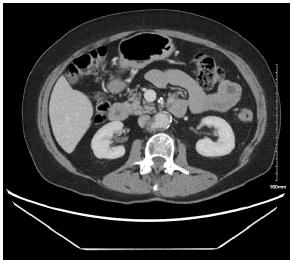
Figure 1: 2.0x1.2cm ulcerated plaque with pseudo-aneurysm arising from the Right lateral margin of the infrarenal aorta.
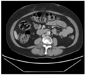
Figure 2: Focal dissection within the infrarenal aorta.
After consultation with the patient and colorectal team, decision was made to proceed with Endovascular Aortic Aneurysm Repair (EVAR) followed by interval colectomy for colon cancer. Patient was sized and thoracic graft (Valiant Medtronic) was used measuring 28mmx100mm.
Procedure
A 28x100mm Valiant thoracic stent graft was introduced through the Right Femoral artery. It was placed precisely below the ostium of the renal arteries. We completed deployment of the graft with slight forward pressure at the distal end to accommodate the distal anatomy.
Upon completion of the deployment, the delivery system was removed. Omni T Flush catheter was brought towards the renal arteries and completion angiography was performed.
Aortogram demonstrated patent Superior Mesenteric Artery (SMA). Both renal arteries were patent, however, Left renal artery had 50% stenosis. Infrarenal aorta appeared to be with significant wall irregularities demonstrating a penetrating ulcer with saccular aneurysm formation (Figure 3). Both iliac arteries, common iliac arteries were patent with some wall irregularities and no evidence of hemodynamically significant disease.
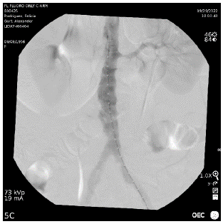
Figure 3: Penetrating ulcer with aneurysm formation.
Final images demonstrated good positioning of the stent in infrarenal aorta with no evidence of extravasation and is below the of ostia of the renal arteries (Figure 4).
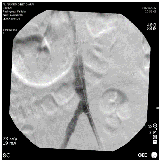
Figure 4: Stent deployment in the infrarenal aorta.
Discussion
Penetrating aortic ulcers were first reported by Shennan in 1934 and are included in the category of acute aortic syndrome: AD, PAU and IMH [1].
PAU originates from a ruptured atherosclerotic plaque causing ulceration in the intima of the aorta. Most commonly it occurs in the DTA and to lesser extent in the infrarenal aorta.
Associated with multiple risk factors, most commonly HTN (94%), smoking and HLD.
Given its risk of progression to focal AD or IMH and rupture, this condition should be managed promptly [4].
There are no set guidelines on when to treat PAUs and practices vary between hospitals. Generally asymptomatic cases of PAU may be managed conservatively with close follow-up [5]. Endovascular repair is the gold standard for complicated PAUs of the DTA [3]. At the level of the AA, a PAU is often managed by open repair.
We describe a case of an actively smoking patient, with high atherosclerotic burden, found to have incidentally a PAU in the infrarenal aorta on CT imaging while being evaluated for colon cancer. After multidisciplinary discussion we proceeded with EVAR of a 2.0x1.2cm ulcerated plaque arising from the Right lateral margin of the infrarenal aorta, complicated by focal dissection.
A 28x100mm Valiant thoracic stent graft was deployed below the renal arteries. Post deployment angiography images demonstrated good positioning of the stent in infrarenal aorta with no evidence of extravasation. Patient was discharged on post-op day 3 and continued her evaluation of colon cancer. Follow up abdominal CT angiography demonstrated exclusion of the ulcer from the aortic lumen without evidence of endoleak.
After a period of recovery of approximately 2 months, patient proceeded with interval open colectomy and primary anastomosis for resection of her cancer.
Conclusion
PAU account for 2-7% of acute aortic syndromes [1]. It’s essential to recognize the presence of coexisting dissection or IMH due to significant increase in risk of rupture. These ulcers could be asymptomatic, like in our case, and most commonly occur in the DTA. Surgical intervention is important especially in case of a complicated ulcer associated with an aneurysm or IMH formation. We describe a case where an endovascular repair was done successfully for an infrarenal complicated penetrating ulcer in a patient recently diagnosed with colon cancer [6].
References
- Kotsis T, Spyropoulos BG, Asaloumidis N, Christoforou P, Katseni K, et al. Penetrating Atherosclerotic Ulcers of the Abdominal Aorta: A Case Report and Review of the Literature. Vasc Specialist Int. 2019; 35: 152-159.
- Murana G, Di Marco L, Fiorentino M, Buia F, Brillanti G, et al. Endovascular treatment of penetrating atherosclerotic ulcers of the arch and thoracic aorta: In-hospital and 5-year outcomes. JTCVS Open. 2022; 10: 12-21.
- Bianchini Massoni C, Ancetti S, Perini P, Spath P, D'Ospina RM, et al. Infrarenal EVAR for Penetrating Aortic Ulcer: A Comparative Study with Abdominal Aortic Aneurysm. Ann Vasc Surg. 2023; 88: 327-336.
- Gabel JA, Tomihama RT, Abou-Zamzam AM Jr, Nekrasov V, Oyoyo UE, et al. Early Surgical Referral for Penetrating Aortic Ulcer Leads to Improved Outcome and Overall Survival. Ann Vasc Surg. 2019; 57: 29-34.
- Salim S, Machin M, Patterson BO, Bicknell C. The Management of Penetrating Aortic Ulcer. Hearts. 2020; 1: 5-13.
- Yoshihiko Tsuji, Yosuke Tanaka, Atsushi Kitagawa, Yutaka Hino, Takanori Taniguchi, et al. Endovascular stent-graft repair for penetrating atherosclerotic ulcer in the infrarenal abdominal aorta. Journal of Vascular Surgery. 2003; 38: 383-388.