
Special Article: Vitamins
Int J Nutr Sci. 2024; 9(1): 1080.
The Exacerbation of Chronic Kidney Disease due to Uncontrolled Vitamin D Supplementation- Case Report
Miroslaw Snit, MD, PhD¹; Dariusz Szczyra, MD¹*; Paulina Ziólkowska²; Michalina Strzedulla, MD, PhD²; Janusz Gumprecht¹
¹Department of Internal Medicine, Diabetology and Nephrology, Faculty of Medical Sciences in Zabrze, Medical University of Silesia, Poland
²Faculty of Medical Sciences in Zabrze, Medical University of Silesia in Katowice, Poland
*Corresponding author: Dariusz Szczyra, MD Department of Internal Medicine, Diabetology and Nephrology, Faculty of Medical Sciences in Zabrze, Medical University of Silesia, 15 Poniatowskiego Str., 40-055 Katowice, Poland. Email: dszczyra@sum.edu.pl.
Received: December 07, 2023 Accepted: January 06, 2024 Published: January 13, 2024
Abstract
Vitamin D plays a vital role in the proper functioning of the human body. It is involved in the regulation of calcium-phosphate metabolism. Currently, the problem of vitamin D deficiency is well-known. Additional oral supplementation is widespread, furthermore the number of cases of vitamin D overdose is increasing. A strong indication of toxicity is hypercalcemia. Excessive supplementation may lead to calcium deposits in tissues and organs and finally lead to damage and conduction disorders. The presented patient used high doses (over 10 000 units per day) of vitamin D for a few months. He was admitted to the hospital with an increased serum creatinine level of 352 μmol/l. The progressive chronic kidney disease was diagnosed, and the replacement renal therapy was considered. After 13 days of treatment, the patient had a good clinical response with the gradual normalization of kidney function and decreased vitamin D and its metabolites serum concentrations. In case of excessive vitamin D intake and symptoms of toxicity, it is essential to monitor the patient for at least 6 months because of the slow release of vitamin D from fat deposits.
Keywords: Vitamin D; Vitamin D toxicity; Vitamin D supplementation; Overdose; Kidney failure; Hypercalcemia
Introduction
Vitamin D is a well-known representative of a fat-soluble vitamin group. It is released, absorbed and transported with dietary fat. The common name “vitamin D” includes, in fact, two significant compounds - ergocalciferol (also known as vitamin D2) and cholecalciferol (the other name is vitamin D3). Vitamin D plays a vital role in the proper functioning of the human body, especially in regulating calcium and phosphate metabolism [1]. Sun exposure and oral intake are the two main sources of the following vitamin. The first includes the natural synthesis of cholecalciferol in the lower layers of the epidermis induced by ultraviolet radiation, whereas oral intake involves diet and conscious supplementation. Vitamin D occurs naturally only in a few comestible products, such as fish, meat, liver and eggs. Consequently, supplementation is an essential issue as vitamin D deficiency is a global problem. More than one billion people worldwide are estimated to suffer from vitamin D deficiency [2]. Due to this, nowadays, a wide range of different supplements containing vitamin D are widely available. However, many of them have an unregulated composition. Moreover, the guidance for their safe application is often insufficient. As a result of both these factors, there is an increased number of cases with excessive supplementation and possible symptoms of vitamin D like apathy, vomiting, abdominal pain, polyuria, polydipsia and dehydration [3, 4]. Treatment includes limiting calcium intake, discontinuing vitamin supplementation, intravenous hydration and intake of the drugs decreasing serum calcium level.
Pathomechanism of Vitamin D Toxicity
In 2008, 3 main hypotheses were proposed regarding the mechanism of Vitamin D Toxicity (VDT). All three involve increased concentrations of a vitamin D metabolite reaching the Vitamin D Receptor (VDR) in the nucleus of target cells and causing gene overexpression: 1. Toxicity is mediated by increased serum concentrations of the active hormonal form, 1,25(OH) 2 D, leading to increased intracellular concentration. 2. 1,25(OH) 2D has a low affinity for Vitamin D Binding Protein (VDBP) and a high affinity for VDR, making it an important ligand for accessing the transcriptional signal transduction machinery. In hypervitaminosis D, the concentrations of various vitamin D metabolites, especially 25(OH)D, are significantly increased, saturating the binding capacity of VDBP, which allows other vitamin D metabolites to enter the cell nucleus. Of the various vitamin D metabolites, 25(OH)D at higher concentrations (a dose-dependent effect) has the strongest affinity for the VDR, so a particular metabolite at elevated serum concentrations stimulates transcription. 3. Ingestion of vitamin D raises the concentration of vitamin D itself and increases the concentration of many other vitamin D metabolites, especially 25(OH)D. In vitamin D hypervitaminosis, the concentrations of vitamin D metabolites, such as vitamin D, 25(OH)D, 24,25(OH) 2 D, 25,26(OH) 2 D and 25(OH)D-26,23 -lactone, increase significantly Abnormally elevated concentrations of vitamin D metabolites exceed the binding capacity of VDBP and cause the release of free 1,25(OH) 2 D; the latter active metabolite enters target cells by diffusion and acts through the vitamin D receptor [14,17].
Guidelines for Vitamin D Supplementation in Poland and Europe
Poland and Central Europe: The initiation of vitamin D supplementation should begin with the determination of vitamin D requirements, the best determinant of which is the serum concentration of the liver metabolite 25(OH)D. According to the Central European guidelines, suboptimal supply is assumed to occur at a serum 25(OH)D concentration of 20-30 ng/ml (50-75 nmol/l). In contrast, maintaining a concentration of 30-50 ng/ml (75-125 nmol/l) was considered to ensure the pleiotropic effect of vitamin D. The finding of higher serum concentrations (above 100 ng/ml, i.e., above 250 nmol/l) may indicate improper supplementation and is an indication for total discontinuation of treatment [21].
According to the above recommendations, 20 to 50 ug/day of vitamin D should be used. [22]
In 2021. The Chief Sanitary Inspectorate (GIS) in Poland regulated the maximum doses of vitamin D in dietary supplements. These are, respectively:
- 2,000 IU (50 μg) for dietary supplements intended for the healthy adult population up to age 75 (01/2021);
- 4,000 IU (100 μg) for dietary supplements intended exclusively for the healthy population of adults over 75 years of age. (01/2021).
Europe: In western, southern European and Scandinavian countries vitamin D supplementation is recommended after the prior determination of serum concentrations of the hepatic metabolite 25(OH)D in 2019. Lips et al., in their publication based on country-specific data, reported that vitamin D requirements for adults (including older adults ranged from 0 to 20 ug/day. [22]
Case Report
67-years old male was admitted to the hospital for diagnosis of rapidly progressive chronic kidney disease (visualized incidentally during laboratory tests performed by the GP who referred the patient to the Nephrology consultation) with unclear aetiology of kidney failure as well as for consideration of the possible need for renal replacement therapy. Previous blood tests revealed increased serum creatinine levels and hypercalcemia. The patient was diagnosed with hypertension, hypertriglyceridemia, liver steatosis, left acoustic neuroma and lichen sclerosis. At the time of admission, he did not report any symptoms and signs. The patient stated that he had been using high doses of vitamin D (over 10 000 units per day for a few months) as the supplemental therapy. The patient was unaware of the threats, and thought he was using the correct dosage. No alcohol use, no herbal agents and no suspected drug use were reported. Physical examination did not reveal any relevant abnormalities. The patient appeared alert, oriented and cooperative. Auscultation showed normal heart sounds as well as normal symmetrical vesicular sounds. No abdominal findings were referred. Vital signs of the patient were also in the normal ranges with 36.6oC body temperature, 75/min. heart rate and 96% blood oxygen saturation without additional oxygen therapy, arterial blood pressure was slightly increased 140/80.
Laboratory tests were performed during the admission. The following results were obtained: serum creatinine level: 352 μmol/l (normal range: 62-106 μmol/l), eGFR: 16.03 ml/min/1.73m2 (normal value >60 ml/min/1.73m2), total vitamin D >100 ng/ml, vitamin D metabolite 25(OH) > 308 ng/ml (concentration >100 ng/ml is considered potentially toxic), vitamin D metabolite 1.25(OH)2: 585 pg/ml (normal range: 25.00-86.50 pg/ml), parathormone: 7.76 pg/ml (normal range: 10-60 pg/ml), HCT: 26,1% (normal range: 42-52%), RBC: 3.21 x10⁶/μl (normal range: 4.7-6 x10⁶/μl), HGB: 9.7 g/dl (normal range: 13.5-17 g/dl), triglycerides: 1.94 mmol/l (normal range <1.7 mmol/l). The arterial blood gases analysis was normal. There were also some electrolyte disturbances such as hyponatremia: 120 mmol/l (normal range: 135-145 mmol/l), hyperkalemia: 5.55 mmol/l (normal range: 3.5-5.1 mmol/l) as well as hypercalcemia - the total calcium level was 3.62 mmol/l (normal range: 2.12–2.62 mmol/l). The serum protein immunofixation was performed and did not reveal the monoclonal gammapathy. The urinalysis also did not show any abnormalities.
The performed12-lead electrocardiography record presented the sinus rhythm with a frequency of 70/min. as well as the left anterior hemiblock, probably caused by high calcium concentration.
The patient underwent a series of medical imaging to extend the diagnostics of the reasons for kidney failure (figure 1, 2). Some of them were interpreted as normal and showed no signs of osteolysis, such as the skull and pelvic radiographs. The chest X-ray reported only minor abnormalities in the lung’s stroma. During the hospitalization, abdominal ultrasonography was also performed revealing slightly higher echogenicity of the liver, pointing to steatosis, significantly elevated echogenicity of the renal cortex and a small cortical cyst (6 mm) in the left kidney.
Because of unclear aetiology of the patient’s symptoms, the renal biopsy was under consideration, however, patient’s informed consent could not be obtained. For this reason, the biopsy was abandoned.
During hospitalization, intravenous fluid therapy was implemented. It consisted of 500 ml 0.9% NaCl with addition of 20 ml of 10% NaCl (on the first day of hospitalization for severe hyponatremia). The aim of that therapy included the protective impact on renal function, the minimalization of hyponatremia and the treatment of hypercalcemia. A good response was obtained progressively (table 1). The treatment also involved the administration of 2000 ml 0.9 % NaCl, Furosemidum 2 x 40 mg i.v., intravenous bisphosphonates (Pamindronate disodium 90 mg/ 500 ml 0,9% NaCl, 3 doses from the 7th to 9th day) and glucocorticosteroids per os (Metyloprednisolone16 mg- 8 mg from 8th day). The aim was to reduce the calcium concentration. The impaired kidney function subsequently led to anaemia, with haemoglobin concentration decreased to 7.1 g/dl. For that reason, blood transfusion was necessary. During hospitalization, the patient did not report symptoms of gastrointestinal bleeding only persistent constipation. After laxative infusion, blood-colored stool was observed (1 episode). He did not agree to gastrointestinal endoscopic diagnostics, with the approval of the patient (initially, he also did not agree), he obtained 4 units of packed red blood cells after crossmatch.
![]()
Day of hospitalization
1
2
3
4
5
6
7
8
9
10
11
12
Cre [μmol/l]
352
-
340
355
-
-
332
-
266
262
237
-
eGFR [ml/min]
MDRD16.0
3-
16.6
915.8
8-
-
17.1
5-
22.1
522.5
425.3
1-
Total serum Ca [mmol/l]
3.62
-
3.37
3.35
-
-
2.76
-
2.62
2.58
-
-
Serum ionized Ca [mmol/l]
-
-
1.64
-
-
1.45
1.41
-
1.28
-
-
-
Vitamin D [ng/ml]
-
-
>100
-
-
-
-
-
>100
-
-
-
Table 1: The results of the patient’s laboratory tests. Abbreviations: Cre-serum creatinine level.
After 11 days of hospitalization, we observed a decrease in serum creatinine to 237 μmol/L, eGFR 25.3 ml/min, serum total calcium was 2.58 mmol/L, and haemoglobin was 12.1 g/dl. High serum vitamin D levels (above the threshold of determination in the hospital laboratory) persisted. On day 13, the patient was released home in stable condition and was advised to avoid vitamin D supplementation and receive regular treatment from a nephrologist. However, he was absent for a follow-up visit.
Summary
Vitamin D is essential for the proper functioning of human organisms. Due to this, the global problem of its deficiency poses a huge medical challenge. The wide range of available supplements seems to resolve that issue. However, the number of cases of vitamin D overdose is increasing. In a retrospective analysis of the data from the National Poison Data System (NPDS), toxic exposure to vitamin D increased from a mean of 196 cases per year from 2000 to 2005, to a staggering mean of 4535 exposures per year from 2005-2011 [16]. One of the strongest markers of vitamin toxicity is hypercalcemia. Acute vitamin D toxicity is usually caused by doses of vitamin D above 10 000 IU/day resulting in serum 25(OH)D concentrations >150 ng/mL. Chronic vitamin D toxicity can potentially occur with the administration of doses above 4 000 IU/day for extended periods. The main goal of treatment during vitamin D toxicity is emergent resuscitation in an unstable patient and correction of hypercalcemia. Vitamin D is stored in adipose tissue, and due to this, it may be slowly released from fat deposits for approximately 18 months. Consequently, organ damage may appear and lead even to death.[3]
A regimen for managing vitamin D poisoning has been proposed in 2022. First of all, all vitamin D and calcium supplements should be discontinued, but the patient's medication list should also be reviewed to adjust any future doses of vitamin D supplements. It is important to avoid excessive bed rest to prevent immobilization-induced hypercalcemia. Use parenteral hydration with 0.9% NaCl. In cases of severe toxicity causing severe hypercalcemia, calcitonin and bisphosphonates can be used. Intravenous calcitonin at a dose of 4 units/kg can be administered, and calcium concentrations repeated after 6-12 hours. Intravenous bisphosphonates can be given at the same time, but they are more beneficial in reducing hypercalcemia in cancer. When choosing therapy, it should be kept in mind that calcitonin can lead to tachyphylaxis, while the effects of bisphosphonates can persist for longer. Calcium levels should be carefully monitored when using these drugs. The use of intravenous corticosteroids is usually reserved for the treatment of vitamin D toxicity associated with granulomatous disease. It lowers plasma calcium levels by decreasing intestinal absorption and increasing urinary calcium excretion. In rare cases, patients may require hemodialysis to get rid of excess calcium due to significant kidney damage. After treatment, patients should be educated frequently to avoid the overuse of vitamin supplements.
The Endocrine Society suggests monitoring 25-hydroxyvitamin D and serum calcium levels in patients receiving high doses of vitamin D [18].
Hypervitaminosis caused by vitamin D should be differentiated from other causes leading to hypercalcemia or diseases mimicking the symptoms of hypervitaminosis as follow: hypercalcemia of malignancy, hypercalcemia of granulomatous diseases, vitamin A toxicity, thyrotoxicosis primary, secondary and tertiary hyperparathyroidism, Paget disease, hypercalcemia caused by prolonged immobilization and milk-alkali syndrome [18].
The described patient used high doses of vitamin D supplementation for several months (over 10 000 units per day) in order to improve quality of life. The patient was unaware of the risks of doing so thinking he was using the correct dosage. Instead, the excessive supplementation led to rapidly progressive chronic kidney disease (in the spring of 2021, the patient had normal serum creatinine levels). The quickly received treatment resulted in the gradual normalization of kidney function and renal replacement therapy was not necessary. During hospitalization, the added difficulty was the patient’s non-compliance, as periodically he did not consent to the proposed treatment and medical tests. He also skipped the follow-up appointments. It is considered that patients, who have overdosed on vitamin D, should be under control for at least 6 months in nephrology and endocrinology outpatient clinic. Furthermore, the serum calcium concentration as well as 25(OH)D concentration should be monitored regularly (at least for patients with symptoms of acute kidney injury, chronic kidney disease, hypercalcemia and hypercalciuria) [3,5].
Physicians should be reminded of and carry out a differential diagnosis of vitamin D toxicity whenever a patient presenting to primary care, specialized outpatient care or a hospital has a positive history of use of oral vitamin D medications and reports symptoms such as persistent vomiting, sensory disturbances, anuria or oliguria, and additional tests can show hypercalcemia, hypercalciuria, an increase in serum creatinine. Prompt diagnosis and implementation of treatment will avoid further kidney damage and consequent renal replacement therapy [19,20].
Author Statements
Supplementary Materials
Table 1: The results of the patient’s laboratory tests. Abbreviations: Cre- serum creatinine level.; Table 2: Implemented treatment.; Figure 1: The patient’ skullradiograph.; Figure 2: The patient’schestradiograph.
![]()
    Day of hospitalization

    1

    2

    3

    4

    5

    6

    7

    8

    9

    10

    11

    12


    Treatment

    i.v. fluid 2000 ml+ furosemidum


    Pamindronate disodium 90    mg/ 500 ml 0.9% NaCl


    Metyloprednisolone 16    mg- 8 mg
Table 2: Implemented treatment.
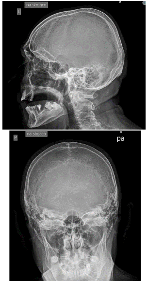
Figure 1: The patient’s skull radiograph.
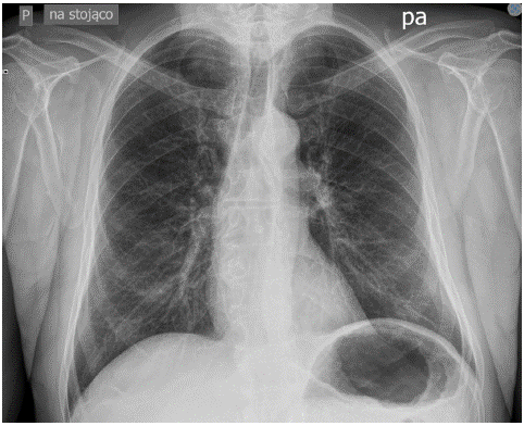
Figure 2: The patient’s chest radiograph.
Author Contributions
All authors have read and agreed to the published version of the manuscript.
Funding
This research received no external funding.
Conflicts of Interest
The authors declare no conflict of interest. The funders had no role in the design of the study; in the collection, analyses, or interpretation of data; in the writing of the manuscript; or in the decision to publish the results.
The patient's consent to publish the clinical case was obtained in writing and can be found in his medical history from his stay in the Nephrology Department.
References
- Kulda V. Metabolizmus vitaminum D [vitamin D metabolism]. Vnitrnilekarstvi. 2012; 58: 400-4.
- Holick MF. The vitamin D deficiency pandemic: approaches for diagnosis, treatment and prevention. Rev Endocr Metab Disord. 2017; 18: 153-65.
- Lim K, Thadhani R. Vitamin D toxicity. Jornalbrasileiro Nefrol: ‘orgaooficial de Sociedades Brasileira e Latino-Americana de Nefrologia. 2020; 42: 238-44.
- Marcinowska-Suchowierska E, Kupisz-Urbanska M, Lukaszkiewicz J, Pludowski P, Jones G. Vitamin D toxicity-A clinical perspective. Front Endocrinol (Lausanne). 2018; 9: 550.
- Chowdry AM, Azad H, Najar MS, Mir I. Acute kidney injury due to overcorrection of hypovitaminosis D: A tertiary center experience in the Kashmir Valley of India. Saudi J Kidney Dis Transpl. 2017; 28: 1321-9.
- Koul PA, Ahmad SH, Ahmad F, Jan RA, Shah SU, Khan UH. Vitamin D toxicity in adults: a case series from an area with endemic hypovitaminosis d. Oman Med J. 2011; 26: 201-4.
- Galior K, Grebe S, Singh R. Development of vitamin D toxicity from overcorrection of vitamin D deficiency: a review of case reports. Nutrients. 2018; 10: 953.
- Garg G, Khadgwat R, Khandelwal D, Gupta N. Vitamin D toxicity presenting as hypercalcemia and complete heart block: an interesting case report. Indian J Endocrinol Metab. 2012; 16: S423-5.
- Guerra V, Vieira Neto OM, Laurindo AF, Paula FJ, Moysés Neto M. Hypercalcemia and renal function impairment associated with vitamin D toxicity: case report. [Jornalbrasileiro de nefrologia: ‘orgaooficial de Sociedades Brasileira e Latino-Americana de Nefrologia]. J Bras Nefrol. 2016; 38: 466-9.
- De Vincentis S, Russo A, Milazzo M, Lonardo A, De Santis MC, Rochira V, et al. How much vitamin D is too much? A case report and review of the literature. Endocr Metab Immune Disord Drug Targets. 2021; 21: 1653-9.
- Stephenson DW, Peiris AN. The lack of vitamin D toxicity with megadose of daily ergocalciferol (D2) therapy: a case report and literature review. South Med J. 2009; 102: 765-8.
- Chakraborty S, Sarkar AK, Bhattacharya C, Krishnan P, Chakraborty S. A nontoxic case of vitamin D toxicity. Lab Med. 2015; 46: 146-9; quiz e31.
- Kumar N. Nutrients and neurology. Continuum (Minneap Minn). 2017; 23: 822-61.
- Chiricone D, De Santo NG, Cirillo M. Unusual cases of chronic intoxication by vitamin D. J Nephrol. 2003; 16: 917-21.
- Graidis S, Papavramidis TS, Papaioannou M. Vitamin D and acute kidney injury: A two-way causality relation and a predictive, prognostic, and therapeutic role of vitamin D. Front Nutr. 2020; 7: 630951.
- Spiller HA, Good TF, Spiller NE, Aleguas A. Vitamin D exposures reported to US poison centers 2000-2014: temporal trends and outcomes. Hum Exp Toxicol. 2016; 35: 457-61.
- Jones G. Pharmacokinetics of vitamin D toxicity. Am J Clin Nutr. 2008; 88: 582S-6S.
- Asif A, Farooq N. Vitamin D toxicity. StatPearls. 2022.
- Bhat JR, Geelani SA, Khan AA, Roshan R, Rathod SG. Vitamin D toxicity due to self-prescription: a case report. J Fam Med Prim Care. 2022; 11: 1561-3.
- Zhang Huanran, Jiang Y, Shi N, Lu YQ. Serum vitamin D levels and acute kidney injury: a systemic review and meta-analysis. Sci Rep. 2022; 12: 20365.
- Rusinska A, Pludowski P, Walczak M, Borszewska-Kornacka MK, Bossowski A, Chlebna-Sokól D, et al. Vitamin D supplementation guidelines for general population and groups at risk of vitamin D deficiency in Poland-recommendations of the polish society of pediatric endocrinology and diabetes and the expert panel with participation of national specialist consultants and representatives of scientific Societies-2018 update. Front Endocrinol (Lausanne). 2018; 9: 246.
- Lips P, Cashman KD, Lamberg-Allardt C, Bischoff-Ferrari HA, Obermayer-Pietsch B, Bianchi ML, et al. Current vitamin D status in European and Middle East countries and strategies to prevent vitamin D deficiency: a position statement of the European Calcified Tissue Society. Eur J Endocrinol. 2019; 180: 23-P54.