
Research Article
Austin J Nucl Med Radiother. 2016; 3(1): 1015.
Comparison of Ankle-Brachial Index and Lower Limb Perfusion Reserve in Patients with Behçet’s Disease
Tan YZ¹*, Kılıç S², Temiz A³, Özdemir S¹, Kurt T4 and Cevizci S5
¹Canakkale Onsekiz Mart University, School of Medicine, Department of Nuclear Medicine, Turkey
²Canakkale Onsekiz Mart University, School of Medicine, Department of Dermatology, Turkey
³Canakkale Onsekiz Mart Üniversty, School of Medicine, Department of Cardiology, Turkey
4Canakkale Onsekiz Mart University, School of Medicine, Department of Cardiovascular Surgery, Turkey
5Canakkale Onsekiz Mart University, School of Medicine, Department of Public Health, Turkey
*Corresponding author: Yusuf Ziya Tan, Canakkale Onsekiz Mart University, School of Medicine, Department of Nuclear Medicine, Turkey
Received: October 16, 2015; Accepted: February 02, 2016; Published: February 04, 2016
Abstract
Objective: The aim of this study was to investigate the Peripheral Vascular Disease (PVD) by comparing Lower Limb Perfusion Reserve (LLPR) and Ankle- Brachial Index (ABI) measurements in patients with Behçet’s Disease (BD).
Materials and Methods: In total, 62 patients (41 women and 21men, with a mean age of 47.26±11.4 years) with and without BD (22 and 40, respectively) were included in the present study. ABIs were measured before study. After 30 times dorsal and plantar flexion was performed to the feet in sitting position to increase blood flow in lower extremity. Tc-99m-sestamibi was injected from antecubital vein. Fifteen minutes after injection statically images were obtained from posterior region at supine position under gamma camera. Perfusion reserves were calculated by drawing region of interest in both calf regions.
Results: While average of PR in BD group was calculated as -3.34±8.7 % it was determined as 8.6±8.5 % in control group. When LLPR averages were compared between groups, a statistically significant decrease was observed in BD group with respect to control group. When ABI <0.9 and ≥ 1.30 was considered abnormal and 0.9<-<1.30 was considered normal and when PR and reference test ABI was compared in both groups sensitivity, specifity, accuracy and p value was determined as 100%, 71,1%, 82.3% and <0.001, respectively.
Conclusion: The results of this study suggest that LLPR calculated with Tc-99m-sestamibi is not superior to ABI in the invention of PVD in BD but it may be further beneficial for this test.
Keywords: Behcet’s disease; Peripheral vascular disease; Ankle-brachial index; 99mTc-Sestamibi; Lower limb perfusion reserve
Abbreviations
BD: Behçet’s Disease; LLPR: Lower Limb Perfusion Reserve; PVD: Peripheral Vascular Disease; BMI: Body Mass Index; MPS: Myocardial Perfusion Scintigraphy; ABI: Ankle Brachial Index; PR: Perfusion Reserve
Introduction
Behçet Disease (BD) is a multisytemic vacuity of unknown origin affecting all size of arteries and veins. Prevalence of vascular Behçet’s disease varies from 3.6% to 24.3% in large series (Venous and arterial involvement together). Furthermore arterial involvement is rare 2.5% in the literature [1-3]. Aneurysm formation is more frequent than occlusion and occlusive forms of BD are not only involving distal parts of the arterial tree. Small vein involvement is responsible from pathological indications and most of the findings. Vascular involvements are especially observed in young men and they are prognostically important findings. Vascular events may involve fatal complications like aneurysms, arterial and venous occlusions [4]. Therefore, it is vital in BD to establish early diagnosis of patients who are under risk of Peripheral Vascular Disease (PVD) and to administer an effective treatment before irreversible organ damages occur [5,6].
In the investigation of PVD, clinical history and simple physical examination as well as invasive and non-invasive diagnosis methods are used also. As noninvasive methods laboratory methods, segmental pressure measurements, Color Doppler USG are used. Nowadays, angiography is considered as golden standard in evaluation of distal vascular bed. However, ABI is used frequently in the investigation of peripheral vascular diseases because it is easy and practical and it contributes to diagnosis with high specifity and accuracy [7].
There are many studies conducted with Thallium-201 and 99mTc sestamibi Scintigraphy in the investigation of PVD scintigraphically [8-10]. Tc-99m-sestamibi disperses in tissues proportionally with blood flow and accumulates by connecting to anionic proteins within mitochondria in the cell. Its involvement is based to mitochondrial activity and cell vitality. Measurement of Perfusion Reserve (PR) calculated in the same session with Myocardial Perfusion Scintigraphy (MPS) is a noninvasive method used in the calculation of leg perfusion that allows evaluation of lower limb perfusion at microcirculation level [11]. When previous literature researches are examined there is no studies found comparing 99m Tc-sestamibi with ABI in investigating PVD in BD.
In this study we intended to investigate the usability of radionuclide methods as a noninvasive method in determination of PVD by comparing LLPR and ABI measurements.
Materials and Methods
Study population
The study was conducted after approval was received from local ethics committee (050.99-153 numbered approval).
In total, 62 patients (41 women and 21men, with a mean age of 47.26±11.4 years) who had no evidence of peripheral arterial disease in their clinical history or Doppler USG were studied. The subjects were divided into two groups, a BD group and a control group. The BD group consisted of 22(15 women, 7 men, mean 42,91±11,9 years). The diagnosis was made according to the international classification criteria of International Study Group for BD was referred to Canakkale Onsekiz Mart University, Division of Dermatology [12]. The control group consisted of 40(14 men and 26 women). Demographic data, systemic diseases and drug usage of all the patients who participated in the study were questioned. The patients with known peripheral vascular disease, lower limb trauma, patients with history of operation and drug usage, patients with complaint of intermittent claudicating, patients with systemic disease such as diabetes mellitus and renal insufficiency, with ages smaller than 18 years for radiation safety, pregnant and suspected pregnancy were excluded from the study.
Consent forms were taken from al the patients participating to the study after detailed information were given about the applications to be performed. Height and weight of each patient was recorded before the study and Body Mass Index (BMI) was calculated. Measurement of Ankle Brachial Index;
ABI was measured according to the methodology proposed by Society of Interventional Radiology (SIR) [13]. Brachial Artery (BA) systolic pressure was measured from both upper arms at supine position with sphygmomanometer before study and the highest values are found. Systolic blood pressure measurements were taken with 8 MHz hand held vascular Doppler from both Dorsalis Pedis Artery (DPA) and Posterior Tibial Artery (PTA) in both lower extremity and highest measurements were determined. ABI was calculated with the below formula with the measurements obtained.
The highest measured DPA and PTA systolic values
ABI: .......................................................................................................
The highest value measured in BA systolic values
ABI: Ankle Brachial Index, DPA: Dorsalis Pedis Artery, PTA: Posterior Tibial Artery, BA: Brachial Artery
Exercise and imaging protocol for lower extremity
MPS study of two days was performed with all the patients enrolled to the study with protocol dipyridamole stress 99mTc sestamibi. The first day 0.56mg/kg/4 minutes of dipyridamole were Injected Intravenously (IV) and pharmacological stress was obtained. Concurrently dorsal and plantar flexion was performed maximum 30 times to both feet to increase blood flow in lower extremity.
Two minutes after dipyridamole infusion, 15mCi (555MBq) Tc- 99m- sestamibi (MIBI, methoxy isobutyl isonitrile) was injected from antecubital vein. Approximately 15 minutes after injection static imaging of 5 minutes was performed from posterior calf region at supine position under gamma camera. Similar imaging was obtained before rest imaging from the patients who came the second day.
Perfusion reserves were calculated by drawing Region of Interest (ROI) on the images obtained from both calf regions after stress and rest.
Calculation of lower limb perfusion reserve
Total counts were calculated by drawing ROI on the static images obtained from posterior calf region after stress and rest. For each leg individually, total count obtained from region of interest drawn after stress ROI was subtracted from total count obtained from region of interest after rest ROI and the result was divided by RROI and multiplied by hundred to obtain percent Perfusion Reserve (%PR) (Figure 1A, Figure 1B).
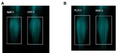
    Figure 1:  Total counts were calculated by drawing Region of Interest (ROI)
on the static images obtained from posterior calf region after stress (A) and
rest (B). The control group of patients with perfusion reserve was calculated
8, 16%.
PR (Right/Left) %= [(Stress ROI- Rest ROI)/ Rest ROI] x100
Myocardial perfusion scintigraphy
Stress Myocardial Perfusion SPECT and Gated SPECT images of the heart were obtained 45 minutes after injection under Gamma Camera. SPECT images were obtained at supine position with low energy high resolution collimator (LEHR) and gamma camera system with dual detector (GE, Infinia model). SPECT images were taken after 99mTc energy peak adjustments were made in gamma camera, within 20% window range, with 64x64 matrix, with 40 seconds at each “frame” stress and 50 scones at “rest” with total 64 projections of 180o from right anterior oblique to left posterior oblique. MPS SPECT and GATED SPECT imaging was obtained on a different day similar to stress imaging at rest.
After stress and rest imaging, MPS SPECT and Gated SPECT imaging’s were examined with computer for cross sections of short axis, horizontal long axis and vertical long axis of heart, regional wall motion and wall thickening. Left ventricular EF % values were calculated.
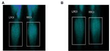
    Figure 2:  In patients with Behcet’s posterior planar images (A=stres, B=rest)
of both legs. Perfusion reserve was calculated -5, 16%.
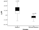
    Figure 3:  Behçet’s patients were found to be lower than in the perfusion
reserve in patients with control groups.
Statistical analysis
The analysis of the data obtained was made with SPSS version 19.0. Compatibility of variables to normal distribution in patient and control group was investigated with Kolmogorov-Smirnov/ Shapiro-Wilk tests. Average, standard deviation, median, minimum, maximum, frequency and percent values were used in the presentation of descriptive data. Chi-square test was used for categorical variables in intergroup comparisons. Cross tables were used to compare sensitivity, specifity, positive and negative predictions and precision level according to ABI. The cases where p value was lower than 0, 05 were considered statistically significant.
Results
Sixty-two patients were included in the study that was convenient for the study from whom MPS was requested for investigating coronary artery disease. Mean age of BD group was 42,91±11,9 years and of control group was 51,52±11 years. Demographic characteristics and results of MPS of the patients were presented in (Table 1). It was determined that prevalence of BD is higher in females than males (%68 vs. %32). No statistically significant difference was found between groups with respect to gender, age distribution, BMI and MPS results (Table 2). While average of perfusion reserve of BD group was -3.34± 8.7%, average of perfusion reserve of control group was calculated as 8.6±8.5%. It was determined that average of perfusion reserve in patients of BD was significantly lower than that of control group (Table 3). When ABI measurements <0.9 and ≥ 1.30 were considered abnormal and measurements 0.9<-<1.30 were considered normal, it was determined that PVD in BD existed more in females when compared with males (69% vs. 31%). However in control group abnormal ABI was detected only in one patient and the other patients had normal ABI values (Table 2).
![]()
    Behcet    disease (n=22)
    Control    group
      (n=40)


    Variables
    n    (%)*
    n    (%)*


    Gender


    Female
    15    (68.2)
    26    (65.0)


    Male
    7 (31.8)
    14    (35.0)


    Age    groups


    20-39
    10    (45.5)
    6 (15.0)


    40-54
    9 (40.9)
    18    (45.0)


    55-64
    2 (9.1)
    9 (22.5)


    >65
    1 (4.5)
    7 (17.5)


    BMI


    <25
    5 (22.7)
    15    (37.5)


    25-30
    11    (50.0)
    19    (47.5)


    >30
    6 (27.3)
    6 (15.0)


    Column    percentages. BMI: Body Mass index.
Table 1:  Demographic characteristics of Patients with Behcet Disease and
control groups.
![]()
    Behcet    disease(n=22)
    Control    group(n=40)


    <0.9
    >0.9-<1.30
    <0.9
    >0.9-<1.30


    n    (%)*
    n    (%)*
    p**
    n    (%)*
    n    (%)*


    Gender


    Female
    9 (69.2)
    6 (66.7)
    0.899
    1 (100)
    25    (64.1)


    Male
    4 (30.8)
    3 (33.3)
    0 (0)
    14    (35.9)


    Age    groups


    20-39
    5 (38.5)
    5 (55.6)
    0.418
    0 (0)
    6 (15.4)


    40-54
    6 (46.2)
    3 (33.3)
    0 (0)
    18    (46.2)


    55-64
    1 (7.7)
    1 (11.1)
    0 (0)
    9 (23.1)


    >65
    1 (7.7)
    0 (0.0)
    1 (100)
    6 (15.4)


    BMI


    <25
    2 (15.4)
    3 (33.3)
    0.398
    1 (100)
    14    (35.9)


    25-30
    7 (53.8)
    4 (44.4)
    0 (0)
    19    (48.7)


    >30
    4 (30.4)
    2 (22.2)
    0 (0)
    6 (15.4)


    MPS


    Normal
    9 (69.2)
    6 (66.7)
    0.899
    0 (0)
    26    (66.7)


    Abnormal
    4 (30.8)
    3 (33.3)
    1 (100)
    13    (33.3)


    Column    percentages, **Chi-square test.
Table 2:  Comparing patients with Behcet’s disease with control group according
to the ABI.
A statistically significant relation was not detected between LLPR and BMI in each group. Ischemia was determined in myocardial perfusion Scintigraphy in 4 out of 9 patients in BD group who were considered positive with regards to PVD by both tests. No statistically significant difference was detected between groups (Table 3).
![]()
    D    (n=22)
    Control    group (n=40)


    LLPR(Mean±SD)


    Gender


    Female
    3.23±9.05
    7.26±8.69


    Male
    -3.59±8.47
    11.05±7.91


    Age    groups


    20-39
    -2.61±9.53
    13.18±1.83


    40-54
    -3.37±9.06
    8.42±8.78


    55-64
    -3.45±7.28
    9.45±7.29


    >65
    -
    3.96±11.53


    BMI


    <25
    1.11±9.39
    4.01±9.10


    25-30
    -3.64±8.60
    12.89±4.62


    >30
    -4.66±9.49
    12.89±4.62


    MP


    Normal
    -2.58±8.25
    7.35±8.42


    Abnormal
    -4.98±9.90
    10.88±8.53


    ABI


    Normal    (0.9-1.3)
    4.50±8.24
    9.19±7.75


    Abnormal    (<0.9)
    -8.78±2.89
    -
Table 3:  Mean and median values of the LLPR parameters in BD and control
groups.
When PR and ABI, our reference test was compared sensitivity, specifity, precision and p value was determined as 100%,77.1%, 82.3% and <0.001, respectively (Table 4).
![]()
    Sensitivity
    Specificity
    Accuracy
    p


    LLPR
    100%
    77.1%
    82.3%
    <0.001


    LLPR:    Lower Limb Perfusion Reserve; ABI: Ankle Brachial Index.
Table 4: Analysis of the LLPR vs. ABI.
Discussion
The early diagnosis of PVD is vital due to its risks. Ischemia may be observed in PVD patients that causes amputation and that increases mortality and morbidity. Although PVD is rare in BD, early diagnosis and appropriate treatment is vital because serious problems may develop in future. In the studies where peripheral vascular bed is investigated in BD it is shown that peripheral vascular disease is detected more in asymptomatic patients than healthy individuals [5].
There are guides for invasive and noninvasive methods as well as clinical history and physical examination in PVD investigation [14]. Angiography is a gold standard for arterial vascular evaluation but it is extremely dangerous for patients with BD because of the puncture induced pseudo aneurysm, thrombosis. In the early diagnosis of PVD, measurement of ABI is stated as a highly practical and noninvasive method providing useful information [7].
In the studies conducted comparatively to investigate the existence of PVD, it is observed that Thallium-201 was used as radiopharmasotics. [8,14-18] it is observed that in recent years 99mTc-sestamibi was used more as radiopharmasotics in evaluation of PVD. Tc-99m-sestamibi is distributed proportionally in tissues. Retention in the tissues depends on mitochondrial activity and cell vitality [19]. One of the important advantages of 99mTc-sestamibi with regards to Thallium 201 is high image quality and minimal redistribution.
In our study, we also used Tc- 99m-sestamibi for PR similar to the study conducted [20]. Although the stress protocol used in our study resembles the protocol used by Celen et al. the significant difference is calculation of perfusion reserves of lower limbs both after rest and after stress. Our purpose was to prevent evaluation errors that might be encountered at different dose and exercise conditions.
Kusmierek et al. stated that exercise test should be conducted at least for 6 minutes in a study they performed two days with protocol with 99mTc sestamibi [11]. Because exercise time might differ from person to person, we performed maximum 30 times dorsal and plantar flexion with a similar method used by Celen et al. to increase blood flow in lower extremity [20].
In the previous studies diabetic and non-diabetic patients were used as patient groups and it was determined that lower limb perfusion reserves were significantly lower in diabetic patients. [3,16] It is apparent that lower limb perfusion reserves shall decrease in diabetic patients in parallel with decrease of vascular dilatation and permeability based on thickening of capillary basal membrane in DM patients. Consequently, in our study we excluded diabetic patients from BD and control group patients since PR and ABI should be evaluated correspondently so that they would not affect our results.
Kownotor et al. [21]. reported that PAD investigation with ABI is required because concurrent existence of Coronary Artery Disease (CAD) and PAD indicates bad prognosis. We did not determine any statistically significant difference between CAD and PAD in BD (p<0.899).
Kaya et al. [22] Determined 56.5% ischemia in BD group in their study. We determined myocardial ischemia in 32% of BD patient group.
Chang et al. [15] Reported a sensitivity of 63% in determining PVD in comparative studies performed with ABI and PR calculated with dipyridamole stress thallium 201 in diabetic patient group. In this study we determined sensitivity as 100% and specificity as 71.1% (Table 4). We attributed this to no diabetic patient in BD and not BD groups, fixed excursive methods, radiopharmaceutical use at the same dose.
Study Limitations
1- Our primary limitation in the study was the inadequacy of BD group in number.
2- The study is an observational study and it does not show cause and effect relation. Hence prospective, randomized studies are required.
Conclusion
In conclusion, although calculation of perfusion reserve performed with dipyridamole stress 99mTc sestamibi is not superior to ABI in the investigation of patients with asymptomatic peripheral vascular disease in BD, it may be a useful additional method to assess regional blood flow.
References
- Sakane T, Takeno M, Suzuki N, Inaba GN. Behcet’s disease. Engl J Med. 1999; 17: 1284-1291.
- Calamia KT, Schirmer M, Melikoglu M. Major vessel involvement in Behçet disease. Curr Opin Rhematol. 2005; 17: 1-8.
- Kuzu MA, Ozaslan C, Koksoy C, Gurler A, Tuzuner A. Vascular involvement in Behcet’s disease: 8-year audit. World J Surg. 1994; 18: 948-953.
- Tursen U, Ulubas B, Kaya TI, Pekdemir H, Ikizoglu G. Cardiac complications in Behcet’s disease. Clin Exp Dermatol. 2002; 27: 651-653.
- Kuzu A, Koksoy C, Ozaslan C, Gurler A, Tuzuner AE. Valuations of peripheral vascular system disorders in vascular symptom- free Behcet’s disease. Cardiovasc Surg. 1996; 4: 381-383.
- Welten GM, Schouten O, Chonchol M, Hoeks SE, Bax JJ, Van Domburg RT, et al. Prognosis of patients with peripheral arterial disease. J Cardiovasc Surg (Torino). 2009; 50: 109-121.
- Dachun Xu, Jue Li, Liling Zou, Yawei Xu, Dayi Hu, Sherry L Pagoto, et al. Sensitivity and specificity of the ankle–brachial index to diagnose peripheral artery disease: a structured review. Vasc Med. 2010; 15: 361.
- Michael E Siegel, Jan K Sıemsen. A new Noninvasive Approach to Peripheral Vascular Disease: Thallium- 201 Leg Scans. Am J Roentgenol. 1978; 131: 827- 830.
- Wolfram RM, Budinsky AC, Sinzinger H. Assessment of peripheral arterial vascular disease with radionuclide techniques. Semin Nucl Med. 2001; 31: 129-142.
- Miles KA, Barber RW, Wraight EP, Cooper M. Appleton DS Leg muscle Scintigraphy with 99Tcm-MIBI in the assessment of peripheral vascular (arterial) disease. Nucl Med Commun. 1992; 13: 593-603.
- Kusmierek J, Dabrowski J, Bienkiewicz M, Szuminski R, Płachcinska A. Radionuclide assessment of lower limb perfusion using 99mTc-MIBI in early stages of atherosclerosis. Nucl Med Rev Cent East Eur. 2006; 9: 18-23.
- David Sacks, Curtis W Bakal, Peter T Beatty, Gary J Becker, John F Cardella, Rodney D Raabe, et al. Position Statement on the Use of the Ankle Brachial Index in the Evaluation of Patients with Peripheral Vascular Disease A Consensus Statement Developed by the Standards Division of the Society of Interventional Radiology(SIR). J Vasc Interv Radiol. 2003; 14: 389.
- Hirsch AT, Haskal ZJ, Hertzer NR, Bakal CW. ACC/AHA 2005 Practice Guidelines for the Management of Patients with Peripheral Arterial Disease. Circulation. 2006; 490-503.
- Segall GM, Lennon SE, Stevick CD. Exercise Whole-Body Thallium Scintigraphy in the Diagnosis and Evaluation of Occlusive Arterial Disease in the Legs. J NucI Med. 1990; 31: 1443-1449.
- Yu-Hong Chang, Chung-Sen Chen, Kuo-Meng Liao, Ming-Hsien Lin. A Comparison of Dipyridamole Stress Lower-Limb Thallium-201 Scintigraphy with Ankle-Brachial Index in Assessing Peripheral Arterial Occlusive Disease of Diabetic Patients. Ann Nucl Med Sci. 2006; 19: 225-230.
- Ching Cheng Lin, Hueisch Jy Ding, Yu Wen Chen, Wen Tao Huang, Albert Kao. Usefulness of thallium -201 muscle perfusion scan to investigate perfusion reserve in lower limb of Type 2 diabetic patients. Journal of Diabetes and Its Complications. 2004; 233-236.
- Duet M, Virally M, Bailliart O, Kevorkian JP, Kedra AW, Benelhadj S, et al. Whole-body (201)Tl Scintigraphy can detect exercise lower limb perfusion abnormalities in asymptomatic diabetic patients with normal Doppler pressure indices. Nucl Med Commun. 2001; 22: 949-954.
- Segal GM, Lang EV, Lennon SE, Stevick CD. Functional Imaging of Peripheral Vascular Disease: A Comparison between Exercise Whole Body Thallium Perfusion Imaging and Contrast Arteriography. J NucI Med. 1992; 33: 1797-1800.
- Wolfram RM, Budisky AC, Sinzinger H. Assessment of peripheral arterial vascular disease with radionuclide techniques. Semin Nucl Med. 2001; 129- 142.
- Celen YZ, Zincirkeser S, Akdemir I, Yilmaz M. Investigation of perfusion reserve using 99Tcm- MIBI in the lower limbs of diabetic patients. Nuclear Medicine Communications. 2000; 21: 817-822.
- Kownotor S, Cambou JP, Caccoub P, Leger P, Luizy F, Herrman MA, et al. Prevalence of unknown peripheral arterial disease in patients with coronary artery disease: data in primary care from the IPSILON study. Arch Cardiovasc Dis. 2009; 102: 625-631.
- Kaya E, Saglam H, Ciftci I, Kulac M, Araca S, Melek M. Evaluation of myocardial perfusion and function by gated SPECT in patients with Behcet’s disease. Ann Nucl Med. 2008; 22: 287-295.
- International Study Group for Behçet’s Disease. Criteria for diagnosis of Behçet’s disease. Lancet. 1990; 335: 1078-1080.