
Research Article
Austin Neurosurg Open Access. 2020; 6(1): 1062.
Surgical Treatment of Spondylodiscitis of the Thoracic and Lumbar Spine
Aleksey Eroshkin¹*, Nikolai Rainov², Dmytro Romanukha¹
1Department of Neurosurgery of Central Hospital of Ministry of Internal Affairs of Ukraine (Central Police Hospital), Kyiv, Ukraine
2MVZ Wirbelsäulenzentrum Taufkirchen, Munich, Germany
*Corresponding author: Aleksey Eroshkin, Head of Department of Neurosurgery of Central Hospital of Ministry of Internal Affairs of Ukraine (Central Police Hospital), 1 Berdychivs’ka Street, Kyiv, 04116, Ukraine
Received: August 05, 2020; Accepted: September 04, 2020; Published: September 11, 2020
Abstract
Background: The incidence of spondylodiscitis (SD) of different origin is increasing in the last decades and its treatment may be difficult and prolonged.
Methods: All patients presented with SD of different origin and with different degrees of pain and/or neurological deficits. All of them underwent standard posterior transpedicular fixation with debridement and decompression of neural structures in the spinal canal. In some cases with significant segmental instability, intervertebral PLIF cages were used in addition to dorsal transpedicular fixation. Clinical outcomes were assessed using functional outcome criteria (ASIA and VAS scales). The sagittal alignment of the affected segments was evaluated preoperatively and postoperatively by measuring the Cobb angle.
Results: 47 patients with SD of different origin underwent posterior transpedicular fixation. PLIF cages were used in addition in 12 cases (26%). 31 (66%) of the patients were males and 16 (34%) females. The average age of the male population was 62.3 ± 4.8 years, and of the female population 58.2 ± 5.1 years. 42 of these patients completed follow-up at 12 months (89.4%).
There was no worsening of clinical symptoms after surgery. Most patients with neurological impairment (ASIA grades A-D) showed a marked regression of neurological symptoms after surgery. Improvement of at least one ASIA grade occurred postoperatively in 25 cases (64.1%). Further improvement by at least one ASIA grade at 1 year follow-up occurred in 6 further cases to a total of 79.5%.
For thoracic, average preoperative Cobb angle was 20.1±9.8°, improving after surgery by a 7.2±5.5°, for lumbar was 5.7±3.3°, and was improved to 1.2±.1.0°.
Conclusions: Instrumented posterior fixation in patients with SD improves neurological outcome, allows early mobilization, and avoids occurrence of spinal deformity. This treatment strategy obviates the higher risks of anterior approaches and staged surgery.
Keywords: Spinal Fixation; Spondylodiscitis; Transpedicular Instrumentation
Introduction
Currently, there is a strong tendency towards increased numbers and severity of inflammatory spine disorders. According to the literature, the annual incidence of spondylodiscitis (SD) of any origin in Europe averages from 0.4 to 5.8/100.000, or 30 per 250.000 persons per year [1-3]. SD predominantly occurs in elderly patients aged over 50 years, with a peak prevalence between 50 and 70 years [4-7].
Treatment of patients with inflammatory thoracic and lumbar spine disorders presents a growing problem in spinal surgery. The use of implants in the infected area of SD is still under discussion [4,6]. In practice, good outcomes mostly follow some sort of surgical intervention at the infection site [8-11]. Apparently this is due at least in part to the immobilization of the segment, which contributes to the speedy repair processes.
The question of the exact method of surgical intervention in patients with SD remains debatable. Some authors prefer posterior fixation [6,11,12], others anterior [13,14], and some a combination of these approaches [15,16]. Each of the methods has its pros and cons. The anterior approach provides excellent access to the site of infection in the intervertebral disc, as well as a stable fixation and immobilization of the affected segment, since the anterior column accounts for most of the supporting function of the spine. However, this approach is technically complex and carries a high risk of contamination of the abdominal cavitiy and the retroperitoneal space after drainage of SD. Dorsal transpedicular spinal fixation allows for immobilization of the damaged vertebral segment and at the same time for a sufficient decompression of the spinal cord and/ or cauda equina through a posterior approach to the spinal canal [17-20]. This surgical approach is less traumatic, which allows for earlier mobilization and rehabilitation of patients. Instrumented dorsal spinal fixation in patients with SD may also have some drawbacks, the main ones being insufficient mechanical stability with resulting deformities, but also loosening, displacement or migration of transpedicular screws [21,22]. Using a combined anterior and posterior approach significantly increases the mechanical stability and creates the maximum possible stabilization in the affected segment. However, it increases the surgical trauma and the duration of surgery, and very often requires staged surgery with two or more sessions, which lengthens the process of recovery and rehabilitation of patients. Therefore, the jury is still out on the most appropriate choice of surgical approach in patients with SD.
The aim of this retrospective study was to summarize the results and to analyze the outcomes of instrumented posterior spinal fixation in patients with SD of the thoracic and lumbar spine.
Patients and Methods
Surgery was carried out by a standard posterior midline approach to the thoracic and/or lumbar spine, combined with debridement and decompression of the spinal canal and its contents, and with hemilaminectomy or laminectomy, foraminotomy and/or facetectomy.
The main indications for surgery were decompression of the spinal cord or the cauda equina and evacuation of the inflammatory focus, which could be an epidural abscess and/or an intervertebral focus, as well as mechanical stabilization of the affected segment. In some cases with gross instability of the spine, intervertebral PLIF cages were placed. A partial or total facettectomy was performed bilaterally.
In all cases, the transpedicular screw system PlatinumTM (Irene, Tianjin, PR China) with polyaxial screws was used. The screw sizes in the lumbar spine were 5.0-6.0 mm x 45-55 mm, and in the thoracic spine 4.0-5.0 mm x 35-55 mm.
Treatment outcomes were evaluated by ASIA scale [23]. Visual analog scale (VAS) was used in assessing pain levels [24]. Radiological follow-up was generally carried out by spinal X-rays in two planes. The sagittal alignment of the affected segments was evaluated preoperatively and postoperatively by measuring the Cobb angle [25]. Final clinical examination was carried 1 year after surgery.
Results
From December 2015 to January 2018, 47 patients with SD underwent surgical treatment at our institution. 31 (66%) of these were males and 16 (34%) females. 5 patients aged 18-20 years, 9 patients aged 21 - 40 years, 22 patients aged 41 - 60 years and 11 patients older than 60 years. The median age of the patient population was 62.3 ± 4.8 years for males, and 58.2 ± 5.1 years for females.
The majority of patients (39 cases, 83%) had neurological deficits of varying degrees (ASIA grades A-D).
17 patients presented with thoracic SD (36.2%), 21 with lumbar SD (44.7%), and 9 with multiple thoracic and lumbar lesions (19.1%). In the thoracic spine, lesions of the Th7/Th8 motion segment occurred most frequently, in 7 cases, while in the lumbar spine the L4/L5 segment was most frequently affected, with 12 cases.
Secondary SD was diagnosed in 12 (25,5%) patients: 7 cases (14.9%) after surgical interventions (in all cases after microdiscectomy); 5 cases (10.6%) with a primary distant inflammation focus, mostly in the paravertebral soft tissues.
Microbiological findings showed the presence of S. aureus in 25 cases (53.2%), M. tuberculosis in 4 cases (8.5%), E. coli in 3 cases (6.4%), Streptococci in 2 cases (4.2%), and no growth of bacteria in 13 cases (27.7%).
VAS score before posterior instrumentation was 6.1±1.8 and fell in the first week after surgery to 3.2±1.5.
According to the ASIA classification, the 47 SD patients were distributed as follows: E - 8 (17%), D - 14 (29.8%), C - 15 (31.9%), B - 6 (12.8%), and A - 4 (8.5%).
Most patients with motor deficits of the lower extremities (ASIA grades A-D) showed a marked regression of neurological symptoms 1 month after surgery. 11 patients from group D moved to group E, 9 patients of group C moved to group D, 3 patients from group B moved to group C, 1 patient from group A moved to group B and 1 patient to group C (Figure 1).
    Figure 1: Bar graph of the changes in ASIA grades prior to surgery and
during follow-up.
No mechanical failures, loosening or migration of the transpedicular screw systems occurred in any patients.
Long-term results at 1 year after surgery were obtained in 42 patients. Five cases were lost to follow-up (10.6%). Six of them (14.3%) initially presented with neurological symptoms and impaired motor function (grade C in the ASIA classification), 3 cases (7.1%) with preserved sensory but not motor function (grade B), 1 case (2.4%) presented with motor and sensory paraplegia (group A).
Five patients from group C of the initial grade A-E cases were lost to follow-up. Two patients from group D moved to group E, 1 patients of group C moved to group D, 2 patients from group B moved to group C, 1 patient from group A moved to group B (Figure 1).
For thoracic, average preoperative Cobb angle was 20.1±9.8°, improving after surgery by a 7.2±5.5°, for lumbar was 5.7±3.3°, and was improved to 1.2±.1.0°.
Illustrative case examples
Case 1: This male patient aged 42 years was admitted to hospital as an emergency with painful SD at Th7/Th8 with spinal epidural abscess at this level, and with spinal cord compression, paraplegia, and incontinence. Emergency surgery was performed with decompression of the spinal cord via a posterior midline approach, drainage of the epidural abscess, and transpedicular instrumented fixation Th6/ Th9. The postoperative period was uneventful. The patient received a course of intravenous antibiotic therapy. Microbiological findings on contents of the abscess showed growth of S. aureus. There was a significant regression of neurological symptoms over the postoperative course. Follow-up examination showed absence of thoracic spinal pain. There was a neurological improvement from ASIA group A to group C. Radiological findings at follow-up showed a preserved sagittal alignment of the thoracic spine and no loosening or breakage of the transpedicular screw system (Figure 2).
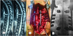
    Figure 2: (a) MRI images of SD at the Th7-8 level with a space-occupying
epidural abscess. (b - c) Intra-operative images.
Case 2: This male patient aged 51 years was admitted with SD at L3/L4 and with severe lumbar and sciatic pain, with a overall VAS score of 9. Elective surgery was performed with dorsal transpedicular fixation at L3/L4, decompression of the spinal canal, and bilateral foraminotomy L3/L4. The postoperative course was uneventful. The patient received a course of intravenous antibiotic therapy. Microbiological findings showed growth of S. aureus. A possible cause of the hematogenous dissemination was the chronic ENT infection. Postoperatively, a significant improvement of lumbar and sciatic pain occurred, with a VAS score of 2. Follow-up examination demonstrated no siginificant lumbar pain. Radiological findings showed a preserved sagittal alignment of the lumbar spine and no loosening or breakage of the transpedicular screw system (Figure 3).
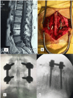
    Figure 3: (a) MRI images of SD at L3-4 level without compression of cauda
equina. b) Intraoperative image. (c - d) Antero-posterior and lateral X-ray
images after surgery.
Case 3: This male patient aged 68 years was admitted with SD L4/L5 and with severe lumbar and sciatic pain with a VAS score of 8. Elective surgery was performed with transpedicular fixation at L3-L5, hemilaminectomy of L3, bilateral foraminotomy of L4/L5 and decompression of the thecal sac. The postoperative course was uneventful. The patient received a course of intravenous antibiotic therapy. Microbiological tests showed no growth of bacteria. Postoperatively, a significant improvement of pain was observed, with a VAS score of 1. Follow-up examination demonstrated no lumbar pain. Radiological findings showed a preserved sagittal alignment of the lumbar spine and no loosening or breakage of the transpedicular screw system (Figure 4).
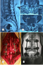
    Figure 4: (a/b) MRI images of SD at L4-5 level with spondylolisthesis and
compression of cauda equina. (c) Intraoperative image. (d) Intraoperative
antero-posterior X-ray image of instrumented fixation.
Discussion
Thoracic and lumbar SD has increased in frequency in the last decades and has become a clinical issue, with different possible treatment pathways used in different institutions [6,9,11,26]. The standard treatment is considered to be conservative, with long periods of immobilization, and is connected with the late occurrence of spinal deformities, which are very difficult to treat.
Many independent studies published in the last decades have shown that instrumented spinal fixation is a safe and effective option in cases with SD [4,19,20]. The risk of chronic infections in need of treatment in patients with instrumented spinal fixation is rather low. No such cases were encountered in our patient population.
The majority of our treatment group (83%) represented the most complex SD patients with neurological deficits. This is in line with published data in the literature. In large samples of SD patients (296 and 250 cases, respectively) with surgical and conservative treatment, the fractions with neurological deficits were 60% and 48%, respectively [4,5]. Such cases usually demand surgery.
In our series, 39 patients presented with neurological deficits and thus urgent surgical decompression was indispensable. These patients had also instability of the thoracic or lumbar spine, where instrumented fixation and stabilization was absolutely indicated. After surgery, most patients 25 (64.1%) improved neurologically, and no patient developed a worsened condition.
Posterior instrumented fixation was performed in all our cases, immediately where indicated, and in an elective manner in less acute cases. One of the main reasons for elective posterior fixation was to avoid delayed spinal deformities and to achieve stability for immediate postoperative mobilization. Intervertebral cages were used in 12 cases (26%) with an inflammatory focus located in the intervertebral disc, which was removed, and/or with signs of instability on preoperative dynamic X-ray in the segment, or with present spondylolisthesis. In our hands, posterior instrumented fixation in the majority of cases was sufficient to achieve and to preserve spinal stability and to avoid late deformity. No failures of the instrumented dorsal fixation used alone or in combination with intervertebral cages were seen in our series.
As it is already well known, infection facilitates arthrodesis even in the absence of ventral intercorporal implants or bone grafts. Some studies suggest that patients with titanium intervertebral cages have greater improvement in sagittal alignment than those without [27], but this could not be confirmed in our study The clinical outcome and the improving Cobb angle of the affected segments at final follow up were the most relevant measures for the success of our approach.
While performing surgery, it was very important to debride inflammatory and necrotic tissue, drain any epidural abscesses, correct or prevent deformity by fixation, and decompress neural structures in the spinal canal.
S. aureus was the most common pathogen with 25 cases (53.2%), which is agreeable with published epidemiological data, followed by M. tuberculosis in 4 cases (8.5%). In 13 patients (27.7%) the pathogen could not be identified. This is also reported in up to one-third of the cases in the published literature [3,28].
No patient had a clinically significant infection of the implanted transpedicular system after surgery.
Our study has some limitations due to its retrospective character and to the limited number of patients. Nevertheless, we concluded that the presence of a dorsal transpedicular implant system does not seem to impair the healing of the infection and the efficacy of intravenous antibiotics therapy. The use of an instrumented dorsal transpedicular fixation system allows for an early mobilization of the patients and avoids complications due to prolonged bed rest. Also, this approach effectively prevents clinically significant spinal deformities, as demonstrated in our patient population.
Conclusions
47 patients with thoracic and lumbar spondylodiscitis underwent dorsal transpedicular instrumented fusion combined with debridement and decompression of neural structures. Long-term results of the treatment were evaluated in 42 patients at 1 year after surgery. Most patients improved neurologically after surgery, and no patient was worsened.
No mechanical failures of the transpedicular screw fixation system occurred. The Cobb angle of the affected spinal segments did not change significantly at follow-up, compared with the value immediately after surgery.
The use of instrumented dorsal transpedicular fusion for the treatment of patients with thoracic and lumbar spondylodiscitis was safe and effective in our hands. It allowed for neurological improvement of the majority of cases and prevented late spinal deformities.
References
- Herren C, Jung N, Pishnamaz M, Breuninger M, Siewe J, Sobottke R. Spondylodiscitis: Diagnosis and Treatment Options. Dtsch Arztebl Int. 2017; 114(51-52): 875-882.
- Fantoni M, Trecarichi EM, Rossi B, Mazzotta V, Di Giacomo G, Nasto LA, et al. Epidemiological and clinical features of pyogenic spondylodiscitis. Eur Rev Med Pharmacol Sci. 2012; Suppl 2: 2-7.
- Sans N, Faruch M, Lapègue F, Ponsot A, Chiavassa H, Railhac JJ. Infections of the spinal column--spondylodiscitis. Diagn Interv Imaging. 2012; 93(6): 520-529.
- Homagk L, Homagk N, Klauss JR, Roehl K, Hofmann GO, Marmelstein D. Spondylodiscitis severity code: scoring system for the classification and treatment of non-specific spondylodiscitis. Eur Spine J. 2015; 25(4): 1012- 1020.
- Pola E, Autore G, Formica VM, Pambianco V, Colangelo D, Cauda R, et al. New classification for the treatment of pyogenic spondylodiscitis: validation study on a population of 250 patients with a follow-up of 2 years. Eur Spine J. 2017; 26(Suppl 4): 479-488.
- Pola E, Taccari F, Autore G, Giovannenze F, Pambianco V, Cauda R, et al. Multidisciplinary management of pyogenic spondylodiscitis: epidemiological and clinical features, prognostic factors and long-term outcomes in 207 patients. Eur Spine J. 2018; 27(Suppl 2): 229-236.
- Homagk L, Marmelstein D, Homagk N, Hofmann GO. SponDT (Spondylodiscitis Diagnosis and Treatment): spondylodiscitis scoring system. J Orthop Surg Res. 2019; 14(1): 100.
- Han L, Keiserrudin MA, Jensen PL Atypical presentation of spontaneous discitis: case report. Surg Neurol. 2004; 61(2): 142-143; discussion 143-144.
- Guerado E, Cerván AM. Surgical treatment of spondylodiscitis. An update. Int Orthop. 2012; 36: 413-420.
- Pola E, Rossi B, Nasto LA, Colangelo D, Logroscino CA. Surgical treatment of tuberculous spondylodiscitis. Eur Rev Med Pharmacol Sci. 2012; 16 Suppl 2: 79-85.
- Yuan S, Ma F, Wang Y, Gong P. Minimally invasive spine surgery in the treatment of pyogenic spondylodiscitis: an initial retrospective series study. Wideochir Inne Tech Maloinwazyjne. 2019; 14(2): 333-339.
- Lee BH, Park JO, Kim HS, Lee HM, Cho BW, Moon SH. Transpedicular curettage and drainage versus combined anterior and posterior surgery in infectious spondylodiscitis. Indian J Orthop. 2014; 48(1): 74-80.
- Linhardt O, Matussek J, Refior HJ, Krudel A. Long-term results of ventrodorsal versus ventral instrumentation fusion in the treatment of spondylitis. Int Orthop. 2007; 31(1): 113-119.
- Pee YH, Park JD, Choi YG, Lee SH. Anterior debridement and fusion followed by posterior pedicle screw fixation in pyogenic spondylodiscitis: autologous iliac bone strut versus cage. J Neurosurg Spine. 2008; 8(5): 405-412.
- Si M, Yang ZP, Li ZF, Yang Q, Li JM. Anterior versus posterior fixation for the treatment of lumbar pyogenic vertebrae osteomyelitis. Orthopedics. 2013; 36(6): 831-836.
- Lin TY, Tsai TT, Lu ML, Niu CC, Hsieh MK, Fu TS, et al. Comparison of twostage open versus percutaneous pedicle screw fixation in treating pyogenic spondylodiscitis. BMC Musculoskelet Disord. 2014; 15: 443.
- Karikari IO, Powers CJ, Reynolds RM, Mehta AI, Isaacs RE. Management of a spontaneous spinal epidural abscess: a single-center 10-year experience. Neurosurgery. 2009; 65(5): 919-923; discussion: 923-924.
- Itzkovich M, Gelernter I, Biering-Sorensen F, Weeks C, Laramee MT, Craven BC, et al. The Spinal Cord Independence Measure (SCIM) version III: reliability and validity in a multi-center international study. Disabil Rehabil. 2007; 29(24): 1926-1933.
- Lin CP, Ma HL, Wang ST, Liu CL, Yu WK, Chang MC. Surgical results of long posterior fixation with short fusion in the treatment of pyogenic spondylodiscitis of the thoracic and lumbar spine: a retrospective study. Spine (Phila Pa 1976). 2012; 37(25): E1572-E1579.
- Tsai TT, Yang SC, Niu CC, Lai PL, Lee MH, Chen LH, et al. Early surgery with antibiotics treatment had better clinical outcomes than antibiotics treatment alone in patients with pyogenic spondylodiscitis: a retrospective cohort study. BMC Musculoskelet Disord. 2017; 18(1): 175.
- Curry WT Jr, Hoh BL, Amin-Hanjani S, Eskandar EN. Spinal epidural abscess: clinical presentation, management, and outcome. Surg Neurol. 2005; 63(4): 364-371; discussion 371.
- Mohi Eldin MM, Ali AM. Lumbar transpedicular implant failure: a clinical and surgical challenge and its radiological assessment. Asian Spine J. 2014; 8(3): 281-297.
- Roberts TT, Leonard GR, Cepela DJ. Classifications In Brief: American Spinal Injury Association (ASIA) Impairment Scale. Clin Orthop Relat Res. 2017; 475(5): 1499-1504.
- Rainov NG, Schneiderhan R, Heidecke V. Triangular titanium implants for sacroiliac joint fusion. Eur Spine J. 2019; 28(4): 727-734.
- Luo M, Li N, Shen M, Xia L. Pedicle screw versus hybrid instrumentation in adolescent idiopathic scoliosis: A systematic review and meta-analysis with emphasis on complications and reoperations. Medicine (Baltimore). 2017; 96(27): e7337.
- Gregori F, Grasso G, Iaiani G, Marotta N, Torregrossa F, Landi A. Treatment algorithm for spontaneous spinal infections: A review of the literature. J Craniovertebr Junction Spine. 2019; 10(1): 3-9.
- Anand N, Cohen RB, Cohen J, Kahndehroo B, Kahwaty S, Baron E. The Influence of Lordotic cages on creating Sagittal Balance in the CMIS treatment of Adult Spinal Deformity. Int J Spine Surg. 2017; 30; 11: 23.
- Patel AR, Alton TB, Bransford RJ, Lee MJ, Bellabarba CB, Chapman JR. Spinal epidural abscesses: risk factors, medical versus surgical management, a retrospective review of 128 cases. Spine J. 2014; 14(2): 326-330.