
Case Report
Austin J Nephrol Hypertens. 2021; 8(2): 1097.
A Rare Culture-Negative Ascites Induced by Clostridium Difficile in a Patient with End-Stage Renal Disease: A Case Report and Literature Review
Alsultan MH¹* and Hassan Q²
1Department of Nephrology, Al Assad and Al Mouwasat University Hospitals, Damascus, Syria
2Department of Nephrology, Al Assad University Hospitals, Damascus, Syria
*Corresponding author: Alsultan MH, Department of Nephrology, Al Assad and Al Mouwasat University Hospitals, Damascus, Syria
Received: August 10, 2021; Accepted: October 07, 2021; Published: October 14, 2021
Abstract
Clostridium difficile infection was identified as the major cause of antibioticassociated diarrhea and cause wide manifestations include asymptomatic, fulminant disease and unusual manifestations such as protein-losing enteropathy. The incidence and severity of healthcare-associated clostridium difficile have been dramatically increased.
A 25-years old male with end-stage renal disease who on hemodialysis complained of nonbloody watery diarrhea and abdominal pain for a month. Also, he had a hospital admission due to secondary peritonitis with negative investigations and was treated with antibiotics with no improvement. Abdominal CT scan revealed a moderate amount of ascites with wall thickening of transverse colon and culture of ascites was negative. A stool examination was positive for clostridium difficile toxins (A+B) and cured by 21 days of oral vancomycin.
A literature review for ascites- induced by clostridium difficile yielded only one patient with end-stage renal disease on hemodialysis. First-line clinicians may not be familiar with such a rare manifestation and may not initially consider it when making differential diagnosis related to secondary peritonitis.
Clostridium difficile should be suspected in all cases of diarrhea in patients with chronic kidney disease and should be considered in the presence of ascites in the context of diarrhea with no obvious source.
Keywords: Clostridium Difficile; Culture-Negative Ascites; End-Stage Renal Disease
Introduction
Clostridium Difficile Infection (CDI) was identified as the major cause of antibiotic-associated diarrhea and colitis after widespread use of antibiotics. There are well-established risk factors for CDI include advanced age, hospitalization, severe illness, chemotherapy and gastric acid suppression. Any antibiotic can predispose to CDI even the perioperative antibiotic prophylaxis, most implicated antibiotics include clindamycin, fluoroquinolones, and broadspectrum penicillins and cephalosporins [1].
The incidence and severity of healthcare-associated CDI have been dramatically increased since 2000. The rate of nosocomial CDI in USA increased from 31 to 61 per 100,000 between 1996 and 2003, and a 20-50% rate of carriage occurs in hospitals and long-term care facilities. The incidence of recurrent and community-associated CDI is also increasing, 36% of patients with community-associated CDI in one multistate study did not receive antibiotics in the 12 weeks prior to diagnosis and up to 25% experience recurrent infection within 30 days of treatment [1].
CDI can cause a wide spectrum of manifestations ranging from an asymptomatic to fulminant disease. Also, unusual manifestations have been reported such as extracolonic involvement and Protein- Losing Enteropathy (PLE), which associates with acute CDI in the absence of fulminant colitis [2].
In this confusing case, a patient with End-Stage Renal Disease (ESRD) developed secondary peritonitis with culture-negative ascites and nearly normal albumin levels. Oral vancomycin induced improvement with no recurrence of diarrhea or abdominal pain and ultrasound showed a significant decrease of ascites.
Case Presentation
A 25-years old male was admitted to the Nephrology Department of Al Assad University Hospital due to non-bloody watery diarrhea (3-5 per day), nausea and vomiting, malaise and abdominal pain for the past month. His past medical history included kidney transplantation 12 years ago. Currently, he had End-Stage Renal Disease (ESRD) with two sessions per week of Hemodialysis (HD) for the past 2 years due to chronic allograft rejection.
Also, two weeks ago, he had hospital admission due to secondary peritonitis with antibiotics treatment (levofloxacin, ceftazidime, metronidazole), but abdominal Computed Tomography (CT) scan and ascites culture were negative.
Physical exam showed diffuse abdominal pain with shifting dullness, diminished breathing sounds in lung bases, diminish heart sounds, blood pressure (BP) 100/70 mmHg, temperature 38oC, and oxygen saturation 96%.
Chest CT scan showed bilateral pleural effusion with massive pericardial effusion (Figure 1A). Echocardiography showed congestive Heart Failure (CHF), Ejection Fraction (EF) 20-25%, large pericardial effusion measured 3.5cm on apical four chambers window, Inferior Vena Cava (IVC) diameter 1.4cm with collapsibility >50% in inspiration and no signs of cardiac tamponade. Abdominal CT scan revealed ascites, which surrounding the liver and in pelvis, with wall thickening of the transverse colon (Figure 2A).
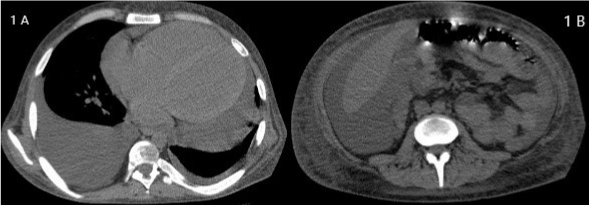
Figure 1: Chest and Abdominal CT. A) Chest; bilateral pleural effusion with massive pericardial effusion. B) Abdomen; ascites around the liver with wall thickening of the transverse colon.
Abdominal and pleural paracentesis was performed. Laboratory findings in Table 1, revealed secondary peritonitis and transudate pleural effusion with negative culture for both. The patient received ceftriaxone and metronidazole for secondary peritonitis and due to a history of inadequate HD, pericardial and pleural effusion were treated with increased HD sessions.
![]()
    Blood
    Pleural fluid
    Ascites


    HB
    7.4
    WBC
    50
    WBC
    2400


    Ur
    65
    RBC
    10
    N%
    80%


    Cr
    11
    Glu
    86
    L%
    20%


    TP
    5.5
    TP
    1.9
    RBC
    1000


    ALB
    3
    ALB
    1.1
    Glu
    75


    LDH
    326
    LDH
    230
    TP
    3.4


    ALT
    5
    Chol
    23
    ALB
    2.1


    AST
    17
    LDH ratio
    0.7
    LDH
    352


    CRP
    12.4
    TP ratio
    0.3
    SAAG
    0.9


    HBS Ag
    neg
    ALB gradient
    1.9
    Culture
    Neg


    Anti HCV
    neg
    Culture
    Neg


    HB: Hemoglobin; Ur: Urea; Cr: Creatinine; AST:    Aspartate Transaminase; ALT: Alanine Aminotransferase; LDH: Lactate    Dehydrogenase; GLU: Glucose; TP: Total Protein; ALB: Albumin; CRP: C-Reactive    Protein; HBS Ag: Hepatitis B Surface Antigen; Anti HCV: Hepatitis C Virus    Antibody; WBC: White Blood Count; N%: Neutrophils Proportion; L%: Lymphocytes    Proportion; RBC: Red Blood Cell Count; Chol: Cholesterol; SAAG: Serum Ascites    Albumin Gradient.
Table 1: Laboratory tests.
This case was confusing due to secondary peritonitis with negative abdominal CT and ascites culture on two occasions. We started the approach from chronic diarrhea. A stool examination was positive for fecal white blood cells and toxins (A+B) for clostridium difficile (C. difficile). By reviewing the literature, we found that severe and rare cases of C. difficile infection may induce culture-negative neutrocytic ascites (CNNA).
Antibiotics were discontinued and started with oral vancomycin (125mg/4 times daily). However, the patient status improved, diarrhea recurred after 10 days. This obligated an increasing dose of vancomycin (250mg/4 times daily) and combined with oral metronidazole (500mg/3 times daily) for total 21 days.
The patient was cured with no recurrence of diarrhea or abdominal pain and ultrasound showed a significant decrease of pericardial, pleural and ascites accumulations.
Discussion
A literature review for ascites induced by Clostridium Difficile Infection (CDI) yielded 16 patients in 12 case reports, Table 2 [3-14]. Previous history of antibiotic was mentioned in 12 patients [3-5,9,11- 14]. Culture-Negative Neutrocytic Ascites (CNNA) was reported in all patients except 3 cases, two of them had culture-positive ascites after abdominal interventions and ascites culture was not performed in the third case [7,8,12]. Only one patient had ESRD with HD [10], but the other 4 patients developed Acute Kidney Injury (AKI) [7-9].
![]()
Author
Age and Primary disease
Antibiotics induced CDI
Ascit/culture
Other symptoms
Diagnosis of CDI
treatment/duration
Guido Poggi et al. [3]
- 54–y
- necrotizing fasciitis
- Ciprofloxacin/clindamycin (IV).
- Ciprofloxacin/ rifampicin (PO).
+/Neg
- Non–bloody, watery diarrhea, fever, abdominal pain.
- Abdominal swelling peripheral edema.
- Pos stool for toxins.
- PO vancomycin (500mg daily/for 5 days)
Christie Masters et al. [5]
- 36-y
- cesarean section
- IV cefazolin
+/Neg
- Abdominal pain, watery diarrhea, fever, nausea, vomiting.
- small pleural effusions
- Pos stool for toxins
- Long taper of po vancomycin and metronidazole
Ali Bukhari et al. [6]
- 46-y
- ND
ND
+/Neg
- Watery diarrhea and abdominal pain.
- C. difficile- pos
ND
George I. Tsourous et al. [13]
- 60-y
- soft tissue infection/possible osteomyelitis
- Po amoxicillin/clindamycin.
+/Neg
- Watery diarrheas, fever, vomiting, abdominal pain.
- Pos stool for toxin A.
- Oral vancomycin (500mg daily/ for 15d)
Yujian Liang et al. [14]
- 6-y
- Severe pneumonia.
- Cephalosporin
+/Neg
- Diarrhea, abdominal distension, nausea, vomiting, shortness of breath.
- Massive hydrothorax.
- Pos PCR stool for toxin B.
- Pos stool culture.
- PO vancomycin for 10 d.
ELI ZUCKERMAN et al. [9]
- 54-y
- AIDS
- Trimethoprim/ sulfamethoxazole
+/Neg
- Abdominal distention pain, pedal edema, weight gain fever, nonbloody watery stools
- AKI
- Autopsy showed PMC.
ND
- 48-y
- Heroin IV usage/URI
- Erythromycin
+/Neg
- Watery stools, abdominal distention
- AKI
- Sigmoidoscopy
- Metronidazole
- 30y
- AIDS
- P. aeruginosa pneumonia
- IV ceftazidim/gentamicin.
+/Neg
- Two episodes of diarrhea: 1st nonbloody watery diarrhea. 2nd cachexia, mild lower extremity edema, ascites.
- Sigmoidoscopy.
- Pos stool culture, toxins.
- 1st metronidazole/ po.
- 2nd vancomycin/ po/15d
- 33-y
- AIDS
- Trimethoprim/sulfamethoxazole
+/Neg
- Diarrhea, increasing abdominal girth, peripheral edema.
- Pos stool culture, toxins.
- Colonoscopy.
- Metronidazole/14d.
- 58-y
- CAP
- Erythromycin
+/Neg
- Nonbloody watery diarrhea, abdominal distention
- Pos stool culture, toxins.
- Sigmoidoscopy
- Metronidazole/21d.
Aman boaz et al. [11]
- 25y
- Dental infection
- Clindamycin.
+/ND
- Green-colored mucoid diarrheas, abdominal pain.
- Pleural effusion.
- Sigmoidoscopy
- PO vancomycin
p.de leeuw et al. [7]
- 50y
- Chronic alcohol abuse/surgical duodenal ulcer
ND
+/Pos*
- No diarrhea
- Abdominal distention
- AKI
- Pos culture of ascites.
- Neg toxins and sigmoidoscopy
- IV metronidazole.
MOHAMMAD ABU HISHMEH et al. [8]
- 39-y
- Liver cirrhosis/splenic embolization
ND
+/Pos*
- Diarrhea, abdominal pain.
- MOF
- Pos blood culture and PCR stool for toxins.
- Po vancomycin / IV metronidazole.
Shweta Garg et al. [10]
- 50-y
- ESRD on HD
- AIDS
ND
+/Neg
- Abdominal distension, abdominal pain, diarrhea
- Pos stool toxins.
- Po vancomycin/IV metronidazole.
- Colonoscopy for vancomycin infusion.
F. Berhane et al. [12]
- 23 y
- Anoxic brain injury
- Linezolid
+/ND
- Fever, productive cough, vomiting, diarrhea, lethargy
- Pos antigen.
- Neg stool toxins A/B.
- Pos stool PCR.
ND
Elie Chouillard et al. [4]
- 47-y
- Adenocarcinoma/ileostomy.
- Cefuroxime
+/Neg
- Abdominal pain, diarrhea, fever.
- Toxin neg
- Stool culture pos
- 48h/IV, 10d/po metronidazole
*Positive ascites culture for CDI and both cases after surgery, 1st case showed no intestinal infection, 2nd case with intestinal infection. IV: Intravenous; Po: per os; AIDS: Acquired Immunodeficiency Syndrome; P. aeruginosa: Pseudomonas aeruginosa; CAP: Community-Acquired Pneumonia; ESRD: End- Stage Renal Disease; HD: Hemodialysis; ND: Non-Defined; Neg: Negative; Pos: Positive; AKI: Acute Kidney Injury; MOF: Multiorgan Failure; C. difficile: Clostridium difficile; PCR: Polymerase Chain Reaction; PMC: Pseudomembranous colitis; d: day; y: year.
Table 2: Summary of case studies with CDI presenting with ascites.
Several studies demonstrated ESRD as a risk factor of CDI [15- 17], this might be multifactorial due to impaired defenses from uremia, repeated hospitalizations and frequent antibiotic use [15].
A Meta-Analysis showed risk ratio of CDI in patients with Chronic Kidney Disease (CKD) and ESRD were 1.95 and 2.63, respectively [17]. Another study demonstrated that more advanced CKD correlated with a higher risk of CDI and patients with dialysis were at the highest risk [16].
The incidence of CDI in ESRD patients was 4.25%, recurrent CDI occurred in 23.6% and associated with increased mortality. Most patients with CDI (over 78%) had a hospital admission in the prior 3 months of CDI diagnosis [16].
Although the patient, in this case, did not receive antibiotics prior to CDI, he had a severe and recurrent infection. This might due to frequent hospital exposer of weekly HD sessions or impaired immunity from uremia. Reviewing records in our department, in the past 3 months, showed CDI-induced diarrhea in another 3 patients with ESRD, who demand hospital admission.
Ascites is a rare manifestation and is not considered a well-known characteristic of severe CDI. Several mechanisms were described to explain ascites induced by CDI, which include Protein-Losing Enteropathy (PLE) secondary to bowel wall inflammation, which allows leakage of albumin into the lumen [2]. Also, transmural colonic inflammation with microperforation and toxin-mediated inflammatory cytokines causing vascular permeability have been described [6].
Herein, the patient had nearly normal albumin and colonic wall thickening, therefore, it is likely that the ascites was a result of transmural colonic inflammation with cytokines-induce vascular permeability.
In the end, first-line clinicians may not be familiar with such a rare manifestation of CDI and may not initially consider it when making differential diagnosis related to secondary peritonitis, which delays the diagnosis as in our case.
Conclusion
In conclusion, CDI should be suspected in all cases of diarrhea in patients with CKD even with no prior antibiotics use. Also, high suspicion of CDI should be considered in the presence of ascites in the context of diarrhea with no obvious source.
References
- JT Lamont, SB Calderwood, et al. Clostridium difficile infection in adults: Epidemiology, microbiology and pathophysiology. Up-to-date.
- JT Lamont, CP Kelly, JS Bakken, SB Calderwood, EL Baron. Clostridium difficile infection in adults: Clinical manifestations and diagnosis. Up-to-date. 5-24.
- Poggi G, Aprile C, Pozzi E. An unusual case of ascites. Int J Case Reports Images. 2013; 4: 32-36.
- Chouillard E, Chouillard MA, Kary NEl, De Simone B, Gumbs AA. Clostridium difficile infection secondary to ileostomy closure. Mini-invasive Surg. 2021; 2021: 9-14.
- Masters C, Mha MBA, Adams SR. Protein-losing Enteropathy and Ascites Associated with Clostridium difficile Infection in a Peripartum Woman. Proc UCLA Healthc. 2014; 18: 1-4.
- Ali Bukhari, Daniel Opris, Amish Patel, Sravya Surapaneni. Culture Negative Neutrocytic Ascites in a Severe Clostridium difficile Infection. Am J Gastroenterol. 2014; 109: 661-664.
- P de Leeuw, Helene de Mot, T Dugernier, J Wautelet, E Bohy. Primary infection of ascitic fluid with Clostridium difficile. J Infect. 1990: 77-80.
- Hishmeh MA, Akbar S, Kumar A. Spontaneous Bacterial Peritonitis and Bacteremia Secondary to Clostridium Difficile Enteritis in Patient with Liver Cirrhosis. Chest. 2019; 156: A1831-A1832.
- ELI ZUCKERMAN GK, CHUNG HA, JEFFREY KAHN BG, KORULA J. Low Albumin Gradient Ascites Complicating Severe Pseudomembranous Colitis. Am Gastroenterol Assoc. 1997: 243-245.
- Shweta Garg, Farzin Rashti, Ekta Gupta, Michael Gold. Pseudomembranous Colitis Presenting with Massive Ascites. Am J Gastroenterol. 2013: 6-7.
- Boaz A, Dan M, Chamzi I, Landau O, Aloni Y, Kyzer S. Pseudomembranous Colitis Report of a Severe Case with Unusual Clinical Signs in a ypung nurse. Dis Colon Rectum. 2000; 14-16.
- Berhane F, Leys L, Moeng L, Thomas A, Poddar V. Atypical Presentation of Clostridium Difficile Infection. Crit CARE CASE REPORTS Infect SEPSIS. 2020; A6921-A6921.
- George I. Tsourous, Leonidas G. Raftopoulos, Eleni E. Kafe, Eleftherios K. Manoleris, Kostas P. Makaritsis SGP. A case of pseudomembranous colitis presenting with massive ascites. Eur J Intern Med. 2007; 18: 328-330.
- Liang Y, He X, Wang T, Chen Y, Huang H, Tang W, et al. Massive Hydrothorax and Ascites as the Primary Manifestation of Infection With Clostridium difficile: A Case Report and Literature Review. Front Pediatr. 2020; 8: 1-6.
- Tirath A, Tadros S, Coffin SL, Kintziger KW, Waller JL, Baer SL, et al. Clostridium difficile infection in dialysis patients. J Investig Med. 2017; 65: 353-357.
- Kim SC, Seo MY, Lee JY, Kim KT, Cho E, Kim MG, et al. Advanced chronic kidney disease: A strong risk factor for Clostridium difficile infection. Korean J Intern Med. 2016; 31: 125-133.
- Phatharacharukul P, Thongprayoon C, Cheungpasitporn W, Edmonds PJ, Mahaparn P, Bruminhent J. The Risks of Incident and Recurrent Clostridium difficile-Associated Diarrhea in Chronic Kidney Disease and End-Stage Kidney Disease Patients: A Systematic Review and Meta-Analysis. Dig Dis Sci. 2015; 60: 2913-2922.