
Research Article
Austin J Musculoskelet Disord. 2023; 10(1): 1062.
Extremity Myxoid Liposarcoma: Prognostic Factors and 20-Year Survival Outcomes
Machado P¹*, Peiró A¹, Trullols L¹, Sebio A1,2, Orellana R¹ and Gracia I1,2
¹Department of Orthopaedic Oncology, Hospital de la Santa Creu i Sant Pau, Barcelona, Spain
²Institutd’Investigació Biomèdica Sant Pau (IIB SANT PAU), Barcelona, Spain
*Corresponding author: Pau MachadoDepartment of Orthopaedic Oncology, Hospital de la Santa Creu i Sant Pau, Barcelona, Spain
Received: December 09, 2022; Accepted: January 17, 2023; Published: January 24, 2023
Abstract
Introduction: Myxoid liposarcoma (MLS) is a rare, malignant musculoskeletal tumour. Due to the rarity of MLS, the factors associated with survival remain only partially understood. The aim of this retrospective study was to identify prognostic factors in patients with extremity MLS treated at our centre.
Methods: We reviewed the clinical records of 60 patients with extremity MLS treated at our hospital from 1997 to 2017. We evaluated patient- and tumour-related characteristics, as well as diagnostic and therapeutic procedures to determine their association with clinical outcomes, including local Recurrence-Free Survival (RFS), Metastasis-Free Survival (MFS) and Overall Survival (OS). Survival data were analyzed with the Kaplan-Meier method. Multivariate analyses were performed with Cox proportional-hazards regression.
Results: Overall survival at 5 years was 91%. On the multivariate analysis, surgical margins were an independent risk factor for MFS (Hazard Ratio [HR]: 10.98, 95% Confidence Interval [CI]:2.59-46.46), local RFS (HR: 9.34, 95% CI: 1.75-49.68), and OS (HR: 27.90, 95% CI: 5.01–155.50) (all p=0.001). Revision surgery was significantly associated (p=0.001) with higher local recurrence rates (HR: 99.13, 95% CI: 6.90–1423).
Conclusions: Surgical margins were an independent risk factor for MFS, local RFS, and OS. The need for revision surgery due to an unplanned excision was strongly associated with an increased risk of local recurrence, but not with MFS or OS. These findings underscore the importance of achieving clear surgical margins, especially in patients undergoing second-look surgery.
Keywords: Myxoid liposarcoma; Surgical Margins; Revision Surgery; Overall Survival; Local Recurrence-Free Survival.
Introduction
Myxoid liposarcoma (MLS) is a rare, malignant musculoskeletal tumour composed of uniform, round to ovoid cells with variable numbers of lipoblasts, set in a myxoidstroma with a branching capillary vasculature. MLS accounts for approximately 20%-30% of all liposarcomas and 5% of all soft tissue sarcomas in adults, without significant sex predilection [1]. MLS typically presents as large, painless masses. Retroperitoneal MLS is usually due to a metastasis (M1) [2]. Multifocal disease, either synchronous or metachronous, indicates the presence of distant soft tissue metastases of monoclonal origin [3]. Diagnosis is supported by the detection of the characteristic chromosomal recurrent translocation, t (12;16) (q13;p11), which is present in > 95% of cases and indicates the aberrant fusion gene FUS-CHOP/DDIT3 [4]. Prognostic factors include age at diagnosis, tumour size and grade, depth, and surgical margins [5].
Currently, the mainstay of treatment for extremity MLS is wide excision surgery. Adjuvant radiotherapy plays an increasingly important role in the treatment of MLS, especially for the control of local recurrence. In fact, radiotherapy, with or without chemotherapy, has become common in the treatment of MLS [6]. Compared to other soft tissue sarcomas, MLS is re sponsive to chemotherapy, particularly anthracycline-based combinations and ifosfamide. In recent years, the combination of surgery, radiotherapy, and chemotherapy has been increasingly used to lower recurrence rates and increase both local control and overall survival (OS) [7]. Nevertheless, it is not clear which clinical and demographic variables have the greatest impact on survival outcomes.
In this context, we reviewed the clinical records of patients treated for extremity MLS at our institution in order to identify the variables that were independently associated with survival outcomes in this patient population.
Materials and Methods
This retrospective study included 60 patients treated for extremity MLS at our hospital (Hospital de la Santa Creu i Sant Pau; Barcelona, Spain) from 1997 to 2017. The study variables were evaluated to identify those that were independently associated with survival outcomes. We did not assess the influence of these variables by group or category. The following variables were included: patient characteristics (age and sex); tumour characteristics (size, location, stage, and histology); diagnostic and therapeutic procedures (type of surgery, margins, complications, neoadjuvant and adjuvant therapies). Survival outcomes were local Recurrence-Free Survival (RFS), Metastasis-Free Survival (MFS), and OS.
We included all patients with a pathological diagnosis of extremity MLS who underwent primary surgery at our centre. All of these patients underwent general examination before treatment, including magnetic resonance imaging (MRI) of the tumour. The diagnosis was based on clinical and imaging data, and confirmed preoperatively by biopsy and postoperatively by pathological examination of the resected tissue.
We also included patients referred from other centres for second-look surgery after an unplanned excision. Thus, the study included two patient profiles: patients who were diagnosed and underwent primary surgery at our hospital (n=45) and patients who underwent second-look surgery at our hospital (n=15). All of these patients had a marginal resection at some part of the tumour and all underwent MRI after the initial surgery, without evidence of macroscopic residual tumour at the surgical site.
Exclusion criteria were: (i) metastasis during initial treatment at our centre, (ii) incomplete clinical, radiographic, and/or pathologic records; (iii) lack of standardized follow-up data; (iv) death from other causes.
Follow-up time was calculated from the initial surgery until the final follow-up examination in the year 2019. All data were obtained from clinical records.
All cases (both primary and second-look patients) were reviewed by the multidisciplinary tumour board to select the most appropriate treatment. In all cases, the primary treatment decision was wide-margin surgery, which was performed in an attempt to completely eliminate the tumour tissue, according to International Union against Cancer (UICC) standards (R0 resection) [8]. In cases in which the tumour was located adjacent to critical structures (e.g., nerves, blood vessels, or bones), a planned marginal surgery was accepted (R1).
Most of the patients (57/60; 95%) underwent pre- or post-operative radiotherapy. However, in three cases (second-look surgery subgroup), radiotherapy was not considered necessary due to the expanded margins.
Chemotherapy was administered as appropriate in accordance with the tumour size, grade, and location. The chemotherapy type and schedule were based on the Spanish Sarcoma Research Group (GEIS) protocol in place at the time of treatment. This protocol consisted of three or five cycles of epirubicin (60mg/m2, days 1-2) or adriamycin (60-75mg/m2) plus ifosfamide (3g/m2, days 1-3) administered every 21 days.
During the postoperative follow-up, the patients were re-examined every 3 months for the first two years, every 6 months from years two to five, and annually thereafter. Local disease control, metastases and survival outcomes were recorded.
Statistical Analysis
All survival data were analysed using the Kaplan-Meier method. Multivariate statistics were performed using the Cox proportional-hazards regression model. Multiple comparisons of specific values between the groups were performed. Analysis of variance and chi-square tests were used for comparisons. Differences were considered statistically significant at p<0.05. The Stata statistical software package (v. 14) was used for data analysis.
Results
General Results
A total of 60 patients (36 men, 24 women) with extremity MLS were included. The mean age was 47.1 years and the mean tumour diameter was 11.5 cm. Most tumours (n=58) were located in the lower extremities. Distribution according to cancer stage (AJCC TNM system) was as follows: stage 1 (36.7%), stage 2 (46.7%), and stage 3 (16.7%).
Wide resection (R0) was achieved in 49 patients and marginal resection (R1) in 11 patients. Most patients (95%) received radiotherapy and 38.3% received chemotherapy (Table 1).
![]()
    Characteristic

    N (%)*


    Age, mean (range)

    47.1 (19-79)


    Sex


    Male

    36 (60)


    Female

    24 (40)


    Tumour location


    Upper extremity

    2 (3.3)


    Lower extremity

    58 (96.7)


    Mean tumour size, cm    (range)

    11.51 (4.5-29)


    Stage


    I

    22 (36.7)


    II

    28 (46.7)


    III

    10 (16.7)


    Surgical margins


    Wide

    49 (81.7)


    Marginal

    11 (18.3)


    Chemotherapy

    23 (38.3)


    Radiotherapy

    57 (95)
Table 1: Clinical and demographic characteristics of the patients (n=60) with primary extremity myxoid liposarcoma.
Overall Survival
The mean follow-up period was 115.1 months (range, 24-264). A total of eight MLS-related deaths (13.3%) were observed during follow-up. Five-year OS was 91.0% (95% Confidence Interval [CI]: 82.3–97.5).Of the 11 patients (18.3%) with an R1 resection, six (54.6%) died from disease-related causes a few years after surgery (Table 2).
On the multivariate analysis, surgical margins were an independent risk factor for OS (Figure 1). R1 resections were significantly associated (p=0.001) with worse OS: Hazard Ratio (HR) = 27.90 (± 24.45) (95% CI: 5.01–155.50).
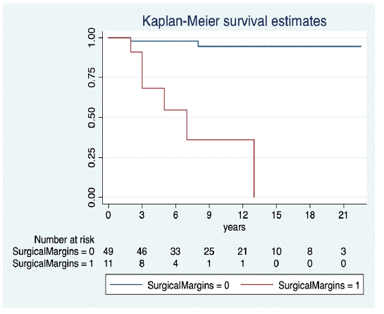
Figure 1: Kaplan-Meier curves showing the effect of surgical resection on overall survival.
On the multivariate analysis, tumour recurrence rates differed significantly between the two types of surgical resection (R0 vs. R1): χ²=5.76, Pr> chi2=0.016, with an HR (± standard deviation [SD]) of 9.34 (± 7.96) (95% CI: 1.75-49.68). A significant between-group difference was also observed for metastasis: χ²=20.64, Pr>chi2=0.001, with an HR of 10.98 (±8.08); 95% CI: 2.59-46.46) (Figure 2).
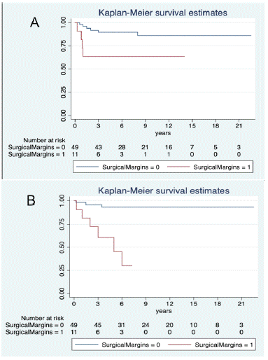
Figure 2: Kaplan-Meier curves showing the effect of surgical resection on tumour recurrence (A) and metastasis (B).
In the primary surgery group (n=45), the resection was considered R0 in 82.2% (n=37) of patients and R1 in 17.8% (n=8). In the second-look group (n=15), 80% of patients (n=12) had an R0 resection and 20% R1 (n=3).
Recurrences were observed in 10 patients, mainly in the second-look group, in which eight patients (53.3%) developed local recurrence, and three of those eight patients developed distant metastases. No association was observed between second-look surgery and metastasis, but there was a significant association between second-look surgery and recurrence; χ²=21.55, Pr>chi2=0.001; HR: 99.13 +/- 134.73 (95% CI: 6.91-1423.03).
On the univariate analysis, five-year local RFS was 52.5% (95% CI: 26.5 - 75) in the second-look group and 86.5% (95% CI: 79.5-100) in the primary surgery group (Figure 3).
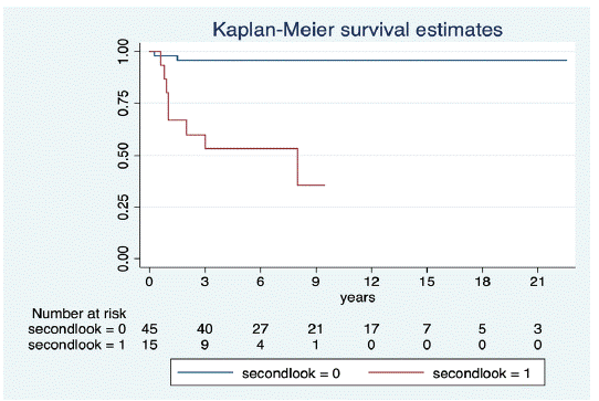
Figure 3: Kaplan-Meier curves showing the influence of second-look surgery on tumour recurrence.
Influence of Radiotherapy and Chemotherapy on Survival Outcomes
Most patients (57/60; 95%) underwent neoadjuvant or adjuvant radiotherapy (60 Gy). The remaining three patients (5%) did not require radiotherapy due to the wide margins achieved in the second-look surgery.
A total of 23 patients (38.3%) received pre- or post-operative chemotherapy (epirubicin or adriamycin plus ifosfamide). Of these, four (17.4%) developed a recurrence, two of whom later developed metastases. Five other patients who received chemotherapy (21.7%) developed metastases without local recurrence and all five later died from the disease.
We found no significant association between chemotherapy or radiotherapy and OS, local recurrence, or metastasis.
Discussion
In this study, we evaluated a series of clinical and demographic variables to determine their influence on survival outcomes in patients who underwent primary or second-look surgery for extremity MLS at our hospital. Overall survival was 91% at 5 years. Only two variables - surgical margins and revision surgery - were significantly associated with survival. Surgical margins were an independent risk factor for MFS (HR: 10.98), local RFS (HR: 9.34), and OS (HR: 27.90). Second-look surgery was associated with significantly higher local recurrence rates (HR: 99.13, p=0.001), although no between-group differences were observed for MFS or OS.
The resection margin is one of the most important factors affecting survival in patients with many cancers, including soft-tissue sarcomas [9]. The recommended treatment for soft-tissue sarcoma is wide-resection surgery; however, this approach is not feasible for some extensive tumours. Importantly, one study found that the recurrence rate in patients with R1 and R0 margins was similar after eight years of follow-up [10]. Nevertheless, Zheng et al. found that wide resections were associated with lower local recurrence rates than marginal resections, but without significant between-group differences in survival outcomes [11]. On the other hand, in a study involving 48 patients, Muratori et al. found that surgical margins were associated with both MFS and OS, but not with local RFS [12].
Although the role of chemotherapy in the treatment of soft tissue sarcoma remains controversial, adjuvant chemotherapy is recommended in high-grade and large MLS tumours, which have shown good chemosensitivity to different chemotherapeutic agents [13]. In our series, 23 patients received chemotherapy (epirubicin and ifosfamide) and seven later died of the disease. Despite the high mortality rate in this subgroup (30.4%), this does not necessarily indicate a lack of efficacy for chemotherapy; rather, it reflects the advanced disease in these patients, which is why they received adjuvant chemotherapy.
Similar to other cancers, the disease stage is an important prognostic factor for soft tissue sarcomas and advanced stages are associated with a worse prognosis [14]. Surprisingly, however, disease stage was not an independent risk factor in our cohort for any of the survival measures. Although the reason for this unexpected finding it not clear, we believe that it is due to the sample size and the staging system (TNM), which resulted in many small subgroups. Our results might have been different if we had classified patients into low- and high-grade disease instead, or used the Enneking staging system.
Although radiotherapy for extremity MLS can be performed in both the pre- and post-operative settings, pre-operative radiotherapy offers several potential advantages, including a lower rate of late complications, lower radiation doses, and the potential to improve tumour resacability [15]. Chung et al. evaluated radiotherapy in 88 patients with marginal resections, reporting a five-year local RFS of 98%, with only two recurrences [16]. In our series, all but three of the patients received radiotherapy. Given that most patients (95%) in our series received radiotherapy, we are unable to draw any definitive conclusion regarding the clinical efficacy of radiotherapy, which would require a control group to permit direct comparisons.
It is estimated that 19% to 53% of patients from other hospitals are referred to specialized sarcoma centres after unplanned excisions. The risk of residual disease in patients who undergo unplanned resections ranges from 24%-60% [17]. Tumour recurrence is 2.2 times more common in these patients than in those who had received appropriate work-up prior to surgery [18]. In most cases, unplanned resection of soft tissue sarcomas leads to incomplete tumour resections [19]. Chandrasekar et al. found that residual tumours were present in approximately 60% of revision surgeries performed to correct insufficient margins [20]. Other studies have found that local relapse and metastasis rates are greater in patients who undergo second-look surgery [21].
Standardized surgery in a specialized cancer centre could benefit patients with extremity MLS. Although our data suggest that chemotherapy and radiotherapy may be useful for MLS, we cannot make any definitive claims in that regard due to the retrospective study design.
This study has several limitations, mainly the retrospective study design, which impedes our ability to evaluate the influence of adjuvant treatments (e.g., chemotherapy), which were prescribed selectively according to the cancer stage. As a result, we cannot reach any definitive conclusions regarding the use of surgery alone or surgery plus chemotherapy. By contrast, an important strength is the large sample size (n=60), which is among the larger cohorts reported to date for this rare cancer type.
Conclusions
Surgical margins were found to be independent risk factor of MFS, local RFS, and OS. By contrast, cancer stage, radiotherapy, and chemotherapy were not independent risk factors for any of the survival outcomes.
In our series, revision surgery to correct insufficient margins due to an unplanned excision was strongly associated with a significant increase in local recurrence, but not with MFS or OS. These findings underscore the importance of achieving clear surgical margins, especially in patients undergoing second-look surgery, who should be referred to a tertiary care centre whenever possible.
References
- WHO Classification of Soft Tissue and Bone Tumours, IARC Press, Lyon, France, 5th edition, 2020.
- Setsu N, Miyake M, Wakai S, Nakatani F, Kobayashi E, et al. Primary retroperitoneal myxoid liposarcomas. Am J Surg Pathol. 2016; 40: 1286-90.
- Antonescu CR, Elahi A, Healey JH, Brennan MF, Lui MY, et al. Monoclonality of multifocal myxoid liposarcoma: confirmation by analysis of TLS-CHOP or EWS-CHOP rearrangements. Clin Cancer Res. 2000; 6: 2788-93.
- CR Antonescu, SJ Tschernyavsky, R Decuseara, DH Leung, Woodruff JM, et al. Prognostic impact of P53 status, TLS-CHOP fusión transcript structure, and histological grade in myxoid liposarcoma: a molecular and clinicopathologic study of 82 cases. Clinical Cancer Research. 2001; 7: 3977-3987.
- LC Moreau, R Turcotte, P Ferguson, Wunder J, Clarkson P, et al. Myxoid/round cell liposarcoma (MRCLS) revisited: An analysis of 418 primarily managed cases. Annals of Surgical Oncology. 2012; 19: 1081-1088.
- Kosela-Paterczyk H, Szumera-Cieckiewicz A, Szacht M, Haas R, Morysinski T, Dziewirski W, et al. Efficacy of neoadjuvanthypofractionated radiotherapy in patients with locally advanced myx oidliposarcoma. Eur J Surg Oncol. 2016; 42: 891-8.
- R Ratan, SR Patel. Chemotherapy for soft tissue sarcoma. Cancer. 2016; 122: 2952-2960.
- Stoeckle E, Coindre JM, Kind M, Kantor G, Bui BN. Evaluating Surgery Quality in Soft Tissue Sarcoma. Recent Results in Cancer Research. 2009: 229–242.
- Kim HS, Lee J, Yi SY, Jun HJ, Choi YL, et al. Liposarcoma: exploration of clinical prognostic factors for risk based stratification of therapy. BMC Cancer. 2009; 9: 205.
- Lemeur M, Mattei JC, Souteyrand P, Chagnaud C, Curvale G, Rochwerger A. Prognostic factors for the recurrence of myxoid liposarcoma: 20 cases with up to 8 years follow-up. Orthop Traumatol Surg Res. 2015; 101: 103-107.
- Zheng K, Yu XC, Xu M, Yang Y. Surgical Outcomes and Prognostic Factors of Myxoid Liposarcoma in Extremities: A Retrospective Study. Orthop Surg. 2019; 11: 1020-1028.
- Muratori F, Bettini L, Frenos F, Mondanelli N, Greto D, Livi L, et al. Myxoid liposarcoma: prognostic factors and metastasic pattern in a series of 148 patients treated at a single institution. Int J SurgOncol. 2018; 2018: 8928706.
- NCCN, “NCNN clinical practice guidelines in oncology. Softtissue sarcoma. NCCN evidence blocks. Version2,” 2017, https://www.nccn.org/professionals/physician_gls/pdf/sarcoma_blocks.pdf
- Nishida Y, Tsukushi S, Nakashima H, Ishiguro N. Clinicopathologic prognostic factors of pure myxoid liposarcoma of the extremities and trunk wall. Clin Orthop Relat Res. 2010; 468: 3041-6.
- Peterson JJ, Kransdorf MJ, Bancroft LW, O’Connor MI. Malignant fatty tumors: classification, clinical course, imaging appearance and treatment. Skeletal Radiol. 2003; 32: 493-503.
- Chung PW, Deheshi BM, Ferguson PC, Wunder JS, Griffin AM, et al. Radio sensitivity translates into excellent local control in extremity myxoid liposarcoma: a comparision with other soft tissue sarcomas. Cancer. 2009; 115: 3254-61.
- HM Umer, M Umer, I Qadir, N Abbasi, N Masood. Impact of unplanned excision on prognosis of patients with extremity soft tissue sarcoma. Sarcoma. 2013: 2013: 498604.
- LG Shapeero, PJL de Visschere, KL Verstraete, B Poffyn, R Forsyth, G Sys, et al. Post-treatment complications of soft tissue tumours. Eur J Rad. 2009; 69: 209-221.
- M Fiore, PG Casali, R Miceli, L Mariani, R Bertulli, L Lozza, et al. Prognostic effect of re-excision in adult soft tissue sarcoma of the extremity. Ann Surg Oncol. 2006; 13: 110-117.
- CR Chandrasekar, H Wafa, RJ Grimer, SR Carter, RM Tillman, A Abudu. The effect of an unplanned excision of a soft-tissue sarcoma on prognosis. J Bone Joint Surg Br. 2008; 90-B: 203-207.
- M Venkatesan, CJ Richards, TA McCulloch, AG Perks, A Raurell, et al. Inadvertent surgical resection of soft tissue sarcomas. Eur J Surg Oncol. 2012; 38: 346-351.