Case Report
J Mol Biol & Mol Imaging. 2014;1(1): 3.
A Rare Presentation of Low-Grade Mucoepidermoid Carcinoma in a Young Male
Zhiling Ding, Xiaoli Lan*, Anand Gungadin, Yueli Tian, Zhijun Pei, Rui An and Yongxue Zhang
Department of Nuclear Medicine, Huazhong University of Science and Technology, China
*Corresponding author: Xiaoli Lan, Department of Nuclear Medicine, Union Hospital, Tongji Medical College of Huazhong University of Science and Technology, Hubei Province Key Laboratory of Molecular Imaging, Wuhan, 430022, No.1277, Jiefang Ave., Wuhan, Hubei Province, P.R.CHINA
Received: August 13, 2014; Accepted: September 14, 2014; Published: September 16, 2014
Abstract
Mucoepidermoid Carcinoma (MEC) is a rare tumor of the lung. In this case, we report an 18-year-old male patient who had an ongoing cough, green expectoration and chest pain for more than 3 months. An irregular nodular lesion with no fluorine-18 ?uorodeoxyglucose (18F-FDG) uptake in the lower lobe of the left lung was found on positron emission tomography/computed tomography (PET/CT). In the diagnostic impression, we made a misdiagnosis of lung infection (pneumonia). Histopathological result however revealed the lesion was a lowgrade MEC. This case was an uncommon and challenging one which should prompt diagnostic imaging specialists to consider this rare malignant disease as one of the differential diagnosis when assessing young adults presenting with a history of repeated cough and a confirmed lung lesion which shows no improvement with standard treatment.
Keywords: Mucoepidermoid Carcinoma; Diagnosis; Computed tomography; Positron emission tomography
Introduction
Mucoepidermoid Carcinoma (MEC) is a rare tumor of the lung, which arises from the minor salivary glands in the tracheo-bronchial tree. Previous reports showed that the lesions commonly present as smoothly oval or lobulated airway masses that are usually located in a segmental bronchus with accompanied obstructive pneumonia or atelectasis on computed tomography (CT) [1]. In this case of an 18-year-old male patient, an irregular nodular lesion with no fluorine-18 fluorodeoxyglucose (18F-FDG) uptake in the lower lobe of the left lung was found on positron emission tomography (PET)/ computed tomography (CT). In the diagnostic impression, we made a misdiagnosis of lung infection (pneumonia). Histopathological result however revealed the lesion was a low-grade MEC.
Case Report
An 18-year-old male patient had an ongoing cough, green expectoration and chest pain for more than 3 months. The patient was diagnosed as having a left basal pneumonia in his local community hospital and antibiotics drugs were administered (the specific drugs used being unknown). Neither the cough nor the chest pain was relieved after treatment. The patient's chest pain sharply increased at rest, upon deep breathing and on exertion. The patient has no history of hemoptysis, fever and night sweats, and denied any past history of smoking, tuberculosis and of exposure to specific substances.
The patient's physical examination was normal. Laboratory tests including blood cell counts were all within normal limits.
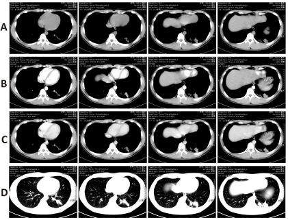
CT images: An irregular nodular lesion at the basal segment of the left lower lobe was found on chest CT. The lesion was about 1.5cm x 2.3cm in size with an average CT value of 22 Hounsfield Unit (HU). An enhanced dual-phase CT scan was performed and the CT values of 69HU and 73HU were recorded (Figure 1).

Figure 1: A series images of CT scan. a, b, c and d indicate CT plain scan, contrast enhanced CT (artery phase), contrast enhanced CT (venous phase) and lung window images, respectively. An irregular nodular lesion at the basal segment of the left lower lobe was found, and the lesion moderately enhanced.
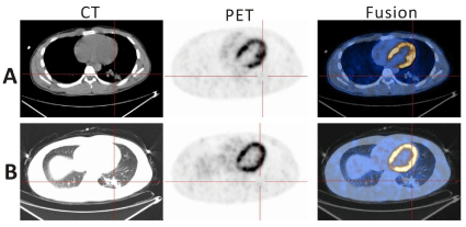
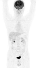
PET/CT images: An irregular nodular lesion was noted with no abnormal 18F-FDG uptake of the corresponding parts in left lower lung (Figure 2). There were no obviously enlarged lymph nodes and abnormal 18F-FDG distribution in the mediastinum, bilateral hilar areas and remaining lung and organs (Figure 3). The primary diagnosis of the lesion was benign, most probably an infectious disease.

    Figure 2:  18F-FDG PET/CT images.
An irregular nodular lesion was noted with no abnormal 18F-FDG uptake of the
corresponding parts in left lower lung (a and b). a and b are the same section
from mediastinal window and lung window, respectively.

    Figure 3:  18F-FDG PET/CT Maximum Intensity Projection (MIP) image which
showed no obvious abnormal uptake in the whole body.
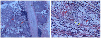
Follow-up: The patient underwent bronchoscopy which showed cauliflower-like growth in the basal segment of the lower left lung. Pathological examination suggested mucoepidermoid carcinoma. Surgical treatment of patient was as follows: a resection of the lower left lung and mediastinal lymph nodes with intercostal nerve freezing. Pathological diagnosis confirmed well-differentiated mucoepidermoid carcinoma in the lower left lung lesion (Figure 4); resected bronchial margins showed no cancer invasion, inspection of five lymph nodes showed no metastatic involvement. The patient was followed up for a period of 30 months. He recovered well postsurgery. Follow-up chest CT scans or X rays were done every 6 months and the reports indicated post-resection changes in the lower left lung area with no residual tumor, metastasis or recurrence.

    Figure 4:  Low-grade mucoepidermoid carcinoma (red arrow in a) shows a
mixture of large amount of mucoid cells (red arrow in b) and epidermis cells
(yellow arrow in b) (H&E staining; a: x40; b: x400).
Discussion
Lung mucoepidermoid carcinoma has an extremely low incidence, accounting for only 0.1% -0.2% of primary lung tumors originating within the minor salivary glands of the trachea or the bronchus. The age group involved, according to reports in the literature, ranges from 3 months to 78 years of age, with more than half of the cases occurring younger than 30 years of age [2]. MEC is classified as high-grade or low-grade on the basis of histological criteria. The vast majority of mucoepidermoid lung carcinomas are low-grade malignancies with an excellent prognosis after surgery. Although the disease is generally considered to be an inactive tumor, it can be locally aggressive and can metastasize to lymph nodes. Reports of MEC as a cause of death have also been recorded [3]. The main clinical manifestations in patients are cough, hemoptysis, wheezing and repeated episodes of pneumonia as a result of bronchial obstruction, but the 9%-28% of patients are asymptomatic. In this case, the young man also had ongoing cough and could not be cured after antibiotics treatment because the bronchial obstruction was caused by the tumor.
Lung MEC has some special features in CT imaging. Firstly, lesions are usually solitary endobronchial nodule or mass with or without obstructive pneumonia or atelectasis [3]. Secondly, a portion of MEC tissues have rich blood vessels; the contrastenhanced CT scan demonstrated that the tumors were significantly strengthened. Thirdly, the tiny sand like calcifications seen in most of the MEC tumors can be easily displayed in CT imaging. Punctate or intratumoral microscopic calcification may be one of the interesting features of lung MEC mass [4]. The incidence of calcification in MEC of the lung was much higher than that of the more common forms of the pulmonary carcinomas (up to 14%). Do P R et.al [5] suggested that the mechanism of calcification in MEC may be a result of a secretory function of the tumor cells. Lastly, lung MEC has low incidence of mediastinal and hilar lymph node metastasis, even in poorly differentiated tumors [4]. However, Li X et.al [6] reported that 2 patients had mediastinal lymph node metastasis, and one of them also had bone metastasis. In this case, no enlarged mediastinal and hilar lymph nodes were found from CT imaging, and five excisional lymph nodes were also certified benign.
Application of 18F-FDG PET/CT imaging in the evaluation for most bronchial tumors is very meaningful but limited due to rarity of MEC. Reports of 12 cases of 18F-FDG PET / CT imaging studies showed maximum selective uptake values (SUVmax) of 0 to 6.2 for well differentiated MEC and of 2.86 to 23.4 for poorly differentiated MEC [1-3,7-9]. Later, Jindal T et al. [10] reported 3 cases of pulmonary lesions with mild uptake of 18F-FDG, which after histopathological examination, were consistent with low-grade MEC in all patients. This led to the conclusion that SUV max could be a histological predicting factor for MEC.
In this case, the irregular nodular lesion with unclear margins is similar to infections changes in CT imaging and different from the nodules or masses mentioned in most of the MEC cases reported before. The lesion had some similar characteristic on CT images as CT features mentioned above. For example, the lesions significantly strengthened on the contrast-enhanced CT, and calcification could be seen in the lesion. However, these features were not specific for malignant tumors. Moreover, no obvious uptake of 18F-FDG was seen in the lesion which in general suggests benign diseases. In addition, the age of the patient does not fall within the high incidence range of malignant tumor of lung. Even though imaging findings in this case led to the given diagnosis of a benign lesion (infectious), it was a misdiagnosis. This case was an uncommon and challenging one which should prompt diagnostic imaging specialists to consider this rare malignant disease as one of the differential diagnosis when assessing young adults presenting with a history of repeated cough and a confirmed lung lesion which shows no improvement with standard treatment.
References
- Ishizumi T, Tateishi U, Watanabe S, Maeda T, Arai Y. F-18 FDG PET/CT imaging of low-grade mucoepidermoid carcinoma of the bronchus. Ann Nucl Med. 2007; 21: 299-302.
- Lee EY, Vargas SO, Sawicki GS, Boyer D, Grant FD, Voss SD. Mucoepidermoid carcinoma of bronchus in a pediatric patient: (18)F-FDG PET findings. Pediatr Radiol. 2007; 37: 1278-1282.
- Jeong SY, Lee KS, Han J, Kim BT, Kim TS, Shim YM, et al. Integrated PET/CT of salivary gland type carcinoma of the lung in 12 patients. AJR Am J Roentgenol. 2007; 189: 1407-1413.
- Kim TS, Lee KS, Han J, Im JG, Seo JB, Kim JS, et al. Mucoepidermoid carcinoma of the tracheobronchial tree: radiographic and CT findings in 12 patients. Radiology. 1999; 212: 643-648.
- do Prado RF, Lima CF, Pontes HA, Almeida JD, Cabral LA, Carvalho YR. Calcifications in a clear cell mucoepidermoid carcinoma: a case report with histological and immunohistochemical findings. Oral Surg Oral Med Oral Pathol Oral Radiol Endod. 2007; 104: e40-44.
- Li X, Zhang W, Wu X, Sun C, Chen M, Zeng Q. Mucoepidermoid carcinoma of the lung: common findings and unusual appearances on CT. Clin Imaging. 2012; 36: 8-13.
- Kinoshita H, Shimotake T, Furukawa T, Deguchi E, Iwai N. Mucoepidermal carcinoma of the lung detected by positron emission tomography in a 5-year-old girl. J Pediatr Surg. 2005; 40: E1-3.
- Yamada T, Chiba W, Yasuba H, Shimada T, Kudo M, Hamada K, et al. [Successful treatment of bronchial mucoepidermoid carcinoma by bronchoplasty]. Kyobu Geka. 2005; 58: 531-536.
- Shim SS, Lee KS, Kim BT, Choi JY, Chung MJ, Lee EJ. Focal parenchymal lung lesions showing a potential of false-positive and false-negative interpretations on integrated PET/CT. AJR Am J Roentgenol. 2006; 186: 639-648.
- Jindal T, Kumar A, Kumar R, Dutta R, Meena M. Role of positron emission tomography-computed tomography in bronchial mucoepidermoid carcinomas: a case series and review of the literature. J Med Case Rep. 2010; 4: 277.