
Case Report
Austin J Microbiol. 2025; 10(1): 1057.
Granulomatous Orchitis Mimicking a Testicular Tumour: A Case Report and Review of the Literature
Okongwu CC1*, Adefidipe AA1, Olaofe OO2, Oladele JO1, Pius J1 and Ewoye EE1
¹Department of Morbid Anatomy and Forensic Medicine, Obafemi Awolowo University Teaching Hospitals Complex, Ile-Ife, Osun State, Nigeria
²Department of Morbid Anatomy and Forensic Medicine, Faculty of Basic Medical Sciences, College of Health Sciences, Obafemi Awolowo University, Ile-Ife, Osun State, Nigeria
*Corresponding author: Chigozie C. Okongwu, Department of Morbid Anatomy and Forensic Medicine, Obafemi Awolowo University Teaching Hospitals Complex, Ile-Ife, Osun State, Nigeria Email: okongwuchigozie1@gmail.com
Received: April 10, 2025 Accepted: April 22, 2025 Published: April 28, 2025
Abstract
Background: Tuberculosis is a bacterial infectious condition induced by the Mycobacterium tuberculosis complex. The World Health Organization reported that 10.6 million cases of tuberculosis occurred in 2022, with a higher prevalence in males than in females. Extrapulmonary tuberculosis accounts for 15% to 20% of all cases. Tuberculous orchitis is a rare form of extrapulmonary tuberculosis affecting the epididymis or testis.
Case Presentation: A 51-year-old black African man presented to the urology clinic on account of right hemiscrotal swelling for five years. The swelling was initially painless and progressively increased in size until four months ago when it became painful and later developed skin ulceration a week before his presentation. The sonographic impression was multifocal right epididymal and testicular masses highly suspicious for malignancy. The initial clinical impression was that of an advanced right testicular tumour with clinical staging of T4NxMx. He was subsequently worked up for a right radical orchidectomy and scrotoplasty. The histopathology report of the resected testicular mass was granulomatous orchitis, which was confirmed to be Mycobacteria tuberculosis infection using GeneXpert.
Conclusion: Early detection, imaging, and histological confirmation are critical for directing treatment and avoiding complications. Although isolated testicular tuberculosis is a rare presentation, it is an important differential diagnosis for testicular masses, especially in endemic countries.
Keywords: Granulomatous orchitis; Autoimmunity; Malignancy; Epithelioid macrophages; Hypogonadism
Abbreviations
MTBC: Mycobacterium Tuberculosis Complex; TNM: Tumour Node and Metastases; ECG: Electrocardiogram; ESR: Erythrocyte Sedimentation Rate; WHO: World Health Organization; AFB: Acid- Fast Bacilli; PLAP: Placental alkaline phosphatase
Introduction
Granulomatous orchitis is an uncommon condition that involves chronic and granulomatous inflammation [1]. Grunberg first reported it in 1926. It is characterized as a non-specific inflammation of the testicles that affects middle-aged and older males and has an unclear cause [2]. Most people believe that it is associated with either autoimmunity, urinary tract infections, or testicular trauma. However, the aetiology of granulomatous orchitis can be broadly categorized into two groups: (a) Infectious, with tuberculosis being the most frequent cause. Leprosy, actinomycosis, syphilis, brucellosis, and some fungal infections are among the other causes. (b) Non-infectious causes encompass inflammatory conditions, including sarcoidosis, trauma, or an immunological response to spermatozoa [2,3]. In rare cases, idiopathic granulomatous orchitis (IGO), an inflammatory testicular illness with an unclear aetiology may occur [1,4].
Tuberculosis (TB) is still a prevalent infection worldwide. This ancient disease is the second most common infectious disease in the world after HIV, infecting millions of people annually and leading to several deaths. It is thought to have infected about one-third of the world's population. In adults with a single infection, tuberculosis is the leading cause of mortality and the ninth most common cause globally [2].
The most prevalent type of tuberculosis (TB) is pulmonary TB. The skeleton, genital tract, and central nervous system are the three most prevalent places where extrapulmonary TB (EP-TB) can occur [5,6]. The organs most commonly impacted in the genitourinary system are the kidneys. It can also affect the prostate, urinary bladder, ureters, urethra, testis, scrotum, epididymis, penis, vas deferens, the uterus, cervix, fallopian tubes, ovaries, vulva, and cervix. Since the urinary tract is more frequently affected than the genital tract, the term "genitourinary TB" has recently been substituted with the term "urogenital TB" (UGTB) [7]. However, isolated testicular TB is quite uncommon, but it often occurs during disseminated TB. Clinically, it most often resembles other testicular lesions, including testicular tumours, infarctions, or even torsion [6].
The typical sonographic findings are usually diffuse hypoechoic intra-testicular lesions that are similar to the appearance of testicular malignancy. Therefore, it can frequently mimic testicular malignancy, resulting in unnecessary orchidectomy if not identified early. In most cases, the diagnosis is made histologically after radical orchiectomy because of the high clinical and sonographic findings that mimic testicular malignancy [1,3].
This case highlights the significance of increasing awareness of TB in the differential diagnosis of testicular lesions and ensuring that appropriate investigations and diagnostic tests are performed to avoid unnecessary orchidectomy. Furthermore, testicular and abdominal ultrasonography combined with a Computed Tomography scan and an ultrasound-guided fine needle aspiration cytology would have served as a management guide in considering the best treatment approach. Herein, we present a case of right hemispheric scrotal masses in a middle-aged black African man.
Case Presentation
A 51-year-old man presented to the urology clinic on account of right hemiscrotal swelling for five years. The swelling was initially painless and progressively increased in size until four months ago when it became painful and later developed skin ulceration a week before his presentation. There was an associated history of weight loss but no history of anorexia, drenching night sweats, or chronic cough. He had right inguinal surgery at the age of 19 months for a right undescended testis. He is a known hypertensive but not on medications.
Examination findings revealed a circumcised penis and scrotal asymmetry with the right greater than the left. The right hemiscrotum revealed an irregularly enlarged testis with multiple nodules infiltrating and fixed to the scrotal base skin covering. There was an overlying ulceration of the involved skin. The right groin also showed a scar, while the right spermatic cord was hard. There were multiple right groin tender lymphadenopathy. There was a normal left testis and spermatic cord. A digital rectal examination also revealed a mildly enlarged prostate with benign feelings.
His tumour markers, including serum alpha-feto protein, beta hCG, and lactate dehydrogenase, were all within normal range. However, there was a markedly elevated PLAP. Scrotal Doppler ultrasound reported a heterogenous parenchymal echotexture with a fairly marginated hypoechoic mass demonstrated in the anterior aspect of its inferior pole measuring 3.8 x 2.1 x 1.3 cm. The rest of the right testicular parenchyma showed multiple subcentimeter hypoechoic lesions diffusely within it. The right epididymal head and body were also enlarged by a similar hypoechoic mass that measured 2.1 x 1.8 cm, 2.5 x 1.3 cm, and 1.2 x 0.9 cm. The sonographic impression was multifocal right epididymal and testicular masses highly suspicious for malignancy (Figure 1). Co-existing with a right haemorrhagic spermatocele (likely secondary to tumour invasion) and a right mild varicocele (Sarteschi grade I). Other laboratory investigations, including full blood count, clotting profile, ESR, chest radiograph, ECG, abdominal ultrasonography scan, and renal function test were all within normal range while his serology test was negative. The initial clinical impression was that of an advanced right testicular tumour with clinical staging of T4NxMx. He subsequently underwent a right radical orchidectomy and scrotoplasty. His immediate postoperative period was uneventful. He was discharged on day 3 after surgery and returned after two weeks for a follow-up visit.
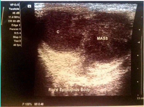
    Figure 1: Scrotal Doppler ultrasound picture showing right testicular
parenchyma with multiple subcentimeter hypoechoic lesions diffusely within.
The right Epididymis is also shown.
The histopathology report from the excised tissue grossly was that of an enlarged right orchidectomy specimen with an attached right spermatic cord as well as the covering scrotal skin. All together measuring 7.0x6.5x3.5cm. The skin was dull and brown in appearance with a focal area of ulcer. This ulcer has a dirty floor and a soft base. The ulcer edges were rough. The specimen was firm. The cut surface shows variably sized and shaped greyish-white masses occupying the majority of the testicular tissue and the adnexal structures with extension into the skin. These masses measure 3.0 cm in its widest diameter (Figure 2). Microscopically was that of a testicular tissue with a distorted architecture due to the presence of numerous granulomas that were composed of a central area of caseous necrosis surrounded by epithelioid macrophages, lymphocytes, foreign body and Langhan’s giant cells, and fibroblast. The remnant seminiferous tubules show hyalinization, some with the thickened basement membrane, and Leydig cell hyperplasia (Figures 3 and 4). The overlying skin shows acanthosis. Ziehl-Neelsen stain (ZN) also showed tubercle bacilli (Figure 5). He was subsequently referred to the microbiology and infectious disease clinic, where a GeneXpert test was done, and it showed a positive result. He has commenced antituberculosis chemotherapy with the first-line therapy that includes rifampin, isoniazid, pyrazinamide, and ethambutol (RIPE) and is currently doing fine at the clinic.
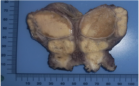
    Figure 2: The cut surface of the orchidectomy specimen shows multiple
greyish-white masses occupying the testes and its adnexa.
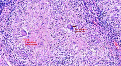
    Figure 3: Histologic sections show granulomatous inflammation with central
caseous necrosis, Langhan’s giant cells, and surrounding lymphocytes
replacing the testicular parenchyma x 40.
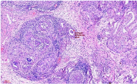
    Figure 4: Histologic section showing granulomatous inflammation
destroying the residual seminiferous tubules. Adjoining tubules also show
hyalinization with thick basement membranes x40
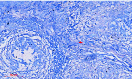
Figure 5: A ZN stain showing a rod-shaped acid-fast bacilli x20.
Discussion
Tuberculosis (?B) is an infectious bacterial condition induced by the Mycobacterium tuberculosis complex (MTBC). The World Health Organization (WHO) reported that 10.6 million cases of tuberculosis occurred in 2022, with a higher prevalence in males than in women. Before the coronavirus disease (COVID-19), tuberculosis (TB) was the most common infectious disease-related cause of death, with HIV/AIDS being the next most common cause [7]. In 2022, an estimated 1.30 million people died from tuberculosis worldwide. 87% of all TB cases globally in 2022 occurred in 30 high-burden countries, with eight countries accounting for two-thirds of the global total: India (27%), Indonesia (10%), China (7.1%), the Philippines (7.0%), Pakistan (5.7%), Nigeria (4.5%), Bangladesh (3.6%), and the Democratic Republic of the Congo (3.0%) [8]. In the same year, WHO reported that a total of 55% of people who developed TB were men, followed by women (33%), and children (ages 0-14) (12%) [8].
While extrapulmonary TB (EP-TB) accounts for 50% of HIVcoinfected patients, it makes up only 15% to 20% of all TB patients [7,9]. TB typically affects the entire male genital tract and is associated with risk factors such as HIV infection, poverty, undernourishment, diabetes, substance misuse, inadequate housing, overcrowded communities, smoking, immunosuppressive medications, and chronic renal illness [6]. However, our index patient did not show any of these risk factors.
In contrast to isolated testicular TB, which is uncommon, genital TB most frequently occurs in the epididymis [10]. Blood and lymphatic spread is the mechanism by which TB occur in the prostate and epididymis. Genital TB may spread to the seminal vesicles, vas deferens, and epididymis from the ejaculatory duct. The testicles are infected by closeness with the epididymis because of the protective function of the blood–testicles barrier [6,10,11]. Because TB orchitis frequently coexists with TB involvement of the lower urinary tract, including the kidneys, it manifests as lower urinary tract symptoms, particularly hematuria and irritative voiding symptoms. Other manifestations include prostatitis, scrotal swelling with or without discharging sinus, and epididymal-orchitis. It is debatable, though, how the TB bacilli moved to the testis [6]. To lessen the likelihood of a delayed diagnosis and complications such as male infertility, hypogonadism, or potential sexual transmission, it is crucial to take tuberculosis into account while making a differential diagnosis of anomalies in the male GU tract [12]. Our case demonstrates a case of isolated testicular TB without lower urinary tract symptoms, which is a rare presentation.
Painless scrotal swelling (observed in 60–70% of cases), pain (30–40%), and, in advanced cases, fistula formation (occurring in approximately 10–15 % of advanced cases) are common symptoms of testicular tuberculosis, which can present clinically in a nonspecific manner, mimicking conditions such as bacterial orchitis, abscesses, or malignancy [13]. Patients should be informed that discharged "cold abscesses" may develop and that they may take weeks or months to heal [12]. Our patient presented with a painful scrotal swelling, which subsequently became ulcerated, forming a discharging abscess.
It will be recalled that granulomatous inflammation may result from infectious or noninfectious aetiologies. Infectious causes are associated with caseating granulomas, which are characterized by a central region of necrosis surrounded by a collar of activated macrophages (epithelioid cells), lymphocytes, giant cells, and fibroblasts. Noninfectious origins produce noncaseating granulomas, which are composed of a collection of epithelioid histiocytes and giant cells with minimal peripheral chronic inflammation [14]. TB is very specific for granulomatous inflammation with caseation, and the most reliable method for identifying testicular TB is still histopathology. Caseating granulomas with core necrosis and with destruction of the seminiferous tubules or the epididymis or interstitium are characteristic of TB, and since Mycobacteria are acid-fast bacilli, they will appear as red bacilli against a blue background on AFB or Ziehl-Neelsen staining in 30–50% of cases [13,14]. Our patient was confirmed to have scattered acid-fast bacilli by this stain.
Furthermore, microbiological tests can help support the diagnosis of urogenital TB in about 60–70% of cases with urine cultures showing Mycobacterium tuberculosis; however, the sensitivity of urine cultures is reduced in isolated genital TB [13]. Similar to our findings.
According to WHO standards, testicular tuberculosis is often treated with a four-drug regimen consisting of 300 mg of isoniazid per day, 600 mg of rifampin per day, 1500–2000 mg of pyrazinamide per day, and 1200 mg of ethambutol per day for two months. Isoniazid and rifampin are then administered for a further four months [11,13].
Testicular TB raises serious fertility issues. Clinicians must think about diagnosing tuberculosis because delays in diagnosis may cause TB to spread or result in infertility, and it may even have more fatal complications [12]. 30-50% of advanced cases had azoospermia, oligospermia, and reduced sperm motility. While bilateral illness, which affects 5–10% of patients, frequently leads to hypogonadism and infertility, unilateral involvement may retain hormonal function. For patients with maintained spermatogenesis, long-term follow-up is advised, and semen analysis is crucial both before and after treatment [13]. This should be considered for patients desirous of fertility preservation. Our index patient did not express a desire for fertility preservation, possibly because he has completed his family.
Conclusion
Early detection, imaging, and histological confirmation are critical for directing treatment and avoiding complications. Although isolated testicular tuberculosis is a rare presentation, it is an important differential diagnosis for testicular masses, especially in endemic countries.
Declarations
Consent for Publication
Written informed consent was obtained from the patient for publication of this case report and any accompanying images. A copy of the written consent is available for review by the Editor-in-Chief of this journal.
Availability of Data and Materials
The tissue blocks are available for future use.
Funding
The work is funded by the authors.
Authors' Contributions
AAA, CCO, JOO, and JP reported the pathologic findings. CCO reported the clinical summary. OOO, AAA, and EEE contributed to the review. All authors were engaged in ensuring the accuracy and coherence of the presented manuscript.
References
- Öztürk Ç, Pasaoglu E, Bölme Savli T. A lesion mimicking malignancy: granulomatous orchitis. Bull Urooncology. 2021: 20: 126–128.
- Liang L, Jiajia W, Shoubin L, Yufeng Q, Gang W, Junjiang L. Granulomatous orchitis: case report and review of the literature. J Int Med Res. 2021; 49: 03000605211003773.
- Nativ O, Badaan S, Artool S, Safouri A, Hoffman A, Zohar Y, et al. Idiopathic granulomatous orchitis: how can we avoid unnecessary orchiectomy? J Clin Urol. 2023; 16: 701–705.
- Benavides-Huerto MA, García-Figueroa J, Chávez-Valencia V, Lagunas- Rangel FA. Idiopathic granulomatous orchitis: a case study. Urol Case Rep. 2024; 54: 102754.
- Mehboob K, Madani T. Isolated tuberculous orchitis presented as epididymoorchitis: an unusual presentation of tuberculosis. Urol Ann. 2022; 14: 189.
- Rishabh S, Krishnamoorthy K, Pratheeban TJ, Mathan E, Hameed OMRS. A rare case of testicular tuberculosis mimicking as testicular malignancy. J Assoc Pulmonologist Tamil Nadu. 2025; 8: 34–37.
- Nakou I, Kotoulas S, Sionidou M, Daios S, Manika C, Hadji-Mitrova M, et al. Two cases of testicular tuberculosis and review of the recent literature. Int J Mycobacteriology. 2024; 13: 225–236.
- Global tuberculosis report 2023. 1st ed. Geneva: World Health Organization. 2023; 1.
- Sharma SK, Mohan A, Kohli M. Extrapulmonary tuberculosis. Expert Rev Respir Med. 2021; 15: 931–948.
- Lamichaney R. Koch’s disease presenting as an isolated testicular mass- an unusual occurance. J Clin Diagn Res [Internet]. 2014.
- Masilamani T, Jayanthi N, Elaiyalwar A, B K, Ganapati S. An Unusual Case Report of Male Genital Tuberculosis. Cureus [Internet]. 2024.
- Bedi N, Rahimi MNC, Menzies S, Kalsi J. Atypical testicular pain. BMJ Case Rep. 2019; 12: e226697.
- Lachkar S, Diouri M, Ibrahimi A, Boualaoui I, Sayegh HE, Nouini Y. Isolated testicular tuberculosis: A case report. Urol Case Rep. 2024; 57: 102869.
- Chavez M, Chen F, Paterson J. Granulomatous epididymo-orchitis, a rare complication of Bacillus Calmette-Guerin immunotherapy for bladder cancer: a case report. North Am J Med Sci. 2024; 17.