Clinical Image
Austin J HIV/AIDS Res. 2014;1(2): 2.
Disseminated AIDS-associated Burkitt’s Lymphoma Demonstrated on FDG PET/CT
Yiyan Liu*
Department of Radiology, New Jersey Medical School, USA
*Corresponding author: Yiyan Liu, Nuclear Medicine Service, Department of Radiology, University Hospital, H-141, 150 Bergen Street, Newark, NJ 07103, USA
Received: October 20, 2014; Accepted: November 25, 2014; Published: November 27, 2014
A 40-year-old woman with history of HIV infection and AIDS had vaginal bleeding. A cervical biopsy suggested Burkitt’s lymphoma. Which imaging modality is best for initial staging? The choice is metabolic imaging F18-fluoro-2-deoxy-D-glucose positron emission tomography/computed tomography (FDG PET/CT). Today FDG PET/CT has gained widespread clinical applications in oncology, and it is accepted as a standard care in most malignancies. FDG is an analog of glucose and is used as a tracer of glycolysis. Malignant tissue and cells often demonstrate increased rate of glycolysis for rapid proliferation, due to increased numbers of glucose transporter protein and increased intracellular hexokinase and phosphofructokinase levels [1,2]. The intensity of FDG is proportional to the rate of glycolysis, therefore is potentially semi-quantitative with standardized uptake value (SUV). FDG PET/CT has a significant advance over traditional images in demonstrating metabolic changes which often precede anatomic changes, and routine whole-body requisition [3,4].
Burkitt’s lymphoma is a highly aggressive B-cell non- Hodgkin lymphoma. The disease is characterized by chromosomal rearrangements involving the c-myc proto-oncogene which results in its inappropriate expression [5]. Is there any difference of incidences of Burkitt’s lymphoma in HIV-infected and non-HIV infected populations? Yes. Burkitt’s lymphoma accounts for only 1-3% of lymphomas in HIV-seronegative adults, but this figure rises to 15-40% for AIDS-related lymphomas [6,7]. Burkitt’s lymphoma is strongly associated with HIV infection, especially in developing countries [8,9].
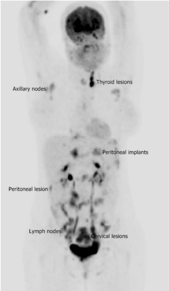
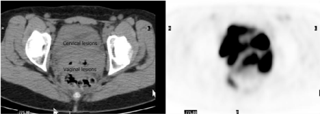
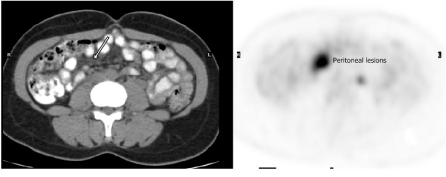
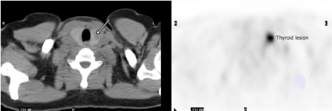
What are findings of FDG PET/CT in the case? As seen in Figure 1-4, there is a bulky cervical mass with intense uptake with parametrial and posterior vaginal invasion. There are multiple sites of lymphadenopathy, the most prominent in the pelvis. There are peritoneal implants. Incidentally noted is FDG avid thyroid lesion. All are consistent with disseminated and stage IV lymphomatous disease.
Figure 1: Maximum intensity projection image of whole-body FDG PET/ CT for initial staging in a 40-year-old woman with history of AIDS and newly diagnosed cervical Burkitt’s lymphoma. There are multiple foci of intense uptake consistent with lymphomatous lesions in the thyroid, axillary/retroperitoneal and pelvic lymphadenopathy, peritoneum, and endocervix.
Figure 2: Axial images of FDG PET/CT in the lower pelvis. There are bulky lesions (the left on the integrated CT) with intense FDG uptake (the right on PET), representing biopsy-confirmed Burkitt’s lymphoma.
Figure 3: Axial images of FDG PET/CT in the mid abdomen. There is a hypodensity lesion (arrow on the left CT) with intense FDG uptake (the right on PET), suggestive of peritoneal implant and carcinomatosis.
Figure 4: Axial images of FDG PET/CT in the lower neck. There is a highly FDG avid (the right on PET) low density lesion (arrow on the left CT) in the left lobe of the thyroid, indicating the lymphomatous involvement of the thyroid.
In Burkitt’s lymphoma, the accurate initial staging is very important since the selection and duration of treatment depends on the accurate staging. Limited studies suggested that FDG PET/ CT is sensitive for the detection of viable disease in Burkitt’s lymphoma [10,11]. All Burkitt’s lesions have high FDG avidity. In the presented case, the disease is disseminated and multiple lesions are demonstrated on FDG PET/CT in different locations, some of which are rare and unexpected such as the thyroid.
References
- Fletcher JW, Djulbegovic B, Soares H, Siegel BA, Lowe VJ, Lyman GH, et al. Recommendations on the use of 18F- FDG PET in oncology. J. Nucl. Med. 2008; 49: 480-508.
- Liu Y, Ghesani NV, Zuckier LS. Physiology and pathophysiology of incidental findings detected on FDG-PET scintigraphy. Seminar of Nuclear Medicine. 2010; 40: 294-315.
- Rohren EM, Turkington TG, Coleman RE. Clinical applications of PET in oncology. Radiology. 2004; 231: 302-332.
- Kapoor V, McCook BM, Torok FS. An introduction to PET-CT imaging. RadioGraphics. 2004; 24: 523-43.
- Armitage JO, Weisenburger DD. New approach to classifying non-Hodgkin’s lymphomas: clinical features of the major histologic subtypes. Non-Hodgkin’s lymphoma classification project. J Clin Oncol. 1998; 16: 2780-2795.
- Spina M, Tirelli U, Zagonel V, Volpe R, Barbare R, Abbruzzese L, et al. Burkitt’s lymphoma in adults with and without human immunodeficiency virus infection: a single institution clinicopathologic study of 75 patients. Cancer. 1998; 82: 766-774.
- Straus DJ. Treatment of Burkitt’s lymphoma in HIV-positive patients. Biomed Pharmacother. 1996; 50: 447-450.
- Steinbrook R. The AIDS epidemic in 2004. N Eng J Med. 2004; 351: 115-117.
- Beral V, Peterman T, Berkelman R, Jaffe H. AIDS-associated non-Hodgkin lymphoma. Lancet. 1991; 337: 805-809.
- Karantanis D, Durski JM, Lowe VJ, Nathan MA, Mullan BP, Georgiou E, et al. F18-FDG PET and PET/CT in Burkitt’s lymphoma. Eur J Radiol. 2010; 75: e68-e73.
- Zeng W, Lechowicz MJ, Winton E, Cho SM, Galt JR, Halkar R. Spectrum of FDG PET/CT findings in Burkitt lymphoma. Clin Nucl Med. 2009; 34: 355-358.


