
Research Article
J Hepat Res. 2014;1(1): 1005.
Non-Alcoholic Steatohepatitis (NASH) Influences the Disease Progression of Chronic Hepatitis C (CH-C): Histopathological Study of Liver Biopsies in 400 Cases with CH-C
Toshiharu Matsumoto1*, Kyoko Fukuhara2, Reiko Yaginuma2, Kenichi Ikejima2, Toshio Morizane3, Yuji Aoki1, Kanako Ogura1, Akihisa Miyazaki2 and Sumio Watanabe2
1Department of Diagnostic Pathology, Juntendo University Nerima Hospital, Japan
2Department of Gastroenterology, Juntendo University, School of Medicine, Japan
3Japan Council for Quality Health Case, Japan
*Corresponding author: Toshiharu Matsumoto, Department of Diagnostic Pathology, Juntendo University Nerima Hospital, 3-1-10 Takanodai, Nerima-ku, Tokyo 177-8521, Japan
Received: July 12, 2014; Accepted: Aug 05, 2014; Published: Aug 07, 2014
Abstract
Aims: The occasional occurrence of non-alcoholic steatohepatitis (NASH) in chronic hepatitis C (CH-C) has been reported, but the influence of NASH on the disease progression of CH-C has not been clarified. The purpose of the present study was to clarify the influence of NASH on the disease progression of CH-C.
Methods and Results: Liver biopsies from 400 cases of CH-C were examined histologically, and the histological evaluation of previous liver biopsies was also performed in selected cases. The association of NASH was observed in 69 cases (69/400, 17%). Among the 21 cases with NASH in an advanced stage, previous liver biopsies were evaluated in four cases, and, in three cases, the progression of stage from the early to advance was confirmed by repeated biopsies. The progression of the stage was induced by both CH-C and NASH equally in two cases, and the progression occurred mainly due to CH-C and partly due to NASH in one case.
Conclusions: The present study confirmed that the association of NASH influences the disease progression of CH-C and the disease progression is caused by both CH-C and NASH equally or CH-C mainly. These new findings are useful for the treatment of CH-C with NASH.
Keywords: Non-alcoholic steatohepatitis; Chronic hepatitis C; Disease progression
Abbreviations
NASH; Non-alcoholic steatohepatitis; CH-C; chronic hepatitis C; HCV, hepatitis C virus
Introduction
Chronic hepatitis C (CH-C) is a chronic liver disease caused by hepatitis C virus (HCV) infection. It shows portal and periportal fibrosis in the initial stage and progresses to bridging fibrosis with lobular distortion and diffuse regenerative nodule formation (cirrhosis) in the advanced stage. The increase of fibrosis reflects the disease progression of CH-C. On the other hand, non-alcoholic steatohepatitis (NASH) is a chronic liver injury with a characteristic liver histology consisting of steatosis, ballooning of hepatocytes, and pericellular fibrosis. Pericellular fibrosis is a primary route for diffuse regenerative nodule formation of cirrhosis in NASH [1]. Therefore, the process of the increase in liver fibrosis resulting in cirrhosis differs between CH-C and NASH.
The occasional occurrence of NASH in CH-C has been reported [2-5], but the influences of NASH on the disease progression of CH-C have not been clarified. Therefore, in the present study, we examined the influences of NASH on the disease progression of CH-C by the histological evaluation of liver biopsies in a large number of cases of CH-C and the histological evaluation of repeated liver biopsies in CH-C cases with NASH in the advanced stage.
Materials and Methods
The liver biopsies from 400 cases of CH-C were examined. The diagnosis of CH-C in the cases was made by the detection of serum HCV-RNA levels using quantitative or qualitative reverse transcription-polymerase chain reaction (RT-PCR) analysis. The liver biopsies were performed in Juntendo Hospital or Juntendo University Nerima Hospital. The examined cases were 400 Japanese adults (229 males and 171 females) with ages ranging from 21 to 78 years (mean: 51 years). None of the cases had a past history taking alcohol.
The liver tissues were fixed in 10% formalin and routinely processed for light microscopy. Tissue sections were stained with hematoxylin-eosin (HE), reticulum, and Azan stains.
In all cases, the aggregation of lymphocytes in the portal tracts, which is not a completely diagnostic finding but a characteristic feature in CH-C, was observed (Figure 1). Histological assessment of CH-C was made employing the New Inuyama classification [6]. In Japan, the New Inuyama classification has been used in numerous cases with CH-C, and the usefulness of the classification for the detail and precise evaluation of the histological changes of CH-C is demonstrated. In the classification, the scores of stage (fibrosis) consist of F0 (no fibrosis), F1 (fibrous portal expansion), F2 (bridging fibrosis), F3 (bridging fibrosis with lobular distortion), and F4 (cirrhosis). The scores of grade (activity of necroinflammatory reaction) consist of A0, A1, A2 and A3.
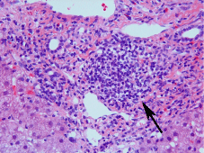
Figure 1: Histology of CH-C. Infiltration of inflammatory cells with the aggregation of lymphocytes (indicating by an arrow) and fibrosis can be seen in the portal area. HE stain.
The association of NASH was evaluated by the histological features of NASH, including steatosis, ballooning of hepatocytes, and pericellular or perisinusoidal fibrosis (Figure 2). Mallory's hyaline was excluded from the evaluation because of the very low incidence of Mallory's hyaline in Japanese NASH cases. The three famous articles for the histological evaluations of NASH have been published [1,7,8]. We think the Brunt's classification is most effective for the comparison of the grade and stage between NASH and CH-C, so we used the classification in the present study. The classification of NASH proposed by Brunt et al. was made using the staging system for fibrosis (S1, S2, S3, and S4) and the necroinflammatory grading system (G1, G2, and G3) [1]. Portal fibrosis is a factor for the evaluation of NASH staging [1]. However, the portal fibrosis associated with the aggregation of lymphocytes is usually produced by HCV (Figure 1), so such fibrosis was excluded from the evaluation of NASH staging.
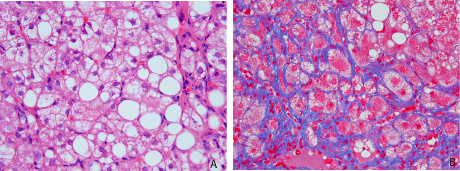
Figure 2: Histology of NASH. A. Steatosis and ballooning of hepatocytes are present. HE stain. B. Pericellular fibrosis is clear on Azan staining.
In CH-C, the cases with F3 (presence of lobular distortion) or F4 (liver cirrhosis) scores belong to the advanced stage of CH-C. Therefore, in the F3 and F4 CH-C cases associated with NASH, previous liver biopsies were examined to evaluate the influence of the association of NASH with the disease progression of CH-C.
The relationship of the association of NASH to the stages and grades of CH-C was examined with chi-square test and likelihood ratio calculation in the CH-C cases with or without NASH. Significance was accepted at P < 0.05.
The current study was approved by the Research Ethics Committee of Juntendo University School of Medicine and Juntendo University Nerima Hospital.
Results
The association of NASH was found in 69 cases (69/400, 17%). The most frequent incidence of NASH in the groups by the scoring system of the stage and grade of CH-C was in the F3 and A3 group (72.7%), followed by F3 and A2 group (37%) (Table 1). In the 69 cases with NASH, 29 cases (42%) showed stage 1 and grade 1 by the scoring system of the stage and grade of NASH (Table 2). Concerning the stage of CH-C with or without NASH, the group with NASH showed higher stage than the group without NASH (P = 0.0057 in the evaluation between the F0 to F1 group and F2 to F4 group) (Table 3). When NASH is recognized, the positive likelihood ratio for the pathological stage of F2 to F4 is 1.76 (95% confidence interval: 1.15- 2.70) against F0 to F1. This means that, when being NASH-positive, the odds of having a more progressed stage is 1.76 times higher than when being NASH-negative.
![]()
    F0
    F1
    F2
    F3
    F4


    A0
    16.2% (6/37)
    29.4% (5/17)
    0% (0/1)
    0% (0/1)
    0% (0/1)


    A1
    7.0% (5/71)
    16.2% (18/111)
    22.2% (4/18)
    0% (0/10)
    0% (0/11)


    A2
    0% (0/5)
    0% (0/14)
    28.6% (10/35)
    37.0% (10/27)
    15% (3/20)


    A3
    0% (0/1)
    0% (0/2)
    0% (0/2)
    72.7% (8/11)
    0% (0/6)
Table 1: Incidence of NASH in each group on evaluating scoring system of stage (fibrosis: F), and grade (activity of necroinflammation: A) of CH-C.
![]()
    S1
    S2
    S3
    S4


    G1
    29
    4
    8
    1


    G2
    8
    5
    7
    1


    G3
    2
    0
    3
    1
Table 2: Stage and grade of NASH in 69 cases.
![]()
    F0 - F1
    F2 - F4


    NASH (+) (n = 69)
    34
    35


    NASH (-) (n = 331)
    224
    107
Table 3: Comparison of stage (fibrosis: F) of CH-C between the groups with and without NASH.
Concerning the grade of CH-C with or without NASH, the group with NASH showed higher grade than the group without NASH (P = 0.0016 in the evaluation between the A0 to A1 group and A2 to A3 group) (Table 4). When NASH is recognized, the positive likelihood ratio for the pathological activity of A2 to A3 is 1.23 (95% confidence interval: 0.81-1.86) against A0 to A1. This means that, when being NASH-positive, the odds of having a higher grade is 1.23 times higher than when being NASH-negative.
![]()
    A0 - A1
    A2 - A3


    NASH (+) (n = 69)
    37
    32


    NASH (-) (n = 331)
    239
    92
Table 4: Comparison of grade (activity of necroinflammation: A) of CH-C between the groups with and without NASH:
In the 69 cases associated with NASH, 21 cases were in the advanced stage (F3 or F4) (Table 1). Among the 21 cases, four cases had previous liver biopsy examination (Table 5). In the three cases (cases 2-4), the duration between the first biopsy and evaluated biopsy was long (12 years in two cases and 18 years in one case), and the stage progressed from the early (F1) to the advanced (F3) stage in all cases (Table 5). In two cases (cases 2 and 4) the progression of fibrosis was caused by both CH-C and NASH equally (Figure 3), and the previous biopsy did not show the association of NASH (Table 5). In one case (case 3), the progression occurred by CH-C mainly and NASH partly and the association of NASH was already found in the previous biopsy (Table 5). In one case (case 1) with a short interval (2 years) between the previous and evaluated biopsy, liver fibrosis was already at an advanced stage in the previous biopsy (Table 5).
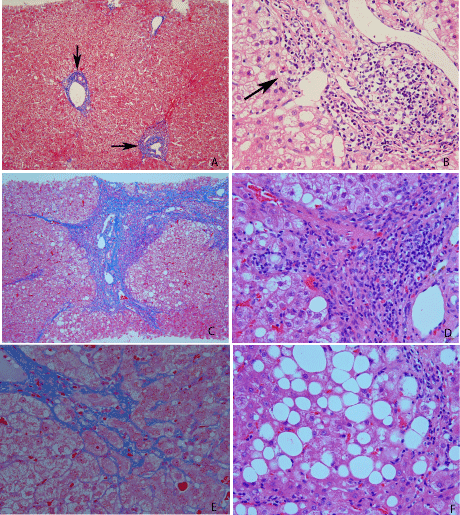
Figure 3: Histology of liver biopsies in case 2. A,B. First biopsy. Histology of CH-C without NASH. A. Fibrosis in portal areas (portal fibrosis; indicated by arrows) is present. Azan stain. B. Inflammatory cell infiltrate with aggregation of lymphocytes and interface hepatitis (indicated by an arrow) are present in the portal area. HE stain. C-F. Second biopsy (9 years after the first biopsy). Histology with admixture of CH-C and NASH. C. Histology of CH-C. Intense fibrosis in portal and periportal areas and bridging fibrosis are present. Azan stain. D. Histology of CH-C. Higher-power view of C. Lymphocytic infiltrate with aggregation is noted in the fibrotic area. HE stain. E. Histology of NASH. Pericellular fibrosis is clear on Azan stain. F. Histology of NASH. Steatosis and ballooning of hepatocytes are present. HE stain.
![]()
Case No
Liver biopsy
NASH
CH-C
Increase of stage (fibrosis)* (Factors relating to the increase)
1
Evaluated
+ (S3, G2)
+ (S3,G1)����������� F3, A2
F3, A2
F3, A2
2
Previous (3 yrs ago)
+ (S3, G2)
+ (S3, G2)
F3, A2
F3, A2
(CH-C and NASH equally)
3
Evaluated
+ (S1, G1)
+ (S3, G2)
F1, A1
F3, A3
(CH-C mainly, NASH partly)
4
Evaluated
+ (S3, G1)
F1, A1
F3, A2
(CH-C and NASH equally)
Table 5: Status of NASH and CH-C in the four cases with repeated liver biopsies.
Discussion
A histological study of the association of NASH in CH-C in a large number of cases has been performed to date in three studies, including the present study, and the present study involves the largest number of examined cases. The incidence of NASH in CH-C ranged from 9 to 18.3% in the studies (Table 6). These results indicate that the incidence of NASH in CH-C is about 10 to 20%.
![]()
    Authors
    Examined cases
    CH-C associated with NASH


    Younossi et al. [2]
    120
    22 (18.3%)


    Bedossa et a[4]
    278
    24 (9%)


    Present study
    400
    69 (17%)
Table 6: Incidence of NASH in CH-C.
The previous two studies [2-4], and the present study showed that patients with NASH had a more advanced stage than those without NASH. In the present study, the likelihood ratio for stage (fibrosis) indicated that, when being NASH-positive, the odds of having a more progressed stage is 1.76 times higher than on being NASH-negative. Thus, the possibility of an increase in fibrosis due to the association of NASH was considered. However, we thought that this hypothesis needs to be investigated by the examination of repeated liver biopsies in the same patients, but such a study had not been published. Thus, we performed the evaluation of liver changes in the cases of CH-C with NASH in repeated liver biopsies. Subsequently, three cases of advanced stages (F3 or F4) of CH-C with NASH showed increasing fibrosis from early (F1) to advance (F3) stages in long intervals (9, 12, and 18 years). This indicated that the association of NASH causes an increase of fibrosis with long intervals. On the other hand, one case with a short interval (2 years) was in an advanced stage (F3) in the first liver biopsy. Thus, the change in liver fibrosis by the association of NASH in short intervals could not be clarified in the present study.
Although the proportion of the increase of fibrosis by CH-C or NASH in CH-C cases with NASH has not been studied, the present study demonstrated that the increase occurred in the same proportion by both CH-C and NASH in two cases, and, in one case, the increase mainly occurred due to CH-C and partly due to NASH. These indicated that the importance of treatment of both CH-C and NASH in CH-C cases with NASH. Our previous study reported that hepatic steatosis is a predictor of a poor response to interferon a-2b and ribavirin therapy in patients with CH-C [9]. So, we think that a new treatment method is needed in patients with CH-C with NASH.
As an interesting finding in this study, the two cases (case no. 2 and 4) with disease progression from early to advanced stages had fibrosis due to both CH-C and NASH, but no association of NASH was found in the initial biopsy. Until now, it has been reported that chronic infection of HCV leads to NASH [10-12]. Thus, in the two cases, HCV induced NASH and disease progression occurred due to the interaction between HCV infection and the NASH state induced by HCV.
In conclusion, the present study confirmed that the association of NASH influences the disease progression of CH-C, and it is the first to demonstrate that the disease progression is caused by both CH-C and NASH equally or CH-C mainly. These findings are useful for the treatment of CH-C with NASH.
Acknowledgement
The authors thank Mr. D. Mrozek (Medical English Service, Kyoto, Japan) for organizing the English revision of this article.
References
- Brunt EM, Janney CG, Di Bisceglie AM, Neuschwander-Tetri BA, Bacon BR. Nonalcoholic steatohepatitis: a proposal for grading and staging the histological lesions. See comment in PubMed Commons below Am J Gastroenterol. 1999; 94: 2467-2474.
- Younossi ZM, McCullough AJ, Ong JP, Barnes DS, Post A, Tavill A. Obesity and non-alcoholic fatty liver disease in chronic hepatitis C. See comment in PubMed Commons below J Clin Gastroenterol. 2004; 38: 705-709.
- Solis-Herruzo JA, Pérez-Carreras M, Rivas E, Fernández-Vázquez I, Garfia C, Bernardos E. Factors associated with the presence of nonalcoholic steatohepatitis in patients with chronic hepatitis C. See comment in PubMed Commons below Am J Gastroenterol. 2005; 100: 1091-1098.
- Bedossa P, Moucari R, Chelbi E, Asselah T, Paradis V, Vidaud M. Evidence for a role of nonalcoholic steatohepatitis in hepatitis C: a prospective study. See comment in PubMed Commons below Hepatology. 2007; 46: 380-387.
- Lok AS, Everhart JE, Chung RT, Kim HY, Everson GT, Hoefs JC. Evolution of hepatic steatosis in patients with advanced hepatitis C: results from the hepatitis C antiviral long-term treatment against cirrhosis (HALT-C) trial. See comment in PubMed Commons below Hepatology. 2009; 49: 1828-1837.
- Ichida F. Tsuji T, Omata M et al. New Inuyama classification; new criteria for histological assessment of chronic hepatitis. Int. Hepatol. Commun. 1996; 6; 112-119.
- Matteoni CA, Younossi ZM, Gramlich T, Boparai N, Liu YC, McCullough AJ. Nonalcoholic fatty liver disease: a spectrum of clinical and pathological severity. See comment in PubMed Commons below Gastroenterology. 1999; 116: 1413-1419.
- Kleiner DE, Brunt EM, Van Natta M, Behling C, Contos MJ, Cummings OW. Design and validation of a histological scoring system for nonalcoholic fatty liver disease. See comment in PubMed Commons below Hepatology. 2005; 41: 1313-1321.
- Yaginuma R, Ikejima K, Okumura K, Kon K, Suzuki S, Takei Y. Hepatic steatosis is a predictor of poor response to interferon alpha-2b and ribavirin combination therapy in Japanese patients with chronic hepatitis C. See comment in PubMed Commons below Hepatol Res. 2006; 35: 19-25.
- Koike K. Hepatitis C as a metabolic disease: Implication for the pathogenesis of NASH. See comment in PubMed Commons below Hepatol Res. 2005; 33: 145-150.
- Koike K, Moriya K. Metabolic aspects of hepatitis C viral infection: steatohepatitis resembling but distinct from NASH. See comment in PubMed Commons below J Gastroenterol. 2005; 40: 329-336.
- Watanabe S, Yaginuma R, Ikejima K, Miyazaki A. Liver diseases and metabolic syndrome. See comment in PubMed Commons below J Gastroenterol. 2008; 43: 509-518.