Case Presentation
Ann Hematol Oncol. 2019; 6(5): 1250.
Three Cases of Autoimmune Hemolytic Anemia following Primary Varicella Infection and Vaccination: Possible Pathogenesis in the Context of Current Information
Biçakci Z1*, Bozkurt HB2 and Olcay L3
1Association Professor, Department of Pediatric Hematology and Oncology, Medicine Faculty, Kafkas University, Kars, Turkey
2Asistant Professor, Department of Pediatrics, Medicine Faculty, Kafkas University, Kars, Turkey
3Professor, Department of Pediatric Hematology and Oncology, Medicine Faculty, Baskent University, Ankara Turkey
*Corresponding author: Zafer Biçakci, Association Professor, Department of Pediatric Hematology and Oncology, Kafkas University, Kars, Turkey, Fax:+904742251430; GSM:+905325137271; E-mail:zaferbicakcib@yahoo.com.tr
Received: March 20, 2019; Accepted: April 26, 2019;Published: May 03, 2019
Abstract
The direct complement-mediated destruction of erythrocytes coated with auto antibodies mainly occurs in the circulation and the liver in autoimmune hemolytic anemia while the antibody-dependent cellular cytotoxicity (NK), cytotoxicity (CD8+ T) and phagocytosis primarily occur in the spleen and lymphoid organs. Since primary varicella infection causes a decrease in the CD4+/CD8+ ratio, CD4+ T cells specific to varicella mainly appear in the Th1 type and produce high levels of interferon-γ (IFN-γ). Th1 cells that produce IFN-γ play a pathogenic role in many autoimmune disorders. IFNγ stimulates the conversion to IgG isotypes and also increases phagocytic functions through the Fcγ receptor of macrophages. We present three patients with autoimmune hemolytic anemia and thrombocytopenia developing secondary to varicella infection and vaccination in a manner similar to Evans syndrome, and discuss the possible pathogenesis in the context of current information. Direct antiglobulin test (IgG and C3d)-positive autoimmune hemolytic anemia and thrombocytopenia were present after primary varicella infection in two of our cases and after varicella vaccination in one case. Growth retardation, decreased CD4+/CD8+ lymphocyte ratio, diffuse large B cell lymphoma and hypergammaglobulinemia were present in case 1; cerebral palsy, growth and development retardation, and decreased CD4+/CD8>sup>+
lymphocyte ratio in case 2; and hypergammaglobulinemia in case 3. Clarifying the pathogenesis of autoimmune hemolytic anemia could facilitate the development of new treatment approaches for this disorder with high morbidity and mortality. Decreasing the effect of INF-γ, which plays an important role in the pathogenesis of autoimmune hemolytic anemia, could be one of these approaches.Keywords: Vaccine; Autoimmune hemolytic anemia; Varicella; Regulator t lymphocyte deficiency
Introduction
Childhood autoimmune hemolytic anemia is a rare and potentially life-threatening disease. Antibodies developing against erythrocytes may lead to early destruction and the development of the disorder. Autoimmune hemolytic anemia can be divided into two as primary and secondary [1]. The development of autoimmune hemolytic anemia requires a general disturbance due to deterioration of the differentiation between self and non-self in the immune system. T-cell mediation has been shown to have a crucial role in the regulation of the humoral immune system [2].
Varicella is a benign disease with a rash and usually affects children. Autoimmune hemolytic anemia is a rare complication [3]. Although primary immunodeficiency and varicella infection have been reported after varicella vaccination, primary immunodeficiency, varicella infection and autoimmune hemolytic anemia after varicella vaccination has been reported in only a single patient [4,5]. The varicella-specific CD4+T lymphocytes that appear during primary infection are predominantly of the T helper 1 (Th1) type and produce high levels of interferon-γ (IFN-γ) supporting the clonal growth of varicella-specific T lymphocytes [6].
Activation of helper CD4+ T lymphocytes is important regarding the autoimmune pathology in rats deficient in IL-2. Th1 lymphocytes producing IFN-γ play a pathogenic role in many autoimmune disorders. IFN-γ stimulates the conversion to IgG isotypes and increases the phagocytic function of macrophages [7].
A decrease in the CD4+/CD8+ lymphocyte ratio in the peripheral blood has been reported in patients with Evans syndrome [8]. A significant increase in the percentage of CD8+ lymphocytes and a significant decrease in the percentage of CD4+ lymphocytes with a decrease in the CD4+/CD8+ ratio has been reported in primary varicella infection [9]. We found a decreased CD4+/CD8+ lymphocyte ratio in two of our cases. This may indicate an imbalance of the immunoregulatory cells (CD4+ T lymphocytes) causing irregular cytokine production. The numerical and functional deficiency of CD4+ T lymphocytes due to varicella could therefore trigger autoimmune hemolytic anemia.
We present three patients with autoimmune hemolytic anemia and thrombocytopenia developing in a manner similar to Evans syndrome secondary to varicella infection and vaccination, and discuss the possible pathogenesis in the context of current information.
Case 1
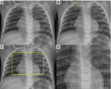
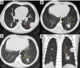
A 14-month-old female patient was administered a measlesmumps- rubella and varicella vaccine at the age of one. She suffered from a varicella infection one month after the vaccine was administered and recovered afterwards. She presented to another hospital due to a widespread rash on her body a month after the varicella infection. The IgA, IgM, IgG and IgE levels were normal but the CD3+ CD4+ lymphocyte value was low and CD3+ CD8+ lymphocyte value high in tests conducted with a suspicion of immunodeficiency when she was ten months old (Table 1). She was referred to our hospital for the investigation of anemia and thrombocytopenia etiology. We found out that her sister and brother had died with high fever when two months old. The physical examination revealed body measurements (height, body weight) under 3% and widespread petechial lesions and varicella marks on the extremities, trunk and back together with splenomegaly (the spleen was 2 cm below the costal margin). The hemogram and other laboratory findings are presented in (Table 1). The peripheral smear revealed 18% atypical lymphocytes, 51% neutrophils, 2% monocytes, 8% rods, 16% lymphocytes, and 5% normoblasts together with macrocytosis, polychromacia and microspherocyte formation in the erythrocytes. The viral (hepatitis A,B,C and etc.) and bacterial serology was negative. Varicella zoster IgM was (-), varicella zoster IgG (+), ANA (-), and Anti dsDNA (-). C3 and C4 levels were normal. The diagnosis was Evans syndrome/ immune hemolytic anemia and 2 mg/kg of prednisolone was started together with an erythrocyte suspension transfusion. The platelets increased to 266 x109/L during follow-up. However, the steroids were discontinued when the patient developed bronchopneumonia on the third day of steroid treatment and an antibiotic (meropenem) was started. Inappropriate anti-diuretic hormone secretion syndrome then developed. Fluid restriction was started with sodium deficit treatment to increase sodium by 10 mEq. Intravenous (iv) Immunoglobulin G (IgG) at a dose of 800 mg/kg was started when the thrombocytopenia recurred. The platelets again increased to normal levels. Areas with increased density were noted in the left lung on chest x-ray (Figure 1). Thoracic computed tomography ordered on the suspicion of a mass revealed a peripheral nodular lesion 17x11 mm in size with its base on the pleura at the level of the left lower lobe superior segment (Figure 2). The patient underwent a pulmonary lymph node biopsy. She was immunohistochemically diagnosed with diffuse large B cell lymphoma. Meanwhile, the patient developed myoclonic seizures followed 5 days later by herpetic keratitis. Meningoencephalitis was considered and acyclovir administered for a month. The seizures were only controlled with multiple antiepileptics. The inappropriate anti-diuretic hormone secretion syndrome, immune hemolytic anemia and thrombocytopenia resolved with medical treatment. The patient’s swallowing function was disrupted after the treatment and feeding via gastrostomy was started. The patient suffered frequent infections treated at the hospital and the positive direct and indirect antiglobulin test results continued. However, a transfusion was not required. The patient did not come for regular follow-up after chemotherapy and was found to have died one year later at home. Informed consent was received from the family for publication.
    Case 1
    Case 2
    Case 3


    Hemoglobin (g/dl)
    7.32
    3.2
    6.7


    Platelet (x103/mm3)
    24
    9
    110


    Leukocyte (x109/L)
    9.6
    4.3
    6.5


    Reticulocytes (%)
    7.8
    10.8
    8.4


    LDH (U/L) (135-225)
    416
    829
    2005


    Total bilirubin (mg/dl) (0-1.2)
    1.62
    2.18
    2.00


    Direct bilirubin(mg/dl) (0-0.3)
    0.76
    0.35
    0.44


    Haptoglobin (g/L) (0.2–5)
    8
    0.01
    0.5


    DAT (IgG)
    +++
    +++
    ++


    DAT (C3d)
    ++++
    ++++
    +++


    I.DAT
    +
    -
    +


    IgG (mg/dl)
      (345–1236)
    1643
    628
    1340


    IgM (mg/dl)
      (43–207)
    224
    129
    237


    IgA (mg/dl)
      (14–159)
    43
    80.4
    93   CD3+ CD4+
      (%31-54)
    %9
    %3
    -


    CD3+ CD8+
      (%10-31)
    %40
    %5
    -


    CD3+CD4+/CD3+CD8+
      (1.2)
    0.225
    0.6
    -


    LDH:    Lactate Dehydrogenase
      DAT: Direct antiglobulin Test
      I.DAT:    Indirect Antiglobulin Test
Table 1: The hemogram results and other laboratory findings are presented.

    Figure 1: The PA lung graphic of the case (A). There were nodular lesions
(yellow arrows) which were seen with unclear edge on the graphic of the
right lung (B). The marked region and enlarged image (C) with yellow frame
showed an increase in density on paramediastinal and paracardiac areas of
the left lung. It is more clearly seen in the left lung than the right lung (D).

    Figure 2: It was observed that thorax CT findings of the case showed
peripheral nodular lesion (red arrow) with irregular borders in the superior
segment of the lower lobe of the left lung and an increase in nodular density
in the right lung (A). It was seen that, there was a nodular lesion with
unclear edges (blue arrow) and in the posterior part of the lung and there
was focal parenchymal density enhancements (B, C) in the lung area near
to anteromedial of this lesion. It was seen another nodular lesion (D) in the
coronal plane of the lower lobe of the left lung (red and blue arrows) and focal
parenchymal density enhancements with unclear edges (yellow arrow) and
supradiyaphragmatic placement in the base of the right lung.
Case 2
A girl aged two and a half years had undergone physical treatment at an external center for cerebral palsy with unknown cause and had a history of varicella 20 days ago. Paleness of the skin and incrusted varicella rashes over the whole body were found at the hospital she presented at for the paleness and fatigue that had started suddenly the previous day and then increased and the accompanying fever (not measured). The patient was thought to be suffering from autoimmune hemolytic anemia and was referred to our hospital. Her body weight was 10 kg (10%) and the height was 90 cm (90%). The skin and mucous membranes were pale and there was a widespread varicella rash on the skin. The liver was found to be 3 cm palpable at the right midcostal line and the lower extremity tonus was bilaterally decreased. The hemogram results and other laboratory findings are presented in (Table 1). The peripheral smear revealed 33% neutrophils, 58% lymphocytes and 9% monocytes together with microspherocytes, poikilocytosis, polychromasia and normoblasts. The viral (hepatitis A,B,C and etc.) and bacterial serology was negative as were ANA and anti dsDNA while the C3 and C4 values were normal. The patient was administered 5 units of erythrocyte suspension (10-20 ml/kg), 2 mg/kg prednisolone and 5 g/kg IVIG. However, these had to be followed by methylprednisolone 20 mg/kg/day for 7 days when there was no response to the previous treatments and the dose was then gradually reduced. The hemoglobin increased to 10.2 g/dl and the platelet count to 399,000/mm3. She was discharged 3 weeks later to continue the treatment as an outpatient. She then presented again with paleness and fatigue that had started 12 days after the discharge. Steroids, IVIG 5 g/kg, 6-mercaptopurine, vincristine, cyclosporine and rituximab were administered this time. The hemoglobin did not increase despite the treatment but the platelet count increased rapidly and did not decrease again, staying at values of 150-250,000/mm3. The patient then experienced a second varicella infection. Zona zoster infection developed along the radial nerve in the right arm during the period when the varicella rashes became incrusted. The patient did not respond to treatment. She developed multiorgan failure and died. Informed consent was received from the family for publication.
Case 3
A four-year-old female patient had suffered a varicella infection about 15-20 days ago. She had been referred with a preliminary diagnosis of autoimmune hemolytic anemia from another hospital where she had presented with increasing jaundice, paleness and fatigue for the last two days. The examination revealed paleness of the skin and incrusted varicella rashes over the whole body. The hemogram and other laboratory findings are presented in (Table 1). The peripheral smear revealed 46% neutrophils, 50% lymphocytes, and 4% monocytes together with microspherocytes, poikilocytosis, polychromasia and normoblasts. The viral (hepatitis A,B,C and etc.) and bacterial serology was negative. The patient was started prednisolone at a dose of 2 mg/kg. The hemoglobin values increased with no hemolysis on follow-up and she was discharged. However, she did not return for follow-up. Informed consent was received from the family for publication.
Discussion
The prevalence of Autoimmune Hemolytic Anemia (AIHA) in adults is approximately 1:100,000 per year but is estimated to be lower in children. Approximately 75% of AIHA cases have warm antibodies and these are always polyclonal. A general irregularity of the immune system due to the deterioration of the differentiation between self and non-self appears to be necessary for the development of autoimmune hemolytic anemia. T-cell mediation has been shown to play a critical role in the regulation of the humoral immune system. The polymorphism of the CTLA-4 (signal substance) gene activating the regulator T lymphocytes (Treg lymphocytes) may lead to a predisposition for autoimmunity [2]
Varicella is a benign disease with rash and usually affects children. The most common complications are bacterial skin infection, pneumonitis, cerebellar ataxia, hepatitis, thrombocytopenia and arthritis. Autoimmune hemolytic anemia is a rare varicella complication [3]. Several autoimmune hemolytic anemia cases after varicella in which the polyspecific direct antiglobulin (IgG and C3b-d), anti-Pr and anti-I cold agglutinin (IgM) tests were positive have been reported [3,10,11].
Varicella infection related to the Oka (vaccine) strain has been reported in adenosine deaminase deficiency, purine nucleoside phosphorylase deficiency, Natural Killer T (NKT) cell deficiency, and combined primary immunodeficiency of unknown etiology [4]. Similarly, autoimmune hemolytic anemia, thrombocytopenia and hypergammaglobulinemia have been reported three weeks after varicella vaccination in a 13-month-old girl with hypomorphic RAG2 mutations (deficiency) [5]. Varicella may lead to further aggravation of cellular immunodeficiency and result in development of symptoms in subjects with cellular immunodeficiency (CD4+/NK lymphocyte) after routine live varicella vaccination because it mainly infects primary CD4+ T lymphocytes.
Children in Turkey are vaccinated with the varicella and measlesmumps- rubella live virus vaccines at the 12th month. Our three cases suffered from direct antiglobulin test positive autoimmune hemolytic anemia and thrombocytopenia that developed after primary varicella infection in two cases and after varicella vaccination in one case. The history and findings revealed sibling death in infancy, growth retardation, decreased CD4+/CD8+ lymphocyte ratio, diffuse large B cell lymphoma and hypergammaglobulinemia in case 1; cerebral palsy, growth retardation and decreased CD4+/CD8+ lymphocyte ratio in case 2; and hypergammaglobulinemia in case 3. These findings also indicate at least one immunological disorder or deficiency in our cases.
A significant increase in the percentage of CD8+ lymphocytes and a significant decrease in the percentage of CD4+ lymphocytes and consequently a decrease in the CD4+/CD8+ ratio have been reported to be present in primary varicella infection [9]. Primary varicella infection also induces CD4+ T lymphocytes specific to the virus producing interleukin-2 (IL-2) and IFN-γ, thought to be Th1 cytokines. Based on the CD4+ T lymphocyte cytokine profiles, IL-2 and IFN-γ as well as IFN-a concentrations were reported to increase in the serum of healthy individuals suffering from varicella infection [6].
A permanent decrease in the CD4+/CD8+ lymphocyte ratio has been found in the peripheral blood of Evans syndrome patients [8]. A decrease in CD4+ and CD8+ lymphocyte levels and CD4+/CD8+ lymphocyte ratio, a full suppression (zero) of the TGF-β level, and an increase in the IL-2, IL-10 and IFN-γlevels were reported in a case of Evans syndrome with IgM deficiency and lymphopenia [8]. The CD4+/CD8+ lymphocyte ratio was found to be 0.225 in our case 1 and 0.6 (decreased) in case 2. We believe that the immune cells (mainly regulator T lymphocytes) may have decreased as the CD4+/CD8+ lymphocyte ratios were decreased in our cases. The CD4+ T lymphocytes specific for VZV predominantly express IFN-γ, IL-2 and TNF-a with a multifunctional phenotype in healthy individuals. However, the percentage of multifunctional lymphocytes (IFN-γ+ IL-2 + TNF-a +) has been found to be lower in parallel with the increase in IFN-γ(single) + lymphocytes. Interestingly, asymptomatic immunosuppressed patients who are generally known to be under increased risk of VZV reactivation have been reported to have significantly lower VZV-specific CD4+ T lymphocyte levels compared to healthy controls [12].
Two different models have been recommended to explain the relationship between lymphopenia and autoimmunity. Normal adult rat auto reactive T lymphocytes are included in both models. A low level of T lymphocytes in peripheral lymphoid organs allow the proliferation of auto reactive T lymphocyte precursors in the “null area” model. A T lymphopenia environment facilitates the interaction of auto reactive T lymphocytes with the professional Antigen Presenting Cell (APC). Such activated auto reactive T lymphocytes may migrate to non-lymphoid organs afterwards and induce organspecific autoimmunity. In the second model, the lymphopenia status is suggested to occur in the selective absence of a critical regulator T lymphocyte population that continuously suppresses the auto reactive T lymphocyte activation [13].
Natural regulator T lymphocytes include T lymphocytes (CD4+CD25+), CD4+ at a rate of 5% [14]. IL-2 is mainly produced by CD4+ T lymphocytes. Most of the regulator CD4+ T lymphocytes express the a chain (CD25) of the IL-2 receptor at a high level [15]. Autoimmune hemolytic anemia develops in rats when the IL-2 and IL-2 receptor a or β chain gene is removed [7]. The presence of low levels of CD3+CD4+ lymphocytes and autoimmune hemolytic anemia in both cases indicate low transforming growth factor-β (TGF-β) and high IFN-γ levels in addition to the IL-2 or IL-2 receptor (a/β) deficiency. Significantly lower values of cells expressing triple cytokines (IFN-γ+IL-2+TNF-a+) and higher (increasing) values of cells expressing single cytokines (IFN-γ (single) +) have been reported after the stimulation of immunosuppressive transplant recipients with varicella (VZV) lysate [12]. We therefore believe that IL-2 levels could have been lower and IFN-γ levels higher in our patients that we thought had an immune irregularity/deficiency. Since the production and life of regulatory T cells are also dependent on IL-2, low IL-2 levels suggest that the regulatory T cell levels may also be low [7]. Autoimmunity has been reported to develop in lymphopenic adult mice in the selective absence of critical regulator T cells that continuously suppress autoreactive T cell activation [13]. The absence of regulator T lymphocytes may lead to inadequate T cell homeostasis, impaired self-tolerance, uncontrolled activation, proliferation of CD4+ T cells, and triggering of autoimmune hemolytic anemia [16].
The most common target for pathogenic anti-erythrocyte antibodies in autoimmune hemolytic anemia is Rhesus (Rh) polypeptides. Pathogenetic antibody production occurs with the activation of T-helper lymphocytes for specific cryptogenic epitopes on the Rh proteins in many cases of autoimmune hemolytic anemia [17]. The active auto reactive CD4+Th1 cells and pathogenic auto antibodies that secrete IFN-γ are therefore specific to the Rh proteins in the erythrocyte membrane in most patients [14]. We believe the positive direct antiglobulin test (IgG) in all three cases and the indirect antiglobulin test in cases 1 and 3 may be the result of IFN-γ production (stimulation of conversion to IgG isotypes).
Erythrocytes sensitized by IgM antibodies are destroyed by the complement system following phagocytosis, after interacting with specific receptors (C3b-CR1 and iC3b-CR3) especially on the liver macrophages (Kupffer cells), either directly by cytolysis or indirectly by the activation of the C3 bound to the erythrocytes and destruction of the fragments (C3b-iC3b) [18]. Kupffer cells express multiple receptors containing anaphylactoxin receptors (C3a and C5a) and complement receptors (CR1, CR3 and CR4). CR3 (CD11b/ CD18) serves as a receptor for erythrocytes opsonized with IgM and facilitates its removal from the circulation. Although binding of erythrocyte-bound C3b to CR1 in Kupffer cells may mediate some of the removal, the interaction between the erythrocyte-bound iC3b and the macrophage CR3 is the basic element of extra vascular destruction of erythrocytes that have probably become sensitized to complement. Removal of such erythrocytes sensitive to complement is believed to be mediated by phagocytosis [18]. Kupffer cells are very sensitive to complement activation products; complement receptors facilitate both the proinflammatory role of Kupffer cells and their critical role in removing pathogens and dead erythrocytes.
IgG is a relatively ineffective initiator of classic complement pathway activation. The removal of erythrocytes sensitized with IgG is provided through FcγRI (CD64) receptor-mediated phagocytosis and sequestration that mediate the cytotoxic activity primarily in the splenic red pulp macrophages (splenic cord) in the absence of complement activation [18,19]. Macrophages express surface receptors for the Fc region of IgG (especially IgG1 and IgG3) with opsonic fragments of C3 and C4 [8]. Transcription of the FcγRI gene and the expression of FcγRI on macrophages are induced by IFN-γ. The antibody isotype that best binds to Fcγ receptors is also partially produced as a result of the IFN-γ mediated isotypes transformation of B lymphocytes. TGF-β is mainly produced from regulatory T lymphocytes and inhibits the proliferation and effectors functions of T lymphocytes together with activation of macrophages [15]. The coexistence of both IFN-γ elevation and low TGF-β and IL-2 values (CD4+ T lymphocyte deficiency) due to varicella could be explained by the phagocytosis due to autoantibody (antierythrocyte antibody) production induced by IFN- γ being the main cause of extracellular hemolysis. Macrophage activation with IFN-γ also increases CR1-mediated phagocytosis. CR1 alone is ineffective in inducing phagocytosis of C3b-coated erythrocytes. However, if the erythrocytes are coated with IgG antibodies that also bind to the Fc receptor at the same time, their ability to induce such phagocytosis increases [15]. This may explain why the anemia and thrombocytopenia are aggravated when patients with autoimmune hemolytic anemia and immunologic thrombocytopenia are exposed to viral infections [20].
The capture of antibody-coated target cells by FcRIII activates NK cells to synthesize and secrete cytokines such as IFN-γ and empties the content of the granules that mediate the killer function of these cells [15]. FcγRIII (CD16) is responsible for phagocytosis, endocytosis and Antibody Dependent Cell-Mediated Cytotoxicity (ADCC), and therefore plays an important role in intravascular hemolysis [18].
In conclusion, since varicella infects CD4+ lymphocytes, it may cause a numerical and functional deficiency in these lymphocytes. This deficiency may potentially trigger autoimmune hemolytic anemia. Autoimmune hemolytic anemia, with or without complement activation, is caused by the increased destruction of erythrocytes by ant erythrocyte auto antibodies. While direct complement-mediated destruction mainly occurs in the circulation and in the liver, ADCC, cytotoxicity and phagocytosis primarily take place in the spleen and lymphoid organs. Direct complement activation or antibody dependent cell-mediated cytotoxicity (mediated by NK FcγRIII) may cause intravascular hemolysis, and IFN-γ may activate both Fcγ and complement receptors and result in extra vascular hemolysis. Intravascular hemolysis is 10 times more active than extra vascular hemolysis [19-21].
The existing medical treatments for autoimmune diseases are inadequate. The treatment is mainly limited to the use of antiinflammatory or immunosuppressive agents. These agents can cause potentially serious toxic effects and provide only temporary improvement. Treatments that decrease the expression of the activating Fcγ receptor (Fcγ RI/III) or increase the expression of the inhibiting Fcγ receptor (Fcγ RIIIB) may be effective. Treatment methods such as using INF-γ antibody, INF-γ receptor antibodies and INF-γ synthesis inhibitors for the INF-γ that increases phagocyte functions through the macrophage Fcγ receptor, in addition to receptor antibodies for CR1 and CR3 receptors, could be developed. Fully elucidating the pathogenesis of autoimmune hemolytic anemia could therefore facilitate the development of new treatment approaches for this disorder with high morbidity and mortality.
References
- Gehrs BC, Friedberg RC. Autoimmune hemolytic anemia. Am J Hematol. 2002; 69: 258-271.
- Berentsen S. Role of Complement in Autoimmune Hemolytic Anemia. Transfus Med Hemother. 2015; 42: 303-310.
- Kumar KJ, Kumar HC, Manjunath VG, Arun V. Autoimmune Hemolytic Anemia due to Varicella Infection. Iran J Pediatr. 2013; 23: 491-492.
- Bayer DK, Martinez CA, Sorte HS, Forbes LR, Demmler-Harrison GJ, Hanson IC, et al. Vaccine-associated varicella and rubella infections in severe combined immunodeficiency with isolated CD4 lymphocytopenia and mutations in IL7R detected by tandem whole exome sequencing and chromosomal microarray. Clin Exp Immunol. 2014; 178: 459-469.
- Dutmer CM, Asturias EJ, Smith C. Late Onset Hypomorphic RAG2 Deficiency Presentation with Fatal Vaccine-Strain VZV Infection. J Clin Immunol. 2015; 35: 754-760.
- Arvin A, Abendroth A. VZV: immunobiology and host response. In: Arvin A, Campadelli-Fiume G, Mocarski E, eds. Human Herpesviruses: Biology, Therapy, and Immunoprophylaxis. Cambridge: Cambridge University Press; 2007.
- Isakson SH, Katzman SD, Hoyer KK. Spontaneous autoimmunity in the absence of IL-2 is driven by uncontrolled dendritic cells. J Immunol. 2012; 189: 1585-1593.
- Karakantza M, Mouzaki A, Theodoropoulou M, Bussel JB, Maniatis A. Th1 and Th2 cytokines in a patient with Evans’ syndrome and profound lymphopenia. Br J Haematol. 2000; 110: 968-970.
- Arneborn P, Biberfeld G. T-lymphocyte subpopulations in relation to immunosuppression in measles and varicella. Infect Immun. 1983; 39: 29-37.
- Friedman HD, Dracker RA. Cold agglutinin disease after chicken pox. An uncommon complication of a common disease. Am J Clin Pathol. 1992; 97: 92-96.
- Terada K, Tanaka H, Mori R, Kataoka N, Uchikawa M. Hemolytic anemia associated with cold agglutinin during chickenpox and a review of the literature. J Pediatr Hematol Oncol. 1998; 20: 149-151.
- Schub D, Janssen E, Leyking S, Sester U, Assmann G, Hennes P, et al. Altered phenotype and functionality of varicella zoster virus-specific cellular immunity in individuals with active infection. J Infect Dis. 2015; 211: 600-612.
- Suri-Payer E, Amar AZ, Thornton AM, Shevach EM. CD4+CD25+ T cells inhibit both the induction and effector function of autoreactive T cells and represent a unique lineage of imunoregulatory cells. J Immunol. 1998;160:1212-1218.
- Ward FJ, Hall AM, Cairns LS, Leggat AS, Urbaniak SJ, Vickers MA, et al. Clonal regulatory T cells specific for a red blood cell autoantigen in human autoimmunehemolytic anemia. Blood. 2008;111:680-687.
- Abbas AK, Lichtman AH, Pillai S. Cytokines. In: Abbas AK, Lichtman AH, Pillai S eds. Cellular and Molecular Immunology. Philadelphia (PA): Saunders. 2007: 267-301.
- Hoyer KK, Kuswanto WF, Gallo E, Abbas AK. Distinct roles of helper T-cell subsets in a systemic autoimmune disease. Blood. 2009;113: 389-395.
- Barker RN, Hall AM, Standen GR, Jones J, Elson CJ. Identification of T-cell epitopes on the Rhesus polypeptides in autoimmune hemolytic anemia. Blood. 1997; 90: 2701-2715.
- Friedberg RC, Johari VP. Autoimmune hemolytic anemia. In: Greer JP, Arber DA, Glader B, eds. Wintrobe’s Clinical Hematology, 13th ed. Philadelphia: Lippincott Williams & Wilkins. 2014: 746-765.
- Packman CH. Hemolytic anemia due to warm autoantibodies. Blood Rev. 2008; 22: 17-31.
- Coutelier JP, Detalle L, Musaji A, Meite M, Izui S. Two-step mechanism of virus-induced autoimmune hemolytic anemia. Ann N Y Acad Sci. 2007; 1109: 151-157.
- Barcellini W. New Insights in the Pathogenesis of Autoimmune Hemolytic Anemia. Transfus Med Hemother. 2015; 42: 287-293.