
Rapid Communication
Ann Hematol Oncol. 2019; 6(1): 1228.
Diagnostic Performance of Baseline 18F-Fluoro-Deoxy- Glucose Positron Emission Tomography/Computed Tomography in Patients with Clinico-Radiological Suspicion of Primary Central Nervous System Lymphoma
Gupta T¹*, Gupta M¹, Purandare N², Puranik A², Rangarajan V², Moiyadi A³, Shetty P³, Epari S4, Pungavkar S5, Krishnatry R¹, Sastri G¹ and Jalali R¹
1Department of Radiation Oncology, Tata Memorial Centre, India
²Department of Nuclear Medicine & Molecular Imaging, Tata Memorial Centre, India
³Department of Neuro-surgery, Tata Memorial Centre, India
4Department of Pathology, Tata Memorial Centre, India
5Department of Imaging & Radio-diagnosis, Global Hospitals, India
*Corresponding author: Tejpal Gupta, MD, DNB, Professor, Department of Radiation Oncology, ACTREC, Tata Memorial Centre, Homi Bhabha National Institute, Kharghar, Navi Mumbai, India
Received: December 03, 2018; Accepted: January 25, 2019;Published: February 01, 2019
Abstract
Background: Morphological features of Primary Central Nervous System Lymphoma (PCNSL) have considerable overlap with other common intra-cranial lesions such as high-grade glioma, brain metastasis, and infection/inflammation on Magnetic Resonance Imaging (MRI) rendering pre-operative diagnosis difficult. We hypothesized that significantly higher uptake in PCNSL compared to other lesions on baseline 18F-Flouro-Deoxy-Glucose Positron Emission Tomography/Computed Tomography (FDG-PET/CT) would allow more accurate and reliable diagnosis.
Methods: Adult patients with a differential diagnosis of PCNSL on conventional neuro-imaging underwent pre-biopsy whole-body FDG-PET/CT after written informed consent followed by planned neuro-surgical intervention for confirmation of diagnosis at a single institute. All pre-treatment FDG-PET/ CT scans were reviewed and interpreted independently by a senior imaging specialist in an unbiased manner, who reported the ‘likely’ diagnosis on FDGPET/ CT blinded to final histo-pathology. Diagnostic performance of baseline FDG-PET/CT was calculated using pathological diagnosis as the reference standard.
Results: Twenty-six of 45 patients were diagnosed as having CNS lymphoma (including 3 with systemic lymphoma), while remaining 19 patients were deemed to have ‘non-lymphomatous’ lesions such as high-grade glioma, brain metastasis, infection/inflammation on pre-treatment FDG-PET/CT. The sensitivity, specificity, positive predictive value, negative predictive value, and overall accuracy of qualitative FDG-PET/CT with 95% confidence interval (95% CI) in the diagnosis of PCNSL (excluding 3 patients with systemic lymphoma) was 77.4% (95% CI=54.6-92.2%); 70% (95% CI=45.7-88.1%), 73.9% (95% CI=58.3-85.2%), 73.7% (95% CI=55.2-86.4%), and 73.8% (95% CI=58-86.1%) respectively.
Conclusions: Baseline FDG-PET/CT has acceptable diagnostic accuracy (adjunctive to MRI) in suspected PCNSL, particularly in patients with deepseated lesions not amenable to a safe neuro-surgical biopsy.
Keywords: Diagnostic accuracy; Brain; FDG-PET/CT; Lymphoma; MRI
Background
Primary Central Nervous System Lymphoma (PCNSL) is a rare and aggressive form of extra-nodal Non-Hodgkin’s Lymphoma (NHL) that arises in the brain, eyes, leptomeninges, or spinal cord in the absence of systemic lymphoma at index diagnosis [1,2]. Multiparametric contrast-enhanced Magnetic Resonance Imaging (MRI) supplemented with spectroscopy, diffusion, and perfusion techniques is the recommended first-line imaging modality for assessment and characterization of intra-cranial lesions including suspected PCNSL [3]. In immuno-competent adults, lesion(s) of PCNSL are typically located in the deep peri-ventricular white matter, show intense solid contrast enhancement without necrosis, and are associated with restricted diffusion and variable peri-focal edema [3,4]. Although, the above-mentioned imaging characteristics are highly suggestive of PCNSL, they cannot reliably and unequivocally differentiate CNS lymphomas from other lesions such as glioblastoma, metastases, and inflammatory or infectious pathologies which may have similar morphology on conventional neuro-imaging [5,6]. Unlike other brain tumors, such as high-grade gliomas or metastases that need maximal safe neuro-surgical resection, often only a biopsy (stereotactic or open) is required in patients with suspected PCNSL for histopathological confirmation of diagnosis. However, even a small biopsy from lesion(s) located deep in eloquent brain may be associated with significant morbidity or be contraindicated in patients with poor performance status. Thus, there is an unmet need for a non-invasive, reliable, and cost-effective adjunctive imaging modality in the initial diagnostic evaluation of patients with suspected PCNSL.
As a contemporary metabolic imaging modality, 18F-Fluoro- Deoxy-Glucose Positron Emission Tomography/Computed Tomography (FDG-PET/CT) has demonstrated excellent clinical utility in the diagnosis, staging, and response assessment in systemic lymphoma [7,8]. Systemic Diffuse Large B-Cell Lymphoma (DLBCL) is typically associated with very high FDG-uptake that is helpful in differentiating it from other histologic entities. We hypothesized that similar to peripheral lesions in systemic lymphoma, FDG-uptake should be sufficiently and significantly higher in PCNSL (being largely DLBCL) compared to other intra-cranial lesions with similar morphology on conventional neuro-imaging allowing accurate and reliable diagnosis. The aim of our study was to assess the diagnostic performance of baseline whole-body FDG-PET/CT as imaging modality (adjunctive to MRI) in patients with a clinico-radiological suspicion of PCNSL.
Materials and Methods
Adult patients with a differential diagnosis of PCNSL on conventional neuro-imaging (as per discussion in the multidisciplinary neuro-oncology joint clinic) were accrued after written informed consent on a prospective observational imaging study at a single institute from October 2014 till July 2017. All patients underwent baseline (pre-biopsy) whole-body FDG-PET/CT scan. This was followed by a planned neuro-surgical intervention either biopsy (stereotactic or open) or resection as deemed appropriate and safe by the neuro-surgeon to provide confirmatory histo-pathological diagnosis. In patients with deep seated lesions wherein biopsy was considered hazardous, and in patients with obvious leptomeningeal enhancement, Cerebrospinal Fluid (CSF) testing for malignant cell cytology (supplemented with flow-cytometry, as and when necessary) was performed to arrive at a cytological diagnosis. The study was duly approved by the Institutional Ethics Committee that functions in accordance with the Declaration of Helsinki. All FDG-PET/CT scans on the study were supported through a competitive intramural research grant.
FDG-PET/CT imaging: After 6-hours of fasting and confirming normal plasma glucose levels (<150mg/dl), 5MBq/kg body-weight of 18-F-FDG was injected intravenously. Static whole-body images (scan volume extending from skull vertex till mid-thigh) were obtained 45-60 minutes post-injection of the radioisotope on dedicated PET/ CT scanners (Discovery ST, GE Healthcare and Astonish TF, Philips Healthcare). PET images were acquired in three-Dimensional (3D) mode on a Time-of-Flight (TOF) system using approximately 8 bed positions (60-90 seconds per bed position) and 50% overlap for the emission study. PET images were reconstructed iteratively using the Row Action Maximum Likelihood Algorithm (RAMLA). The corresponding CT with 80-100ml of non-ionic intravenous contrast was acquired at 120 kV and set to auto mA as tube current. Attenuation correction of the TOF acquired data was done using post-acquisition correction algorithms. Dead-time correction and decay correction was also applied as appropriate. Region of Interest (ROI) was placed on the tumor using image slices corresponding to maximum FDG-tracer uptake. Maximum Standardized Uptake Value (SUVmax), a commonly used semi-quantitative measure of FDGuptake was expressed as a ratio of the maximum tissue radioactivity concentration ‘c’ of FDG in the selected ROI at time point ‘t’ (MBq/ kg) and decay corrected amount of injected FDG (MBq) divided by patient’s body weight (Kg).
All pre-treatment FDG-PET/CT scans were reviewed and interpreted independently by a senior imaging specialist in an unbiased manner, who reported the ‘likely’ diagnosis on FDG-PET/ CT blinded to the final histo-pathology. The diagnostic performance of baseline FDG-PET/CT was calculated with histological/cytological diagnosis as reference standard using standardized indices including sensitivity, specificity, Positive Predictive Value (PPV), Negative Predictive Value (NPV), and overall accuracy along with their 95% Confidence Intervals (95% CI).
Results
A total of 55 patients were consented prior to any neurosurgical biopsy/resection on our prospective observational imaging study assessing the diagnostic performance of pre-treatment FDG-PET/ CT in patients with a clinic-radiological suspicion of PCNSL. One patient withdrew consent and another patient died even prior to PET imaging, leaving 53 patients with baseline whole-body FDG-PET/ CT. Diagnosis was confirmed histologically and/or cytologically in 49 patients (3 patients did not undergo biopsy due to the presence of deep-seated lesions and 1 died shortly after FDG-PET/CT awaiting biopsy confirmation). Three patients who had undergone prebiopsy FDG-PET/CT at an outside centre and one patient with an incidentally detected FDG-avid lung mass on whole-body FDGPET/ CT were excluded from the calculation of metrics of diagnostic accuracy thereby leaving 45 patients that constitute the study cohort. The study work-flow is depicted in (Figure 1), while baseline patient and disease characteristics are described in (Table 1).
![]()
    Characteristics
    Number (%)


    Age    distribution (in years)


    Median
    42 years


    Range
    18-72 years


    Gender distribution


    Males
    26 (58%)


    Females
    19 (42%)


    Eastern Co-operative Group (ECOG) Performance Status    (PS)


    PS: 0-1
    20 (45%)


    PS: 2-3
    25 (55%)


    Focality    of disease


    Unifocal    disease
    21 (47%)


    Multi-focal    disease
    24 (53%)


    Distribution    of histological diagnosis


    Lymphoma    (includes 3 systemic lymphoma)
    25 (55.6%)


    Glioblastoma/Anaplastic    Astrocytoma
    15 (33.4%)


    Brain    metastasis
    01 (02.2%)


    Granulomatous    inflammation
    02 (04.4%)


    Non-specific    inflammation
    02 (04.4%)
Table 1: Clinical characteristics of the study cohort (N=45).
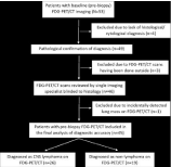
    Figure 1:  Standards for Reporting of Diagnostic Accuracy (STARD)-type
diagram depicting flow of participants through the study.
Twenty-six of 45 patients were diagnosed as having CNS lymphoma on the pre-treatment FDG-PET/CT imaging including 3 patients with synchronously detected systemic lymphoma. Twenty of them were subsequently pathologically confirmed as lymphoma on histology/cytology (true positives). The remaining 6 patients (false positives) were diagnosed as having high-grade glioma (n=3), brain metastasis (n=1), or non-specific inflammation (n=2) on brain biopsy. (Figure 2) is an illustration of a patient diagnosed as PCNSL on baseline FDG-PET/CT, but subsequently proven to be glioblastoma on biopsy (false positive). The histo-pathological characteristics of both patients with non-specific inflammation was not consistent with ‘steroid-treated lymphoma’, hence they were considered as ‘indeterminate’ and kept on close clinico-radiological observation without any active anti-lymphoma treatment. Serial interval imaging showed lack of progression strengthening the diagnosis of ‘non-lymphomatous’ pathology. Nineteen patients were deemed to be non-lymphomas on baseline FDG-PET/CT. Fourteen of these 19 patients were subsequently confirmed as non-lymphomas on histo-pathology (true negatives). The remaining 5 patients turned out to be lymphoma on biopsy/cytology (false negatives). An illustrative example of a patient with no appreciable FDG-uptake on PET/CT which eventually turned out to be PCNSL on CSF cytology and flow cytometry (false negative) is provided in (Figure 3). The sensitivity, specificity, PPV, NPV, and overall accuracy (with 95% CI) of qualitative FDG-PET/CT imaging based on visual interpretation by an imaging specialist in the diagnosis of PCNSL was 77.4% (54.6-92.2%); 70% (45.7-88.1%), 73.9% (58.3-85.2%), 73.7% (55.2- 86.4%), and 73.8% (58-86.1%) respectively (Table 2). Similar metrics in the diagnosis of any CNS lymphoma (including 3 patients with synchronously detected systemic lymphoma) are also depicted in (Table 2).
![]()
    Diagnostic metrics
    PCNSL
    Any CNS lymphoma*


    Sensitivity (95%CI)
    77.4% (54.6-92.2%)
    80.0% (59.3-93.2%)


    Specificity (95% CI)
    70.0% (45.7-88.1%)
    70.0% (45.7-88.1%)


    Positive Predictive Value (95% CI)
    73.9% (58.3-85.2%)
    76.9% (62.4-87.0%)


    Negative Predictive Value (95% CI)
    73.7% (55.2-86.4%)
    73.7% (54.9-86.6%)


    Overall Accuracy (95% CI)
    73.8% (58.0-86.1%)
    75.6% (60.5-87.1%)


    FDG-PET/CT:    Flouro-Deoxy-Glucose Positron Emission Tomography/Computed Tomography; PCNSL:    Primary Central Nervous System Lymphoma; CNS: Central Nervous System; CI:    Confidence Interval
      *Any CNS    lymphoma includes additional 3 patients with synchronously detected systemic    lymphoma.
Table 2: Diagnostic performance of baseline FDG-PET/CT in patients with
suspected PCNSL.
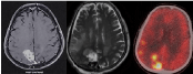
    Figure 2:  Axial post-contrast T1-weighted (a) and T2-weighted (b) MRI
showing solid contrast enhancing lesion in the right parietal periventricular
location with smaller satellite nodules (hypointense on T2) in the adjacent
parietal lobe with associated perilesional edema raising a differential
diagnosis of Primary Central Nervous Lymphoma (PCNSL) versus multifocal
glioblastoma. Fused axial FDG-PET/CT image (c) showed intense
focal FDG-uptake (SUVmax=34.3) in the lesion (intense hypermetabolism)
which was interpreted as PCNSL. Biopsy from the lesion supplemented with
immunohistochemistry established the diagnosis of glioblastoma, ruling out
any possibility of lymphoma (false positive).
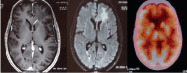
    Figure 3:  Axial post-contrast T1-weighted (a) and T2-FLAIR (b) images
showing an ill-defined enhancing lesion in the left frontal periventricular
region with disproportionate perilesional edema in adjacent brain parenchyma
of uncertain etiology. Fused axial FDG-PET/CT image (c) showed no
appreciable FDG-uptake in the lesion (hypometabolism) suggesting ‘nonlymphoma’.
Malignant cell cytology supplemented with flow-cytometry on
a cerebrospinal fluid sample led to the final diagnosis of primary central
nervous system lymphoma (false negative).
Discussion
Presently, multi-parametric MRI remains the recommended first-line imaging modality in the diagnostic work-up of any suspected CNS lesion including PCNSL [3,4]. Although PCNSL has some classical morphologic features on conventional neuroimaging [4], none of them are pathognomonic and often overlap with other common entities (high-grade glioma, brain metastases, and infective/inflammatory lesions). In addition, several patients present with atypical imaging findings (mild patchy enhancement or nonenhancement, cortical location, and diffuse infiltration) rendering it further difficult to reliably differentiate it from other CNS lesions. Given its varied morphology, imaging diagnosis of PCNSL can be quite difficult and challenging [5,6].
Recent advancements in imaging technology allow noninvasive assessment of tumor micro-environment such as hypoxia, angiogenesis, cellularity, proliferation, and metabolism. FDG-PET/ CT that evaluates lesions on the basis of glucose metabolism is the most widely used functional imaging modality for diagnosis, staging, response assessment, and surveillance in contemporary clinical oncology practice. Tumor tissues in high-grade lymphomas including CNS lymphoma have high cellular density with accelerated glucose metabolism resulting in significantly high FDG-avidity and concentration. A growing body of evidence supports the use of FDGPET/ CT in systemic lymphoma for staging at initial diagnosis, early interim response assessment after 1-2 cycles of chemotherapy, and post-treatment monitoring following completion of therapy [7,8]. However, there is sparse and conflicting data on the diagnostic performance of baseline FDG-PET/CT in patients with suspected PCNSL.
Many studies [9-12] have reported good diagnostic utility of FDG-PET/CT in differentiating PCNSL from morphologically similar lesions on conventional neuro-imaging such as highgrade gliomas and brain metastases. CNS lymphomas generally demonstrate high FDG-avidity with a mean SUVmax about 2.5 times higher than the average uptake in the normal uninvolved gray matter. Some studies [13-15] have even provided Receiver Operating Characteristics (ROC)-defined cut-offs of SUVmax and Tumor/ Normal Tissue (T/N) ratio with acceptably high accuracy in the diagnosis of PCNSL. However, few authors [16,17] have reported substantial overlap of SUVmax and T/N ratios between PCNSL and other non-lymphomatous CNS lesions questioning its incremental value in the diagnostic evaluation of patients with suspected PCNSL. Morphological appearance of CNS lymphoma appears to be an important parameter in its detection on FDG-PET/CT. Patients with typical radiological findings on MRI show strong FDG-avidity in most cases; however, PCNSL with atypical findings on MRI such as non-enhancing lesions or diffuse infiltration show low detection rates on FDG-PET/CT [18].
Given the low incidence of PCNSL, prior studies have been retrospective with small sample size leading to weak inferences. To overcome some of these limitations, Zou et al [19], pooled data from 8 studies involving 129 patients in a systematic review and meta-analysis of the diagnostic value of FDG-PET/CT in immunocompetent adults with PCNSL. The pooled sensitivity and specificity of FDG-PET/ CT in the diagnosis of PCNSL was 0.88 (95% CI=0.80-0.94) and 0.86 (95% CI=0.73-0.94) respectively. Similarly, the pooled Positive Likelihood Ratio (PLR) was 3.99 (95% CI=2.31-6.90) and Negative Likelihood Ratio (NLR) was 0.11 (95% CI=0.04-0.32). The pooled Diagnostic Odds Ratio (DOR) was 33.40 (95% CI=10.40-107.3). Area Under the Curve (AUC) of the summary ROC curve was 0.92 and Q index (where sensitivity equals specificity) was 0.85 respectively, suggesting high diagnostic accuracy of FDG-PET/CT.
The performance of qualitative FDG-PET/CT imaging for the diagnosis of CNS lymphoma in our study though acceptable was somewhat lesser than previously reported [19]. Neuro-surgical biopsy still remains the gold-standard in the diagnosis of PCNSL and cannot be replaced by MRI or FDG-PET/CT either singly or in combination. However, FDG-PET/CT can be considered to have good diagnostic utility as an imaging modality [20] adjunctive to MRI in cases where histo-pathological diagnosis is either not possible or considered risky due to deep-seated eloquent location of the lesion and/or patient’s poor performance status.
Strengths and limitations: The major strength of our study is its prospective nature, relatively large sample size compared to smaller studies reported previously, and sound methodology. All FDG-PET/ CT scans were reviewed by a single dedicated imaging specialist blinded to the histo-pathological diagnosis. Patients where histopathology was deemed ‘indeterminate’ on brain biopsy were also included in the analysis as false positives to give a more conservative estimate of diagnostic accuracy. Despite being the only prospective study till date, it was associated with certain caveats and limitations. Our calculation of diagnostic performance was case-based and not a lesion-based analysis. We did not assess the confounding effect, if any, of pre-imaging and pre-biopsy corticosteroids on interpretation and diagnosis. Imaging diagnosis was primarily based on qualitative PET features without using any semi-quantitative parameters like SUVmax or T/N ratios, that could have further improved the accuracy. Finally, we included only patients who had a clinico-radiological suspicion of PCNSL on conventional neuro-imaging which could have potentially introduced some selection bias.
Conclusion
Baseline FDG-PET/CT has reasonably good and acceptable diagnostic accuracy (adjunctive to MRI) in suspected PCNSL, particularly in patients with deep-seated lesions not amenable to a safe neuro-surgical biopsy which still remains the gold-standard for the diagnosis of PCNSL.
References
- Rubenstein J, Ferreri AJ, Pittaluga S. Primary lymphoma of the central nervous system: epidemiology, pathology and current approaches to diagnosis, prognosis and treatment. Leuk Lymphoma. 2008; 49: 43-51.
- Melani C, Roschewski M, Wilson WH. Advances in the Treatment of Primary CNS Lymphoma. Clin Lymphoma Myeloma Leuk. 2018; 18: S106-S109.
- Buhring U, Herrlinger U, Krings T, Thiex R, Weller M, Kuker W. MRI features of primary central nervous system lymphomas at presentation. Neurology. 2001; 57: 393-396.
- Partovi S, Karimi S, Lyo JK, Esmaeili A, Tan J, DeAngelis LM. Multimodality imaging of primary CNS lumphoma in immunocompetent patients. Br J Radiol. 2014; 87: 20130684.
- Baraniskin A, Deckert M, Schulte-Altedorneburg G, Schlegel U, & Schroers R. Current strategies in the diagnosis of diffuse large B-cell lymphoma of the central nervous system. Br J Hematol. 2011; 156: 421-432.
- Chiavazza C, Pellerino A, Ferrio F, Cistaro A, Soffietti R, Ruda R. Primary CNS Lymphomas: Challenges in Diagnosis and Monitoring. Biomed Res Int. 2018; 3606970.
- Kostakoglu L. 18F-Flourodeoxyglucose Positron Emission Tomography in the Management of Lymphomas. Clin Lymphoma Myeloma Leuk. 2008; 8: 273-276.
- Cronin CG, Swords R, Truong MT, Vishwanathan C, Rohren E, Giles FJ, et al. Clinical utility of PET/CT in lymphoma. AJR Am J Roentgenol. 2010; 194: W91-W103.
- Karantanis D, O’Eill BP, Subramaniam RM, Witte RJ, Mullan BP, Nathan MA, et al. 18F-FDG PET/CT in primary central nervous system lymphoma in HIVnegative patients. Nucl Med Commun. 2007; 28: 834-841.
- Makino K, Hirai T, Nakamura H, Murakami R, Kitajima M, Shigematsu Y, et al. Does adding FDG-PET to MRI improve the differentiation between primary cerebral lymphoma and glioblastoma?. Observer performance study. Ann Nucl Med. 2011; 25: 432-438.
- Das K, Mittal BR, Vasistha RK, Singh P, Mathuriya SN. Role of 18F-fluorodeoxyglucose Positron Emission Tomography scan in differentiating enhancing brain tumors. Indian J Nucl Med. 2011; 26: 171-176.
- De-Bonilla-Damia A, Fernandez-Lopez R, Capote-Huelva FJ, de la Cruz- Vicente F, Egea-Guerrero JJ, Borrego-Dorado I. Role of 18F-FDG PET/CT in primary brain lymphoma. Rev Esp Med Nucl Imagen Mol. 2017; 36: 298-303.
- Purandare NC, Puranik A, Shah S, Agrawal A, Gupta T, Moiyadi A, et al. Common malignant brain tumors: can 18F-FDG PET/CT aid in differentiation? Nucl Med Commun. 2017; 38; 1109-1116.
- Gupta T, Gupta M, Rangarajan V, Purandare N, Sastri G, Menon H, et al. Clinical utility of FDG-PET/CT for diagnosis and staging in patients with primary central nervous system lymphoma: prospective single arm study. Neuro-Oncol. 2017; 19: iii107–iii108.
- Zhou W, Wen J, Hua F, Xu W, Lu X, Lin B, et al. 18F-FDG PET/CT in immunocompetent patients with primary central nervous system lymphoma: differentiation from glioblastoma and correlation with DWI. Eur J Radiol 2018; 104: 26-32.
- Rosenfeld SS, Hoffman JM, Coleman RE, Glantz MJ, Hanson MW, Schold SC. Studies of primary central nervous system lymphoma with fluorine-18- fluorodeoxyglucose positron emission tomography. J Nucl Med. 1992; 33: 532-536.
- Kawai N, Zhen HN, Miyake K, Yamamaoto Y, Nishiyama Y, Tamiya T. Prognostic value of pretreatment 18F-FDG PET in patients with primary central nervous system lymphoma: SUV-based assessment. J Neuro-Oncol. 2010; 100: 225-232.
- Albano D, Bosio G, Bertoli M, Giubbini R, Bertagna F. 18F-FDG PET/CT in primary brain lymphoma. J Neuro-Oncol. 2018; 136: 577-583.
- Zou Y, Tong J, Leng H, Jiang J, Pan M, Chen Z. Diagnostic value of using 18F-FDG PET and PET/CT in immunocompetent patients with primary central nervous system lymphoma: A systematic review and meta-analysis. Oncotarget. 2017; 8: 41518-41528.
- Yamaguchi S, Hirata K, Kaneko S, Kobayashi H, Shiga T, Kobayashi K, et al. Combined use of 18F-FDG PET and corticisteroid for diagnosis of deepseated primary central nervous system lymphoma without histopathological confirmation. Acta Neurochir (Wien). 2015; 157: 187-194.
Citation: Gupta T, Gupta M, Purandare N, Puranik A, Rangarajan V, Moiyadi A, et al. Diagnostic Performance of Baseline 18F-Fluoro-Deoxy-Glucose Positron Emission Tomography/Computed Tomography in Patients with Clinico-Radiological Suspicion of Primary Central Nervous System Lymphoma. Ann Hematol Oncol. 2019; 6(1): 1228.